-
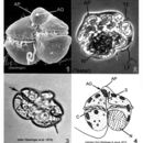
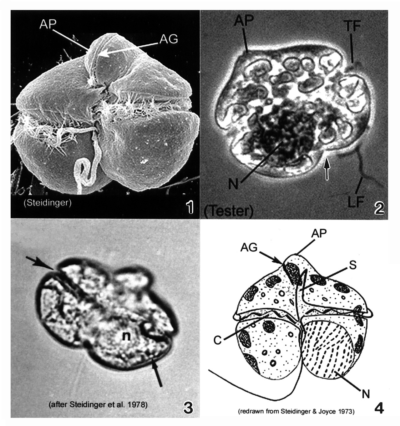
Plate 22. Gymnodinium breve. Fig. 1. SEM: ventral view. Cell small, wider than long, dorso-ventrally flattened. Cell nearly square in outline; prominent apical process (AP) directed ventrally. Apical groove (AG) present on apical process, adjacent to sulcus. Figs. 2-3. LM. Fig. 2. Dorsal view: large nucleus (N) in hypotheca. Transverse (TF) and longitudinal (LF) flagella present. Hypotheca bilobed (arrow). Fig. 3. Ventral view: displaced cingulum (large arrow) and lipid globule (small arrow). Fig. 4. Line drawing. Cingulum (C) displaced, descending. Long sulcus (S) extends to apex of cell.
-
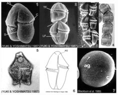
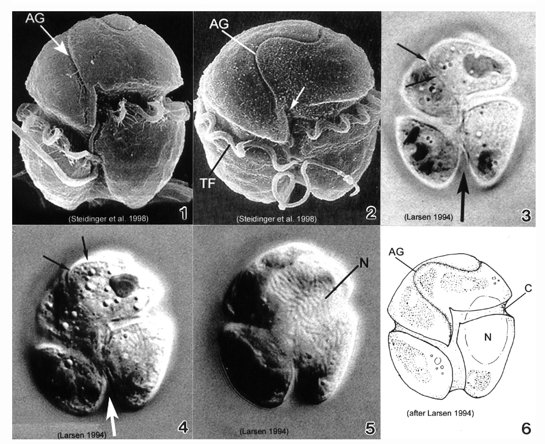
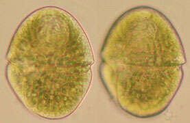
Plate 23. Gymnodinium catenatum. Figs. 1-3. SEM: ventral view. Fig. 1. Cell small, elongate-ovoid with slight dorso-ventral compression. Conical apex; rounded and notched antapex. Cingulum (C) excavated; sulcus (S) long. Distinctive horse-shoe shaped apical groove (AG) encircles apex. Fig. 2. Two cell chain; attachment point visible (arrow). Premedian cingulum displaced 2X its width. Longitudinal (LF) and transverse (TF) flagella visible. Fig. 3. Chain cells with anterior-posterior compression. Terminal cell slightly longer. Thecal surface rugose to smooth (Blackburn et al. 1989). Figs. 4-5. LM. Fig. 4. Chain-formation (Yuki and Yoshimatsu 1987). Fig. 5. Single cell. Conical epitheca with concave to flat apex. Bilobed hypotheca (arrow). Fig. 6. Line drawing. Fig. 7. SEM: cyst with microreticulations. ag=apical groove; c=cingulum
-
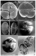
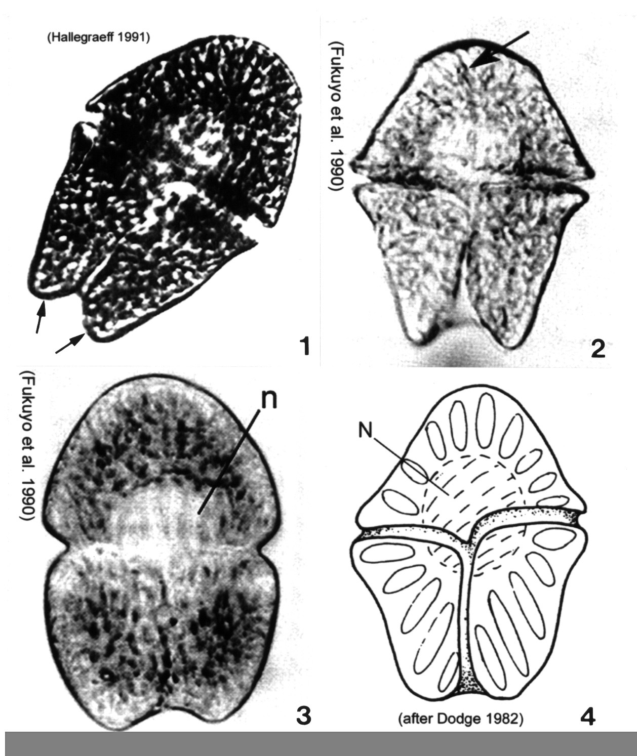
Plate 24. Gymnodinium mikimotoi. Figs. 1-4. SEM. Fig. 1. Ventral view: cell small, broadly oval to almost round. Epitheca slightly smaller than hypotheca. Characteristic straight apical groove (AG). Cingulum (C) deep, displaced 2 times its width. Sulcus (S) slightly invades epitheca (arrowheads). Hypotheca notched by widening sulcus (arrow). Fig. 2. Dorsal view: apical groove extends to dorsal side of epitheca creating slight indentation at the apex (arrowhead). Hypotheca bilobed (arrow). Fig. 3. Apical view of apical groove (arrow)(after Fukuyo et al.). Fig. 4. Cell compressed dorso-ventrally (after Fukuyo et al.). Figs. 5-7. LM. Fig. 5. Cingulum displaced 2 times its width (arrows)(from Larsen & Moestrup 1989: fig. 16g). Fig. 6. Large nucleus (N) in left lobe of hypotheca. Fig. 7. Vegetative division. Division plane oblique.
-
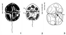
Plate 25. Gymnodinium pulchellum. Figs. 1-2. SEM: ventral view. Fig. 1. Cell small and broadly oval. Cingulum wide, displaced 1-1.5 X its width. Deeply excavated sulcus creates lobed hypotheca. Conspicuous undulating apical groove (AG). Fig. 2. Well-developed apical groove: reverse S-shape. Transverse flagellum (TF) housed in cingulum. Sulcus slightly invades epitheca with finger-like projection (arrow). Figs. 3-5. LM: ventral view. Figs. 3-4. Apical groove distinguishable (small arrows). Chloroplasts and pyrenoids present. Lobed hypotheca (large arrow). Fig. 5. Large elliptical nucleus (N) in left central part of cell. Fig. 6. Line drawing. C=cingulum
-
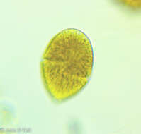
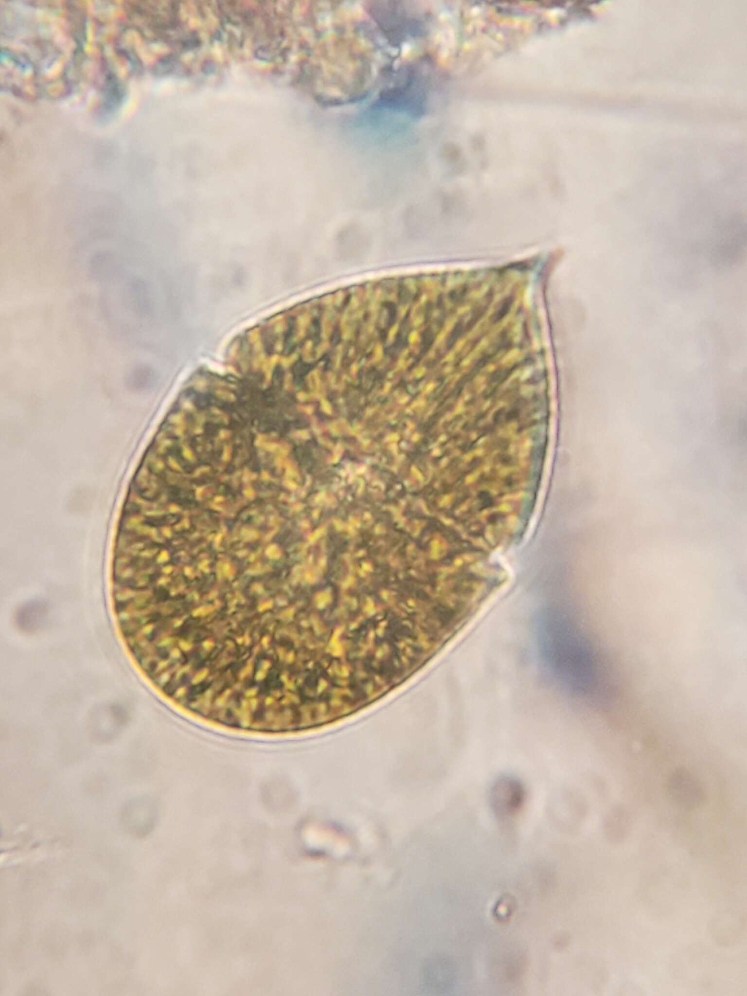
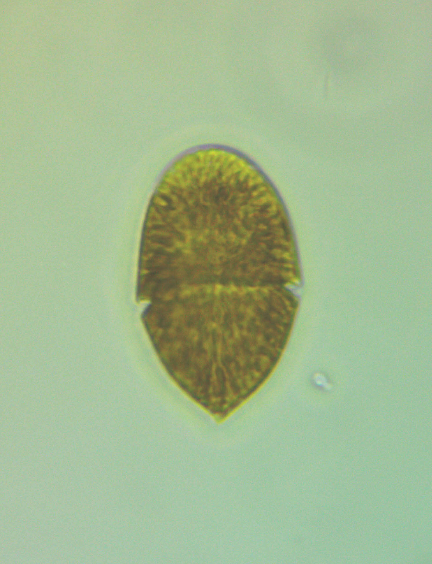
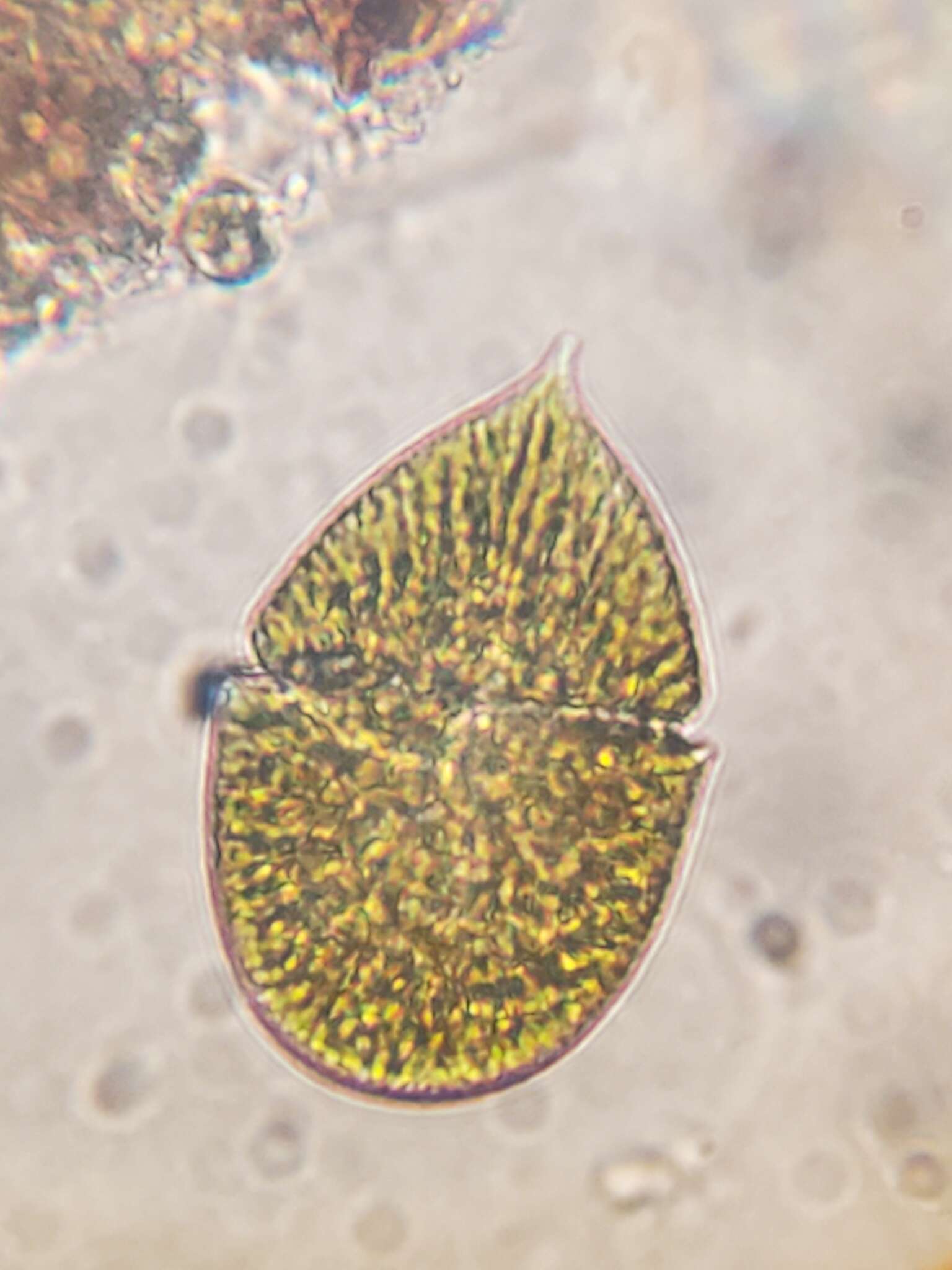
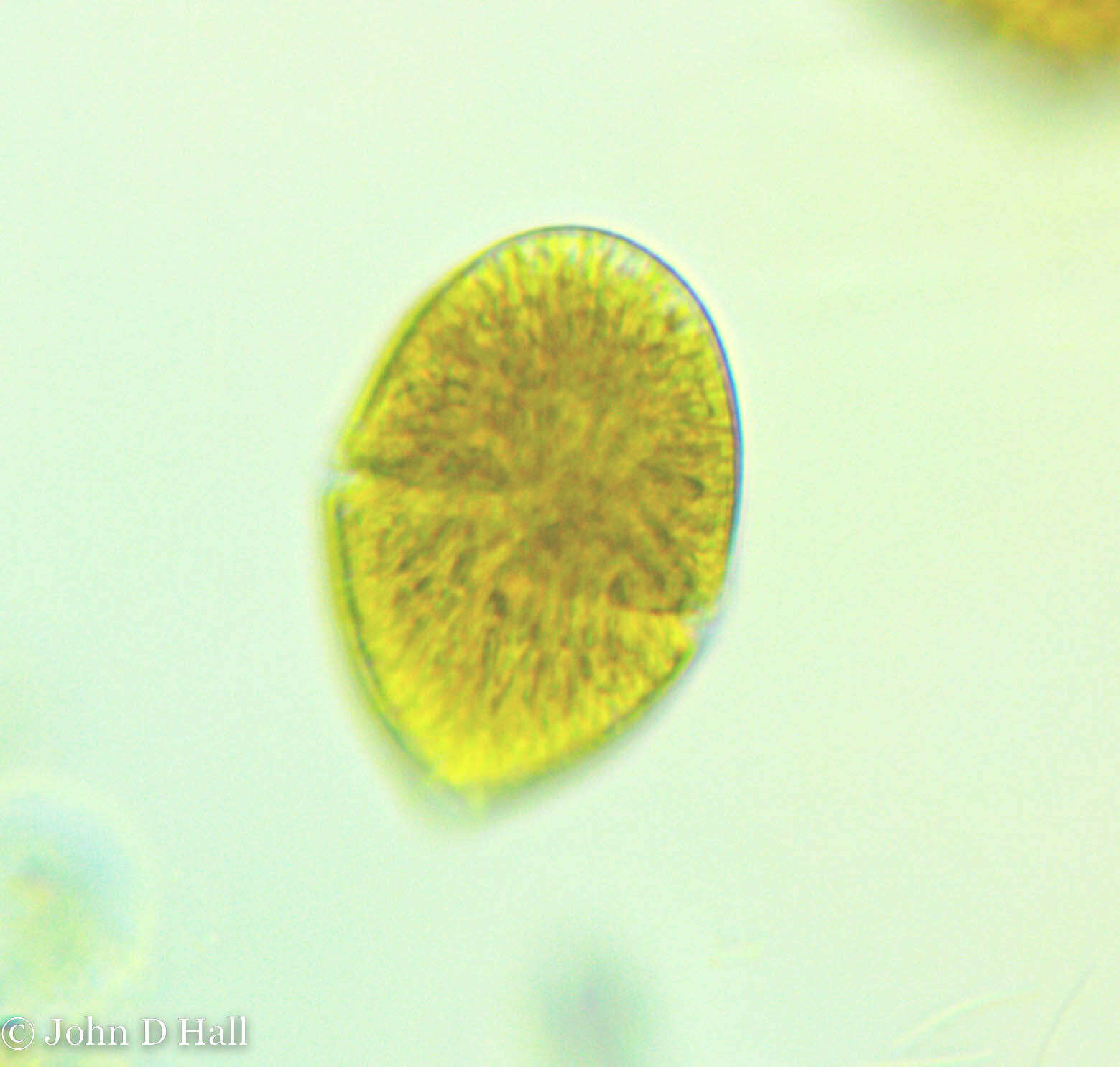
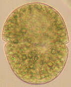
Plate 26. Gymnodinium sanguineum. Figs. 1-3. LM. Cell large, pentagonal, and slightly dorso-ventrally flattened. Cells vary in shape and size. Fig. 1. Ventral view. Epitheca and hypotheca nearly equal in size: epitheca conical, hypotheca bilobed (arrows). Fig. 2. Ventral view. Deep cingulum median, displaced 1-2 times its width. Sulcus deeply notches hypotheca. Apical groove present (arrow). Fig. 3. Cell deeply pigmented; central nucleus (n). Fig. 4. Line drawing. Spindle-shaped chloroplasts radially arranged.
-
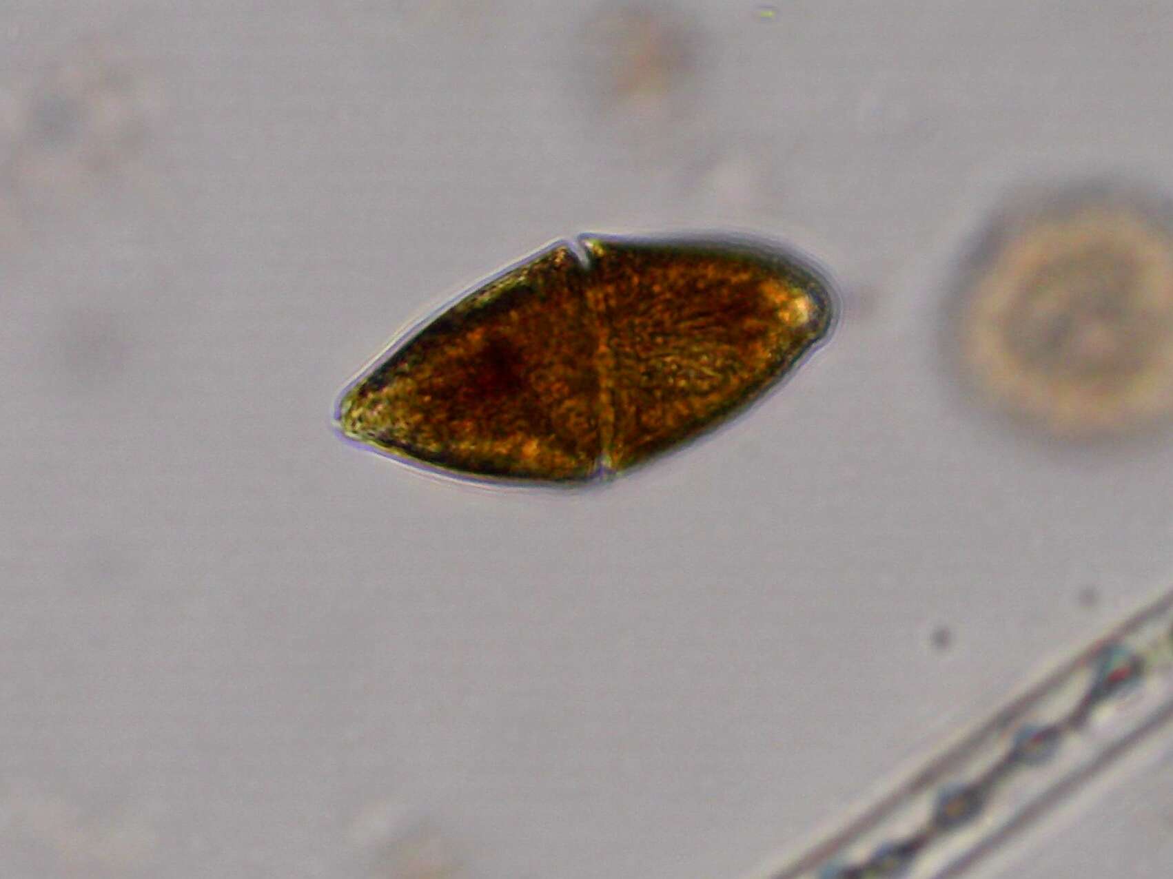
Plate 27. Gymnodinium veneficum. Figs. 1-3. Line drawings. Fig. 1. Ventral view: cell small and ovoid. Epitheca slightly pointed, without apical groove. Cingulum deep and displaced 1-2 times its width. Fig. 2. Dorsal view: large central nucleus (N). Two to eight irregular chloroplasts present (C). Fig. 3. Sigmoid sulcus slightly invades epitheca (arrow).
-
-
-
-
-
-
-
-
-
[taxonomy:genus=Gymnodinium]
Date:
23 Aug 2011
Location:
Small lake in Kent Ridge Park. Water margin with vegetation, brown sediment with organic detritus. Tadpoles resting nearby.
Microscope:
Bright-field with closed condenser aperture.
Camera:
Nikon D7000
Collector:
Brandon Seah
Scale:
20830 pixels/mm = 20.8 pixels/µm
-
Description:
Gymnodinium Stein 1883 sp. (agile?); Gymnodiniaceae, Gymnodiniales, Dinophyceae, Dinoflagellata (Dinophyta) English: North-West
Black Sea, coastal waters, at a depth of 0.5 metre Русский: Северо-Запад
Чёрного моря, прибрежные воды, на глубине 0,5 м. Date: 5 August 2007. Source: Own work. Author:
Minami Himemiya.
-
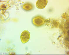
Longitude (deg): -1.2. Latitude (deg): 51.4. Longitude (deg/min): 1° 10' W. Latitude (deg/min): 51° 20' N. Vice county name: Berks. Vice county no.: 22. Country: England. Identified by: Malcolm Storey. Comment: in puddle. Category: microscope photograph. Photographic equipment used: Pixera Pro tethered low-resolution digital camera with Meiji microscope using CS adaptor and x.7 projection eye-piece.
-
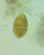
Longitude (deg): -1.2. Latitude (deg): 51.4. Longitude (deg/min): 1° 10' W. Latitude (deg/min): 51° 20' N. Vice county name: Berks. Vice county no.: 22. Country: England. Identified by: Malcolm Storey. Comment: in puddle. Detail to note: flagellum toward bottom right (faint). Category: microscope photograph. Photographic equipment used: Pixera Pro tethered low-resolution digital camera with Meiji microscope using CS adaptor and x.7 projection eye-piece.
-
Longitude (deg): -1.2. Latitude (deg): 51.4. Longitude (deg/min): 1° 10' W. Latitude (deg/min): 51° 20' N. Vice county name: Berks. Vice county no.: 22. Country: England. Identified by: Malcolm Storey. Comment: in deep puddle. Category: microscope photograph. Photographic equipment used: Pixera Pro tethered low-resolution digital camera with Meiji microscope using CS adaptor and x.7 projection eye-piece.
-
Longitude (deg): -0.7. Latitude (deg): 51.3. Longitude (deg/min): 0° 50' W. Latitude (deg/min): 51° 20' N. Vice county name: Berks. Vice county no.: 22. Country: England. Associated species: Sphagnum. Identified by: Malcolm Storey. Comment: in Sphagnum in bog pool. Category: microscope photograph. Photographic equipment used: Pixera Pro tethered low-resolution digital camera with Meiji microscope using CS adaptor and x.7 projection eye-piece.
-
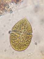
Longitude (deg): -0.7. Latitude (deg): 51.3. Longitude (deg/min): 0° 50' W. Latitude (deg/min): 51° 20' N. Vice county name: Berks. Vice county no.: 22. Country: England. Associated species: Sphagnum. Identified by: Malcolm Storey. Comment: in Sphagnum in bog pool. Detail to note: flagellum on right view. Category: microscope photograph. Photographic equipment used: Pixera Pro tethered low-resolution digital camera with Meiji microscope using CS adaptor and x.7 projection eye-piece.
-
Longitude (deg): -0.7. Latitude (deg): 51.3. Longitude (deg/min): 0° 50' W. Latitude (deg/min): 51° 20' N. Vice county name: Berks. Vice county no.: 22. Country: England. Associated species: Sphagnum. Identified by: Malcolm Storey. Comment: in Sphagnum in bog pool. Category: microscope photograph. Photographic equipment used: Pixera Pro tethered low-resolution digital camera with Meiji microscope using CS adaptor and x.7 projection eye-piece.
-
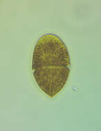
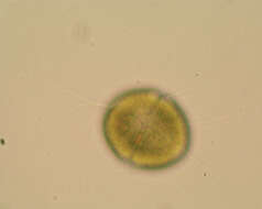
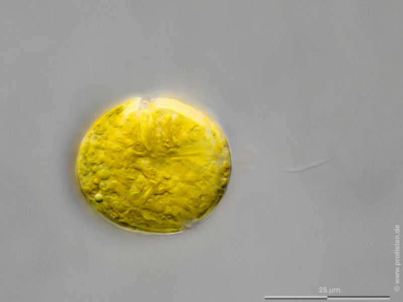
Gymnodinium spec. Scale bar indicates 25 µm. The specimen was gathered in a tiny freshwater pond called Suploch on the island of Hiddensee (Baltic Sea, Germany). Sampling date 9/2022. The image was built up using several photomicrographic frames with manual stacking technique. Images were taken using Zeiss Standard with Olympus OM-D M5 MKII. Image under Creative Commons License V 3.0 (CC BY-NC-ND). Place name: Pond Suploch, Hiddensee (Germany) Latitude: 54.538638 Longitude: 13.097802 Multiebenen-Abbildung, manuell gestapelt. Der Messbalken markiert eine Länge von 25 µm. > Probe aus dem Suploch, einem kleinen Süßwasserteich auf der Insel Hiddensee. Datum der Aufsammlung: 9/2022. Mikrotechnik: Zeiss Standard, Kamera: Olympus OM-D M5 MKII. Creative Commons License V 3.0 (CC BY-NC-ND). For permission to use of (high-resolution) images please contact postmaster@protisten.de.