-
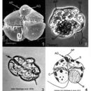
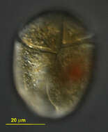
Gymnodinium fuscum,. common freshwater dinoflagellates, with greatly folded radiating plastid, red granules near middle or in posterior half of the cell. With equatorial groove - the cingulum more or less running around the middle of the cell, the longitudinal groove - the sulcus - not strongly developed. This is the type species of the genus Gymnodinium. Differential interference contrast.
-
Gymnodinium fuscum,. common freshwater dinoflagellates, with greatly folded radiating plastid, red granules near middle or in posterior half of the cell. This image focusses on the surface to show the equatorial groove - the cingulum, and the longitudinal groove - the sulcus. This is the type species of the genus Gymnodinium. Differential interference contrast.
-
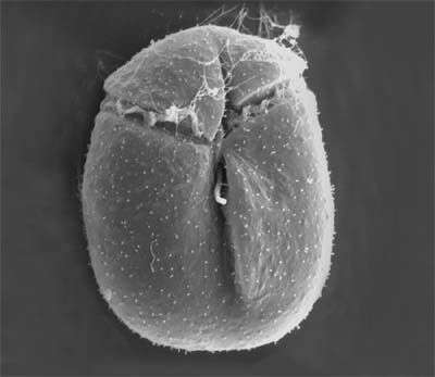
Cells oval from the ventral side, dorso-ventrally flattened. Length 20 - 28 microns, width 10 - 20 microns, length to width ratio 1.3 - 2.0. Epicone triangular, curved anteriorly, deflected to the left. Cingulum beginning 0.2 of the cell length from the apex, distal end 2 - 4 microns below and to the right of the proximal. Sulcus beginning just to the right of the mid-ventral line, initially deep and wide (2 - 3 microns), becoming less distinct as it nears the posterior of the cell. Two pusules present, each approximately 1 microns diameter, one below the origin of the cingulum, the to the right of the origin of the sulcus. Longitudinal flagellum arising in a pocket just to the left of and below the origin of the sulcus. Nucleus in the posterior part of the hypocone, round to oval, approximately 10 microns diameter. Plastids yellow-brown, in strands radiating from the central pyrenoid, 4 -5 microns diameter . Asexual division occurring in hyaline covered cysts, in which either two or three daughter cells may be formed.
-
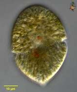
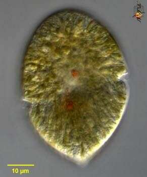
A gymnodinioid dinoflagelate, identified by Shauna Murray, isolated from sandy sediments and photographed by Bob Moore and Dan Lahr. The nucleus with very dense genetic material is very evident in his picture.
-
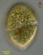
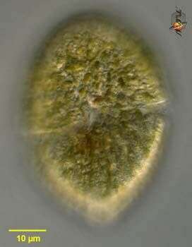
A optical slice showing some of the grooves in this gymnodinioid dino. Identifyed by Shauna Murray, Isolated and photographed by Bob Moore and Dan Lahr.
-
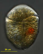
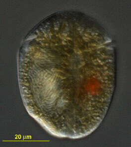
Evident grooves on this vicious predator. Identified by Dr. Murray, isolated by Bob Moore, picture by Dan Lahr.