-
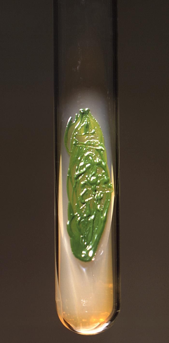
This 1971 image depicted a Sabourauds dextrose agar slant culture, which had cultivated a colony of Chlorella sp. algal organisms.Created: 1971
-
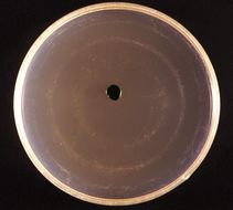
This 1971 image depicted a frontal view of a Petri dish culture in which a small coloney of Chlorella algal organisms had been cultivated.Created: 1971
-
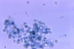
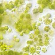
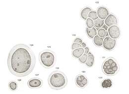
At a magnification of 1125X, this photomicrograph revealed the presence of a number of Chlorella sp. algal organisms.Created: 1971
-
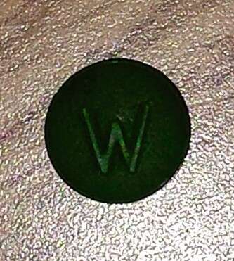
Description: English: Chlorella in pill form. Date: 28 February 2015, 16:53:28. Source: Own work. Author:
Alexsautographs.
-
Description: English: Chlorella in pill form. Date: 28 February 2015, 17:01:31. Source: Own work. Author:
Alexsautographs.
-
Description: Nederlands: C.sorokiniana. Date: 9 September 2020. Source: Own work. Author:
Garnhami.
-
-
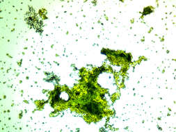
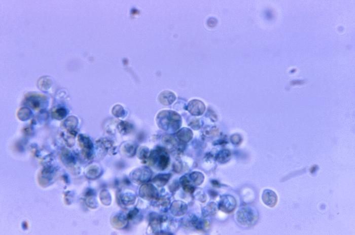
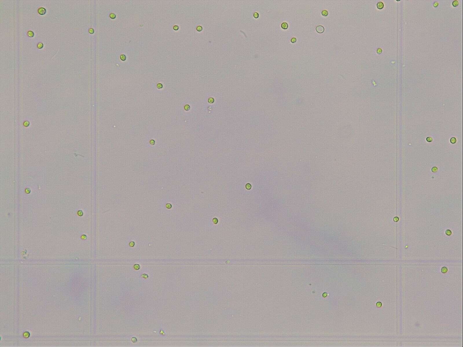
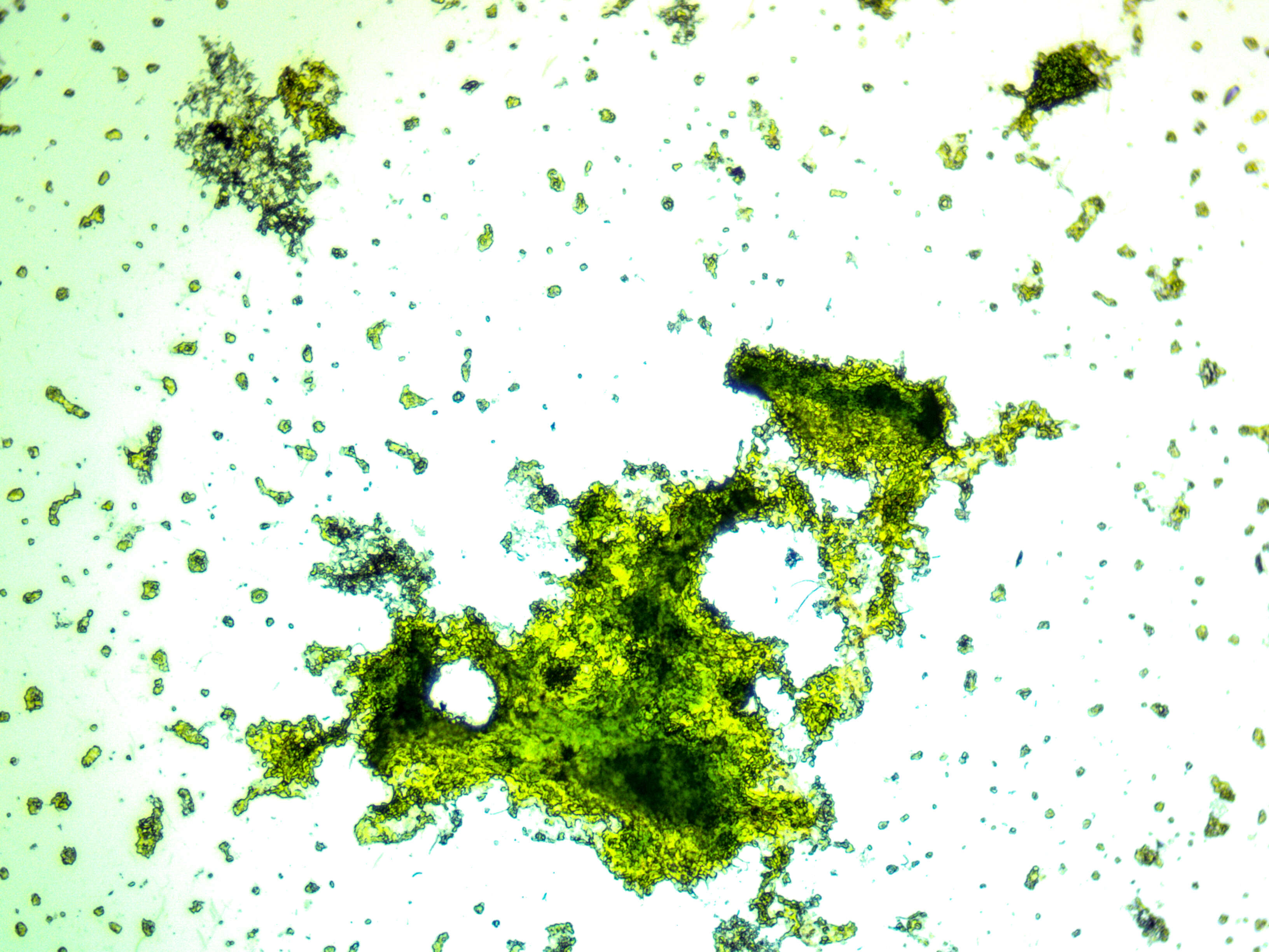
Description: English: Chlorella in culture with cells showing numerous daughter cells indicating rapid growth of these cells under light microscopy. Date: 14 January 2013. Source: United States Geologic Survey. Author: Barry H. Rosen.
-
Blake T. Hovde, Seth A. Steichen, Shawn R. Starkenburg, Judith K. Brown
Wikimedia Commons
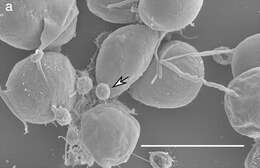
Description: English: Scanning Electron Micrograph of Chlorella sorokiniana and attached Vampirovibrio chlorellavorus cells. Image of a sample collected from Arizona test site at 10 000 × magnification with V. chlorellavorus indicated by the white arrow.Scale bar is displayed in white representing 5.0 μm. Date: 17 July 2019. Source: Fig. 1a at
https://onlinelibrary.wiley.com/doi/10.1111/pre.12392 Vampirovibrio chlorellavorus draft genome sequence, annotation, and preliminary characterization of pathogenicity determinants Phycological Research Vol. 68, No. 1 p. 23-29,
doi:10.1111/pre.12392 . Author: Blake T. Hovde, Seth A. Steichen, Shawn R. Starkenburg, Judith K. Brown. Other versions:.
-
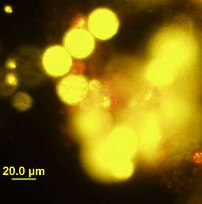
Description: English: Chlorella in culture with cells showing numerous daughter cells indicating rapid growth of these cells - under epifluorescence microscopy, with the red color indicative of chlorophyll. Date: 14 January 2013. Source: United States Geological Survey. Author: Barry H. Rosen.
-
-
-
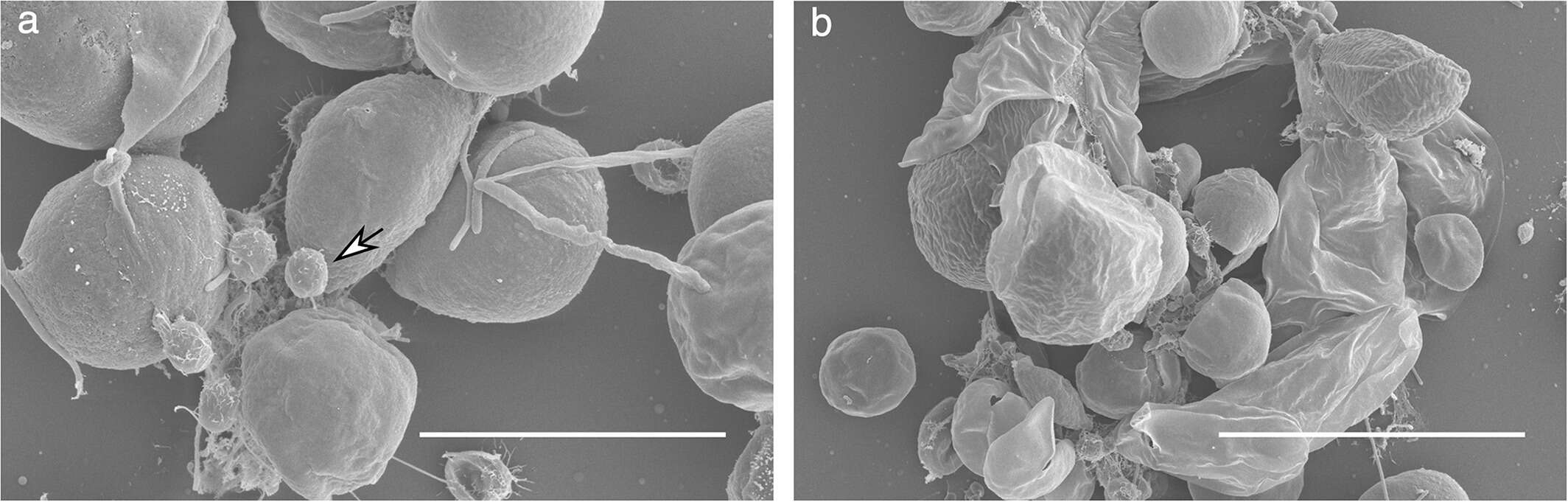
Blake T. Hovde, Seth A. Steichen, Shawn R. Starkenburg, Judith K. Brown
Wikimedia Commons
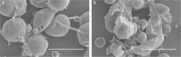
Description: English: Scanning Electron Micrographs of Chlorella sorokiniana and attached Vampirovibrio chlorellavorus cells. Images of samples collected from Arizona test site at (a) 10 000 × magnification with V. chlorellavorus indicated by the white arrow and (b) 5000 × magnification.Scale bars are displayed in white representing (a) 5.0 μm and (b) 10.0 μm. Date: 17 July 2019. Source: Fig. 1 at
https://onlinelibrary.wiley.com/doi/10.1111/pre.12392 Vampirovibrio chlorellavorus draft genome sequence, annotation, and preliminary characterization of pathogenicity determinants Phycological Research Vol. 68, No. 1 p. 23-29,
doi:10.1111/pre.12392 . Author: Blake T. Hovde, Seth A. Steichen, Shawn R. Starkenburg, Judith K. Brown. Other versions:
This file has an extracted image:
File:PhycRes-pre12392-fig-0001a-m-Vampirovibrio-chlorellavorus.jpg.
.
-
-
-
Description: English: Chlorella vulgaris grown on the walls of the bottle for watering domestic flowers Русский: Хлорелла обыкновенная выращенная на стенках бутылки для полива домашних цветовEnglish: Photo taken on Levenhuk 320 Plus with m800 plus camera. Date: 25 April 2021, 13:53:50. Source: Own work. Author:
Shipelin.
-
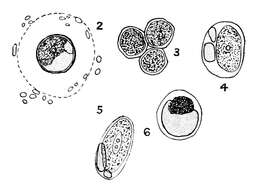
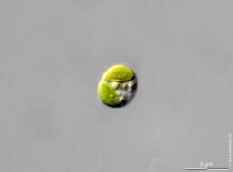
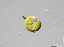
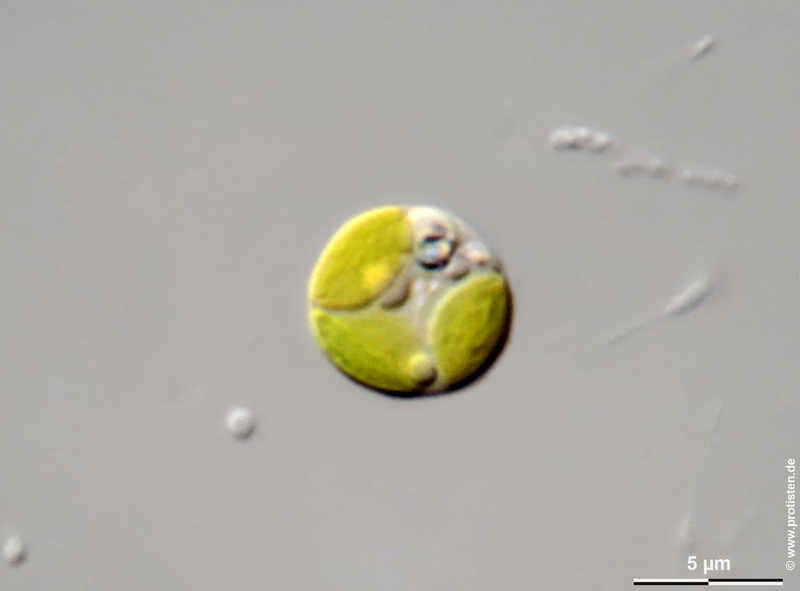
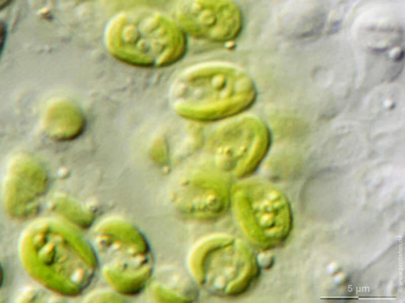
Chlorella spec. Chlorella as symbionts in Hydra viridissima. Nucleus, cup shaped chloroplast, oil droplet. Please press the MORE button for skipping to the annotated version. Scale bar indicates 5 µm. Sample from a tropical freshwater aquarium. Sampling date 1/2023. Images were taken using Zeiss Axioplan with Olympus OM-D M5 MKII. Image under Creative Commons License V 3.0 (CC BY-NC-SA). Place name: Tropical freshwater aquarium Latitude: 54.3018013 Longitude: 10.07120132 Chlorella als Symbionten in Hydra viridissima. Zellkern, becherförmiger Chloroplast, Öltröpfchen. Bitte drücken Sie die Schaltfläche MORE, um zur kommentierten Version zu gelangen. Der Messbalken markiert eine Länge von 5 µm. Probe aus einem Süßwasseraquarium. Datum der Aufsammlung: 1/2023. Mikrotechnik: Zeiss Axioplan, Kamera: Olympus OM-D M5 MKII. Creative Commons License V 3.0 (CC BY-NC-SA). For permission to use of (high-resolution) images please contact postmaster@protisten.de.
-
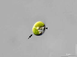
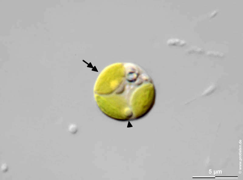
Chlorella spec. Chlorella as symbionts in Hydra viridissima. Nucleus (arrow), cup shaped chloroplast (double headed arrow), oil droplet (arrowhead). Scale bar indicates 5 µm. Sample from a tropical freshwater aquarium. Sampling date 1/2023. Images were taken using Zeiss Axioplan with Olympus OM-D M5 MKII. Image under Creative Commons License V 3.0 (CC BY-NC-SA). Place name: Tropical freshwater aquarium Latitude: 54.3018013 Longitude: 10.07120132 Chlorella als Symbionten in Hydra viridissima. Zellkern (Pfeil), becherförmiger Chloroplast (Pfeil mit Doppelkopf), Öltröpfchen (Pfeilkopf). Der Messbalken markiert eine Länge von 5 µm. Probe aus einem Süßwasseraquarium. Datum der Aufsammlung: 1/2023. Mikrotechnik: Zeiss Axioplan, Kamera: Olympus OM-D M5 MKII. Creative Commons License V 3.0 (CC BY-NC-SA). For permission to use of (high-resolution) images please contact postmaster@protisten.de.
-
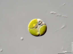
Chlorella spec. Chlorella as symbionts in Hydra viridissima. Nucleus, cup shaped chloroplast, oil droplet. Please press the MORE button for skipping to the annotated version. Scale bar indicates 5 µm. Sample from a tropical freshwater aquarium. Sampling date 1/2023. Images were taken using Zeiss Axioplan with Olympus OM-D M5 MKII. Image under Creative Commons License V 3.0 (CC BY-NC-SA). Place name: Tropical freshwater aquarium Latitude: 54.3018013 Longitude: 10.07120132 Chlorella als Symbionten in Hydra viridissima. Zellkern, becherförmiger Chloroplast, Öltröpfchen. Bitte drücken Sie die Schaltfläche MORE, um zur kommentierten Version zu gelangen. Der Messbalken markiert eine Länge von 5 µm. Probe aus einem Süßwasseraquarium. Datum der Aufsammlung: 1/2023. Mikrotechnik: Zeiss Axioplan, Kamera: Olympus OM-D M5 MKII. Creative Commons License V 3.0 (CC BY-NC-SA). For permission to use of (high-resolution) images please contact postmaster@protisten.de.
-
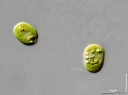
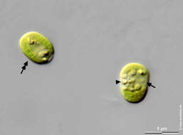
Chlorella spec. Chlorella as symbionts in Hydra viridissima. Nucleus (arrow), cup shaped chloroplast (double headed arrow), oil droplet (arrowhead). Scale bar indicates 5 µm. Sample from a tropical freshwater aquarium. Sampling date 1/2023. Images were taken using Zeiss Axioplan with Olympus OM-D M5 MKII. Image under Creative Commons License V 3.0 (CC BY-NC-SA). Place name: Tropical freshwater aquarium Latitude: 54.3018013 Longitude: 10.07120132 Chlorella als Symbionten in Hydra viridissima. Zellkern (Pfeil), becherförmiger Chloroplast (Pfeil mit Doppelkopf), Öltröpfchen (Pfeilkopf). Der Messbalken markiert eine Länge von 5 µm. Probe aus einem Süßwasseraquarium. Datum der Aufsammlung: 1/2023. Mikrotechnik: Zeiss Axioplan, Kamera: Olympus OM-D M5 MKII. Creative Commons License V 3.0 (CC BY-NC-SA). For permission to use of (high-resolution) images please contact postmaster@protisten.de.
-
Chlorella spec. Chlorella as symbionts in Hydra viridissima. Nucleus, cup shaped chloroplast, oil droplet. Please press the MORE button for skipping to the annotated version. Scale bar indicates 5 µm. Sample from a tropical freshwater aquarium. Sampling date 1/2023. Images were taken using Zeiss Axioplan with Olympus OM-D M5 MKII. Image under Creative Commons License V 3.0 (CC BY-NC-SA). Place name: Tropical freshwater aquarium Latitude: 54.3018013 Longitude: 10.07120132 Chlorella als Symbionten in Hydra viridissima. Zellkern, becherförmiger Chloroplast, Öltröpfchen. Bitte drücken Sie die Schaltfläche MORE, um zur kommentierten Version zu gelangen. Der Messbalken markiert eine Länge von 5 µm. Probe aus einem Süßwasseraquarium. Datum der Aufsammlung: 1/2023. Mikrotechnik: Zeiss Axioplan, Kamera: Olympus OM-D M5 MKII. Creative Commons License V 3.0 (CC BY-NC-SA). For permission to use of (high-resolution) images please contact postmaster@protisten.de.
-
Chlorella spec. Chlorella as symbionts in Hydra viridissima. Nucleus (arrow), cup shaped chloroplast (double headed arrow), oil droplet (arrowhead). Scale bar indicates 5 µm. Sample from a tropical freshwater aquarium. Sampling date 1/2023. Images were taken using Zeiss Axioplan with Olympus OM-D M5 MKII. Image under Creative Commons License V 3.0 (CC BY-NC-SA). Place name: Tropical freshwater aquarium Latitude: 54.3018013 Longitude: 10.07120132 Chlorella als Symbionten in Hydra viridissima. Zellkern (Pfeil), becherförmiger Chloroplast (Pfeil mit Doppelkopf), Öltröpfchen (Pfeilkopf). Der Messbalken markiert eine Länge von 5 µm. Probe aus einem Süßwasseraquarium. Datum der Aufsammlung: 1/2023. Mikrotechnik: Zeiss Axioplan, Kamera: Olympus OM-D M5 MKII. Creative Commons License V 3.0 (CC BY-NC-SA). For permission to use of (high-resolution) images please contact postmaster@protisten.de.


