-
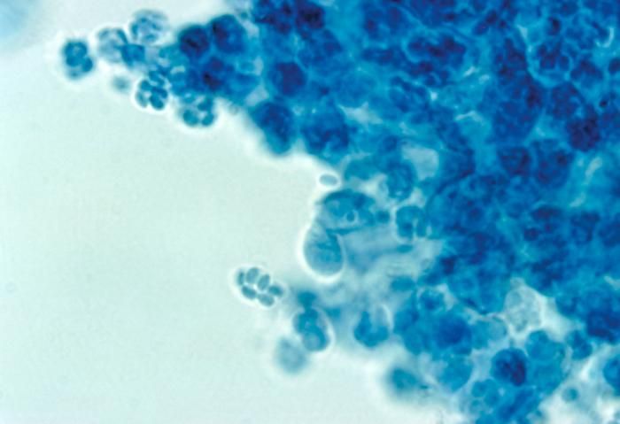
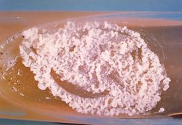
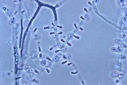
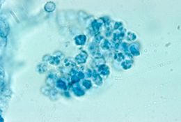
This is a slant culture growing the fungus Paracoccidioides brasiliensis during its yeast phase.Created: 1963
-
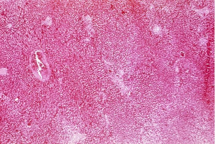
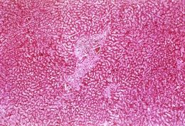
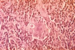
This photomicrograph reveals the histopathologic chronic inflammatory changes associated with paracoccidioidomycosis.Created: 1963
-
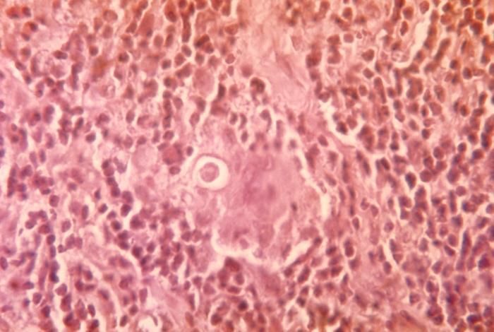
This photomicrograph reveals the histopathologic chronic inflammatory changes associated with paracoccidioidomycosis.Created: 1963
-
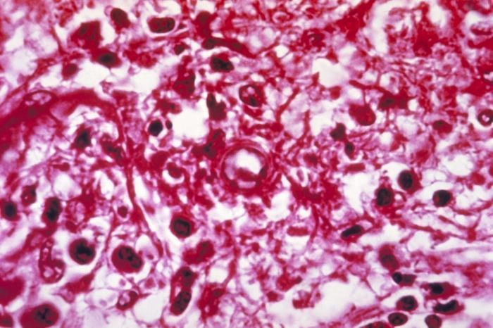
Note the histopathologic changes seen in paracoccidioidomycosis. A P. brasiliensis conidium is visible at center.Created: 1963
-
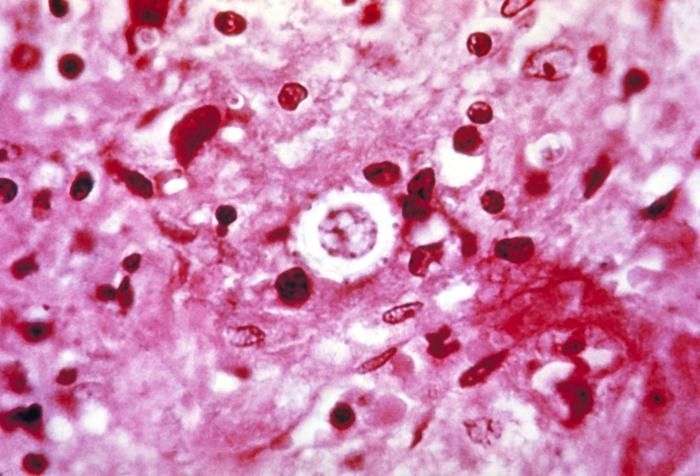
This micrograph depicts the histopathologic changes associated with paracoccidioidomycosis due to P. brasiliensis.Created: 1963
-
This micrograph depicts the histopathologic changes associated with paracoccidioidomycosis due to P. brasiliensis.Created: 1963
-
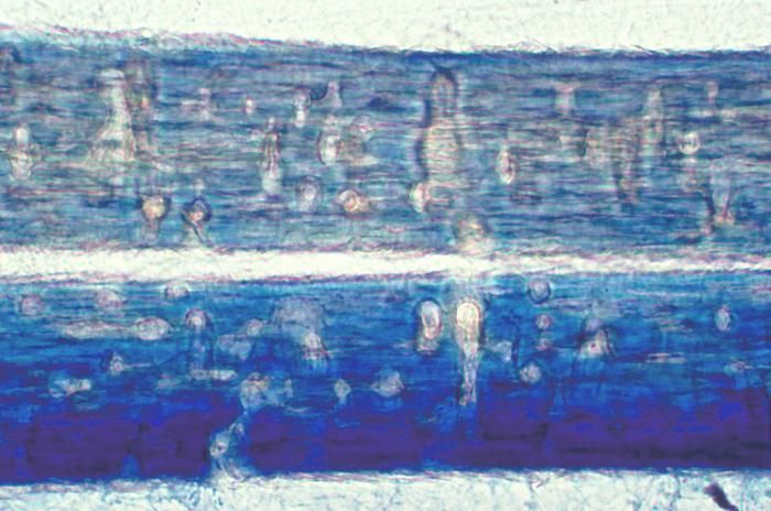
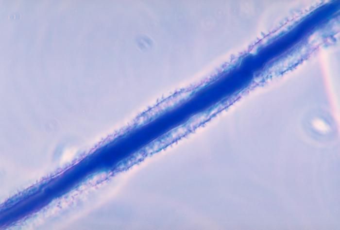
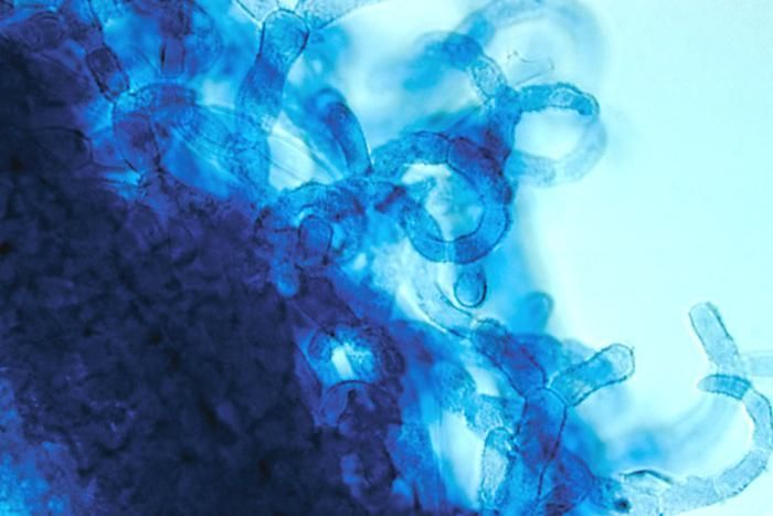
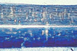
This magnified image reveals hair perforations caused by the fungus Trichophyton mentagrophytes.Created: 1968
-
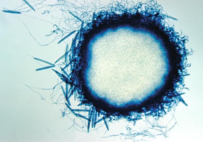
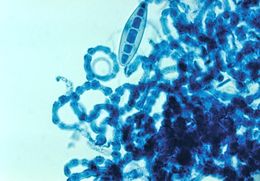
This image depicts the edge of an abortive cleistothecium of Trichophyton ajelloi, formerly Keratinomyces ajelloi.Created: 1961
-
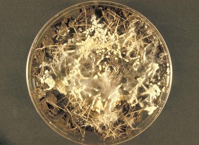
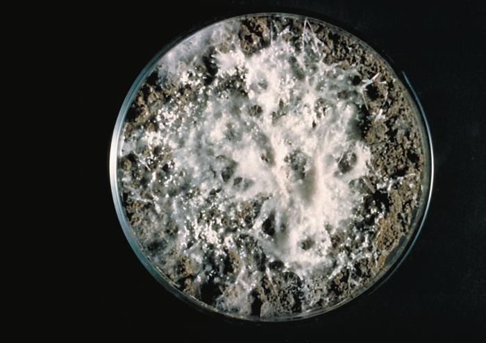
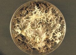
This image shows a soil hair plate culture growing the fungus Microsporum gypseum.Created: 1973
-
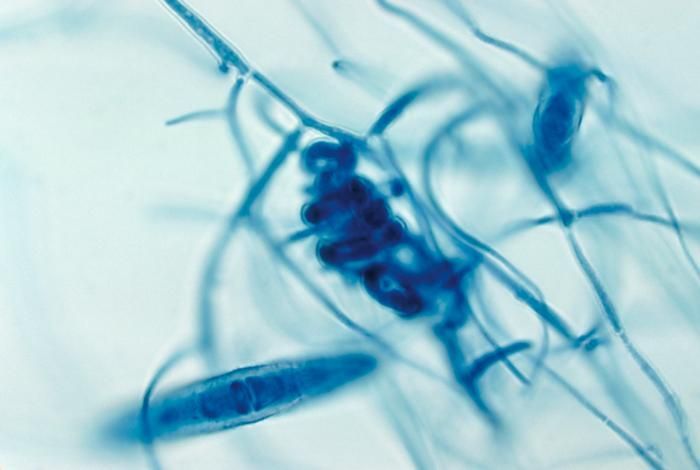
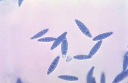
This photomicrograph shows the spindle-shaped macroconidia of the fungus Microsporum gypseum.Created: 1964
-
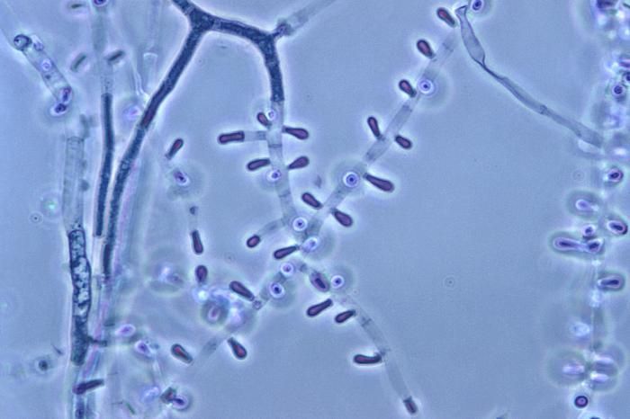
Magnified 1000X, this photomicrograph revealed some of the ultrastructural morphology exhibited by this dermatophytic fungus, Trichophyton soudanense. Of particular interest in this image are the macroconidia that were budding from the multiseptate conidiophores.Created: 1974
-
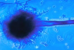
This photomicrograph depicts the appearance of a rough conidiophore of the fungus Aspergillus flavus.Created: 1963
-
This photomicrograph depicts the appearance of a conidiophore of the fungus Aspergillus flavus.Created: 1963
-
This photomicrograph shows the cleistothecium of the fungus Arthroderma grubyi, formerly Nannizzia grubyia.Created: 1961
-
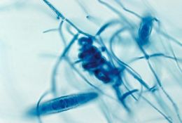
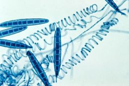
This micrograph reveals spirals and macroconidia from the edge of an immature cleistothecium of the A. grubyi fungus.Created: 1961
-
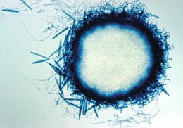
This photomicrograph shows a mature cleistothecium of the fungus Arthroderma grubyi, formerly Nannizzia grubyia.Created: 1961
-
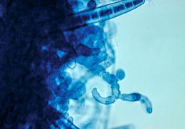
This is the edge of a mature cleistothecium of the fungus Arthroderma grubyi, formerly Nannizzia grubyia.Created: 1961
-
This image shows the cleistothecium of the fungus Arthroderma grubyi, formerly known as Nannizzia grubyia.Created: 1961
-
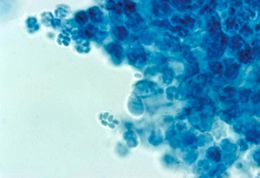
This photomicrograph shows the asci and ascospores of the fungus Arthroderma grubyi, formerly Nannizzia grubyia.Created: 1961
-
This photomicrograph shows the asci and ascospores of the fungus Arthroderma grubyi, formerly Nannizzia grubyia.Created: 1961
-
These Arthroderma grubyi cleistothecia were grown on a soil and hair plate culture; formerly Nannizzia grubyia.Created: 1961
-
-
-