-
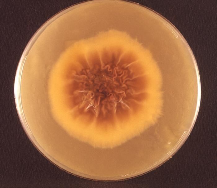
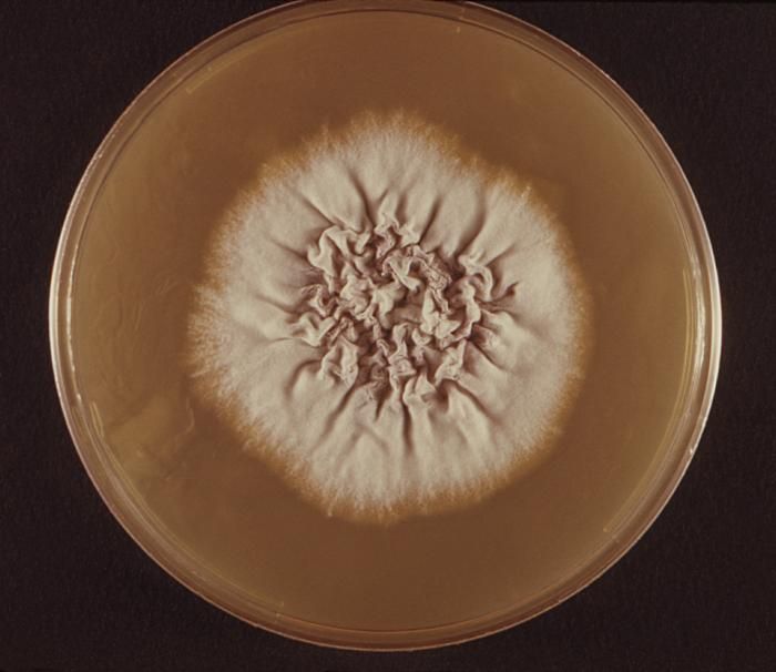
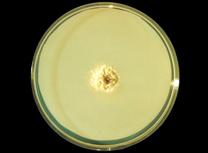
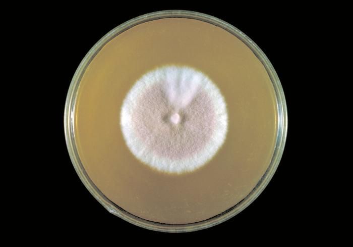
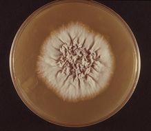
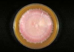
This photograph depicts the reverse view of a Petri dish within which fungal colonies of Trichophyton rubrum var. granulare had been cultured. From the front, (see PHIL 10540) the colonial morphology, which in the case of T. rubrum is said to be waxy, glaborous, i.e., flat to cottony, and display from a frontal perspective, a white to bright yellowish-beige, and even a red-violet coloration. From this reverse view, the colonies display a coloration that is a light yellowish to brown, or a reddish brown.Created: 1972
-
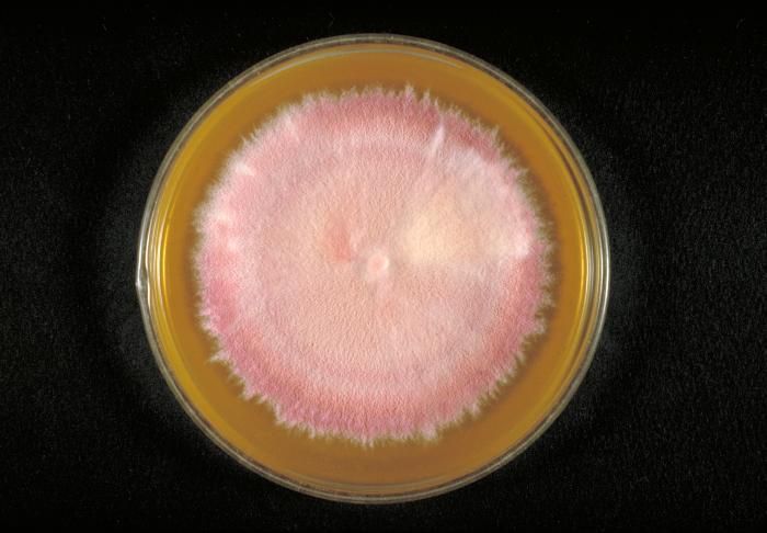
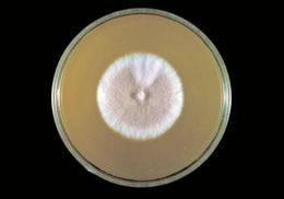
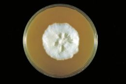
This photograph depicts the frontal view of a Petri dish within which fungal colonies of Trichophyton rubrum var. granulare had been cultured. Revealed is the colonial morphology, which in the case of T. rubrum is said to be waxy, glaborous, i.e., flat to cottony, and display from a frontal perspective, a white to bright yellowish-beige, and even a red-violet coloration. From the reverse (see PHIL 10541) or from the back, the colonies display a coloration that is a light yellowish to brown, or a reddish brown.Created: 1972
-
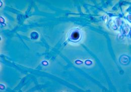
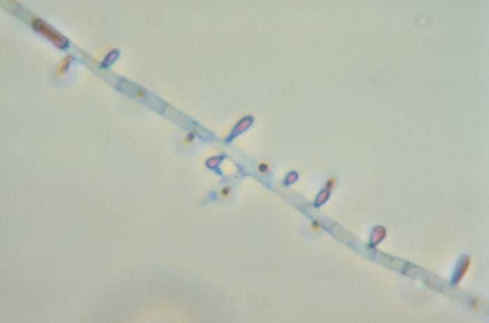
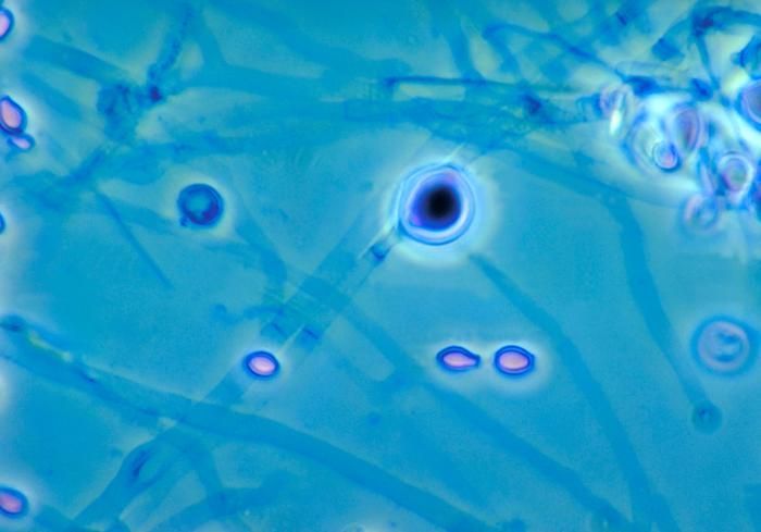
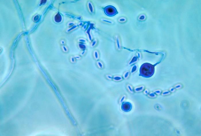
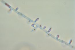
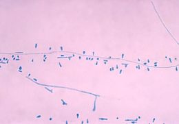
This photomicrograph reveals the microconidia of the fungus Trichophyton rubrum.Created: 1973
-
This photomicrograph reveals the microconidia of the fungus Trichophyton rubrum.Created: 1973
-
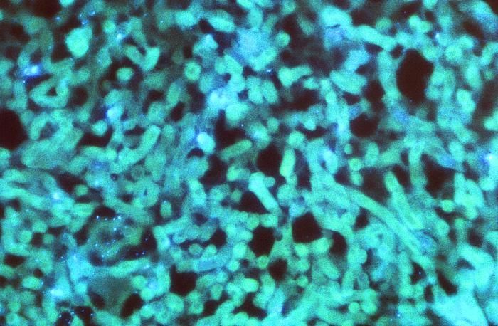
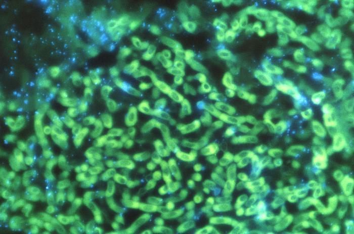
Magnified 562X this photomicrograph, stained using a fluorescent antibody (FA) staining technique (NOT stained using a Candida conjugate) revealed the presence of Aspergillus sp. organisms, in a case of aspergillosis.What is Aspergillus?Aspergillus is a fungus (or mold) that is very common in the environment. It is found in soil, on plants and in decaying plant matter. It is also found in household dust, building materials, and even in spices and some food items. There are lots of different types of Aspergillus, but the most common ones are Aspergillus fumigatus and Aspergillus flavus. Some others are Aspergillus terreus, Aspergillus nidulans, and Aspergillus niger.Created: 1972
-
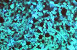
Magnified 562X this photomicrograph, stained using an Aspergillus conjugate fluorescent antibody (FA) staining technique, revealed the presence of Aspergillus sp. organisms, in a case of aspergillosis.What is Aspergillus?Aspergillus is a fungus (or mold) that is very common in the environment. It is found in soil, on plants and in decaying plant matter. It is also found in household dust, building materials, and even in spices and some food items. There are lots of different types of Aspergillus, but the most common ones are Aspergillus fumigatus and Aspergillus flavus. Some others are Aspergillus terreus, Aspergillus nidulans, and Aspergillus niger.Created: 1972
-
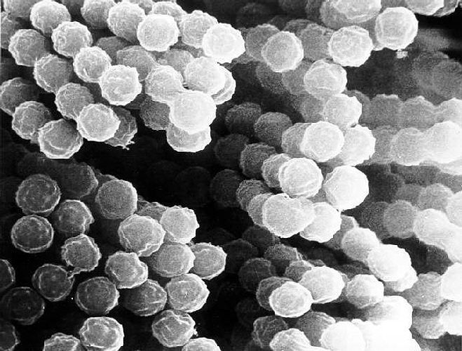
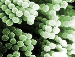
Scanning Electron Micrograph of Aspergillus species. See PHIL 9998 for a colorized version of this image.Created:
-
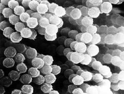
Scanning Electron Micrograph of Aspergillus species. See PHIL 9998 for a colorized version of this image.Created:
-
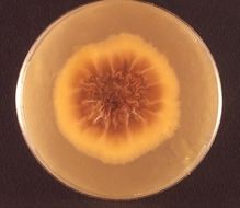
This was a Sabouraud's dextrose agar plate culture of the anthrophilic dermatophyteTrichophyton yaoundei at wk. 6.Created: 1962
-
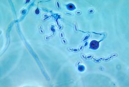
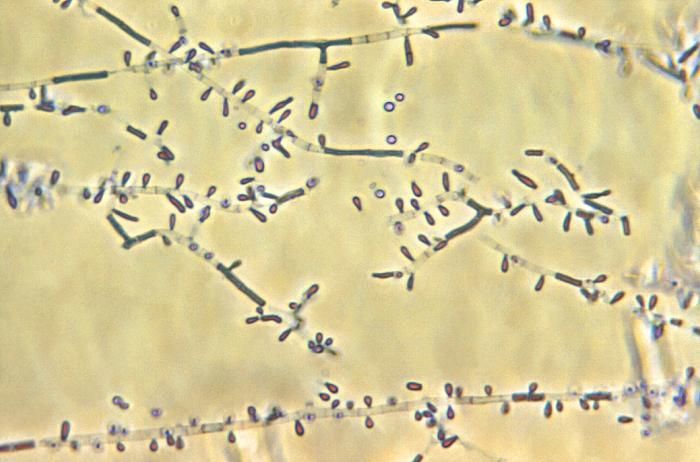
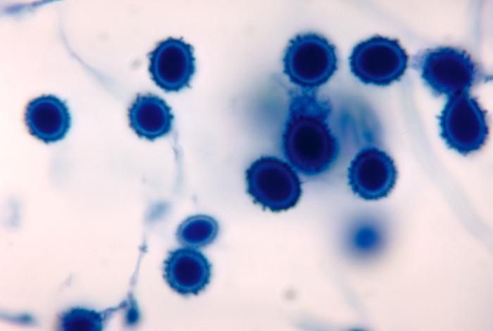
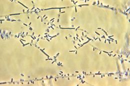
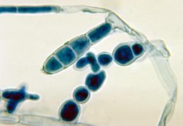
This photomicrograph reveals a number of macroconidia of the dermatophytic fungus Epidermophyton floccosum.Created: 1972
-
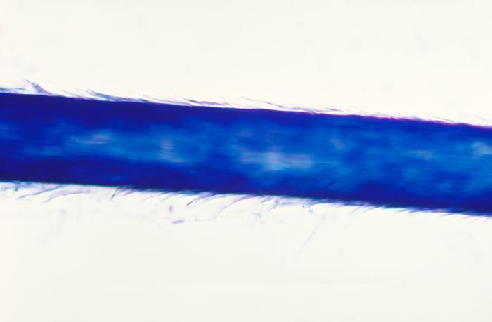
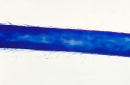
This slide was created during a hair perforation test for the zoophilic fungus Microsporum equinum.Created: 1978
-
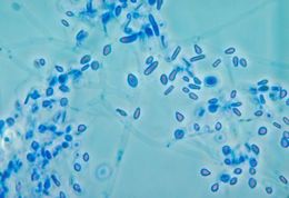
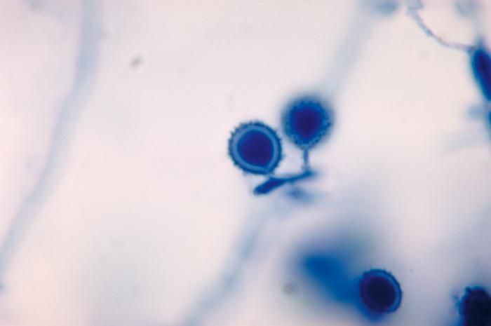
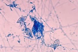
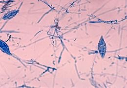
This photomicrograph shows the macroconidia of the zoophilic fungus Microsporum equinumCreated: 1978
-
This photomicrograph shows the microconidia of the zoophilic fungus Microsporum equinum.Created: 1978
-
This photomicrograph depicts the macroconidia of the zoophilic fungus Microsporum equinum.Created: 1978
-
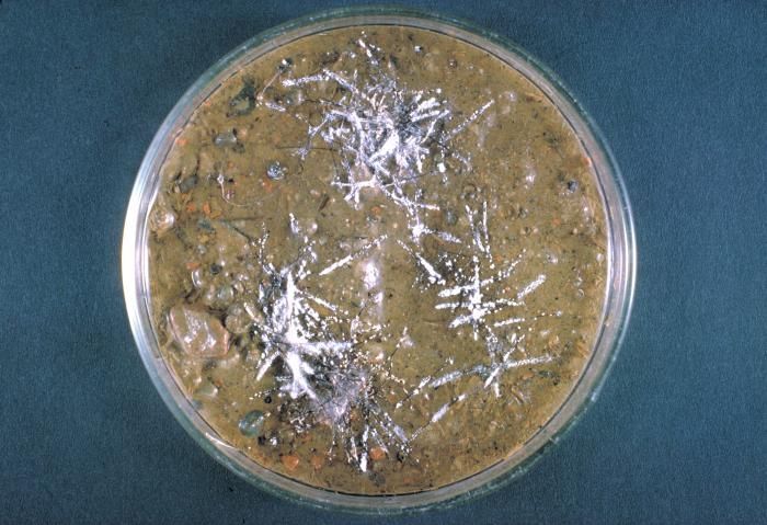
His hair-plate culture is growing the fungus Trichophyton terrestre.Created: 1963
-
This Sabourauds dextrose agar plate culture is growing T. terrestre fungus, rose-pigmented strain x231.Created: 1963
-
This Sabourauds dextrose agar plate culture is growing T. terrestre fungus, white strain x231, day 12.Created: 1963
-
This Sabourauds dextrose agar plate culture is growing T. terrestre fungus, strain x231 producing a red pigment.Created: 1963
-
This photomicrograph depicts fungal spores and adiaspores of what may be an Emmonsia sp. or Chrysosporium sp..Created: 1964
-
This photomicrograph depicts fungal spores and adiaspores of what may be an Emmonsia sp. or Chrysosporium sp..Created: 1964
-
This photomicrograph depicts fungal spores of what may be an Emmonsia sp. or Chrysosporium sp..Created: 1964
-
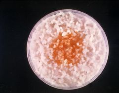
This image shows a culture of Microsporum audouinii growing poorly on boiled polished rice grains.Created: 1962
-
This is a highly magnified photomicrographic view of a number of Ctenomyces serratus conidia.Created: 1971
-
This is a highly magnified photomicrographic view of a number of Ctenomyces serratus conidia.Created: 1971