-
-
-
-
-
-
-
-
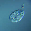
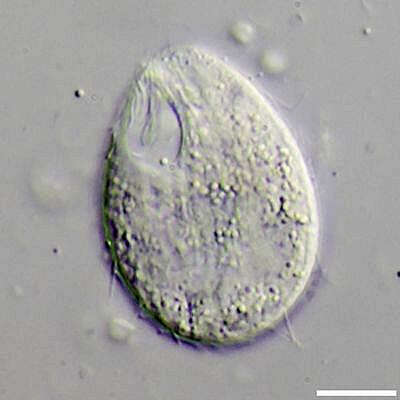
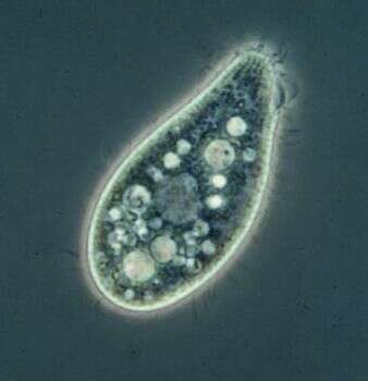
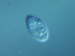
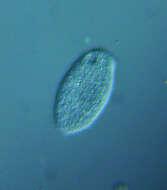
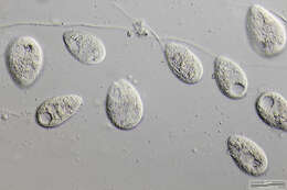
Tetrahymena pyriformis Scale bar indicates 10 µm. From the culture collection of University Duisburg-Essen, AG Boenigk, original culture from University Wuppertal. This image was taken using Zeiss Axioplan with Canon EOS 600D.Image under Creative Commons License V 3.0 (CC BY-NC-SA). Place name: Protist culture of University Duisburg-Essen (Germany) Latitude: 51.463807 Longitude: 7.005321 Multiebenen-Abbildung, manuell gestapelt. Der Messbalken markiert eine Länge von 10 µm. Aus der Kulturensammlung der Uni Duisburg-Essen AG Boenigk; diese Kultur stammt von der Uni Wuppertal. Mikrotechnik: Zeiss Axioplan, Kamera: Canon EOS 600D. Creative Commons License V 3.0 (CC BY-NC-SA). For permission to use of (high-resolution) images please contact postmaster@protisten.de.
-
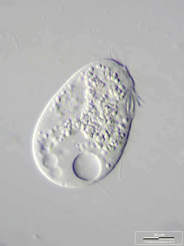
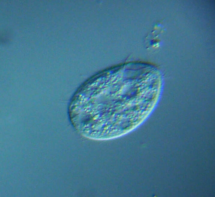
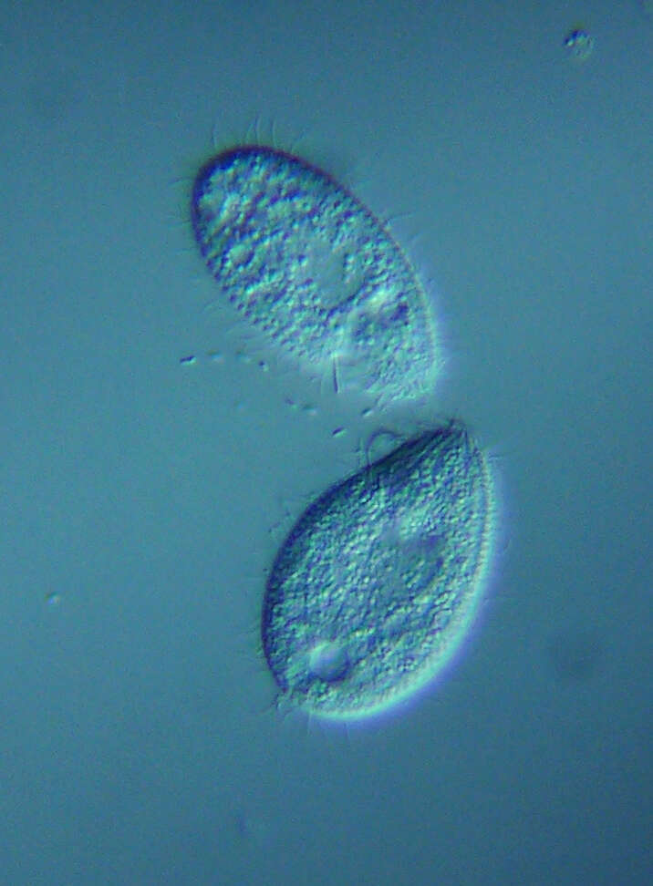
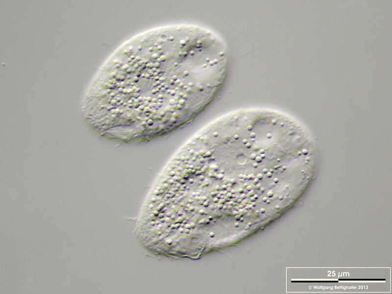
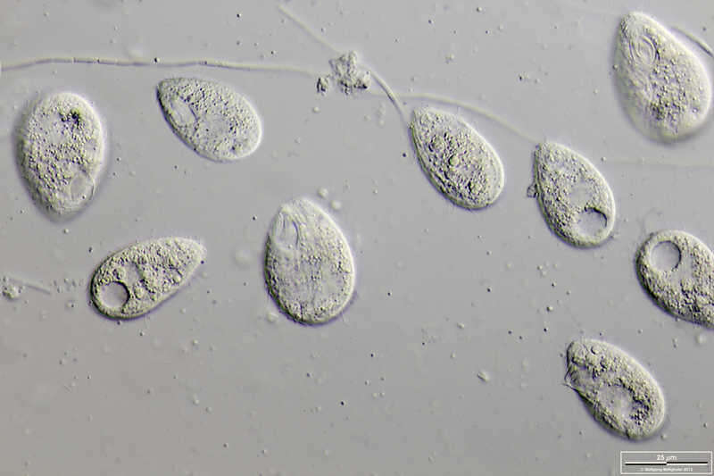
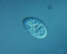
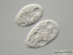
Tetrahymena pyriformis Scale bar indicates 25 µm. From the culture collection of University Duisburg-Essen, AG Boenigk, original culture from University Wuppertal. This image was taken using Zeiss Axioplan with Canon EOS 600D.Image under Creative Commons License V 3.0 (CC BY-NC-SA). Place name: Protist culture of University Duisburg-Essen (Germany) Latitude: 51.463807 Longitude: 7.005321 Multiebenen-Abbildung, manuell gestapelt. Der Messbalken markiert eine Länge von 25 µm. Aus der Kulturensammlung der Uni Duisburg-Essen AG Boenigk; diese Kultur stammt von der Uni Wuppertal. Mikrotechnik: Zeiss Axioplan, Kamera: Canon EOS 600D. Creative Commons License V 3.0 (CC BY-NC-SA). For permission to use of (high-resolution) images please contact postmaster@protisten.de.
-
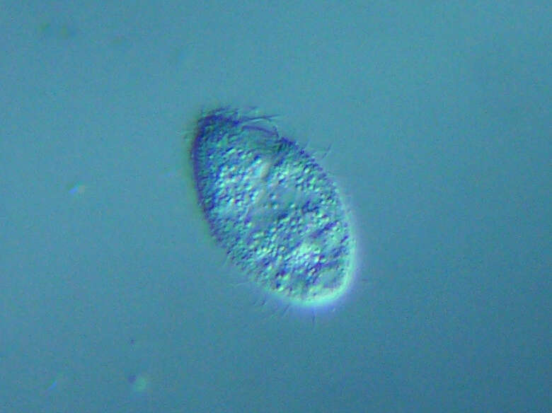
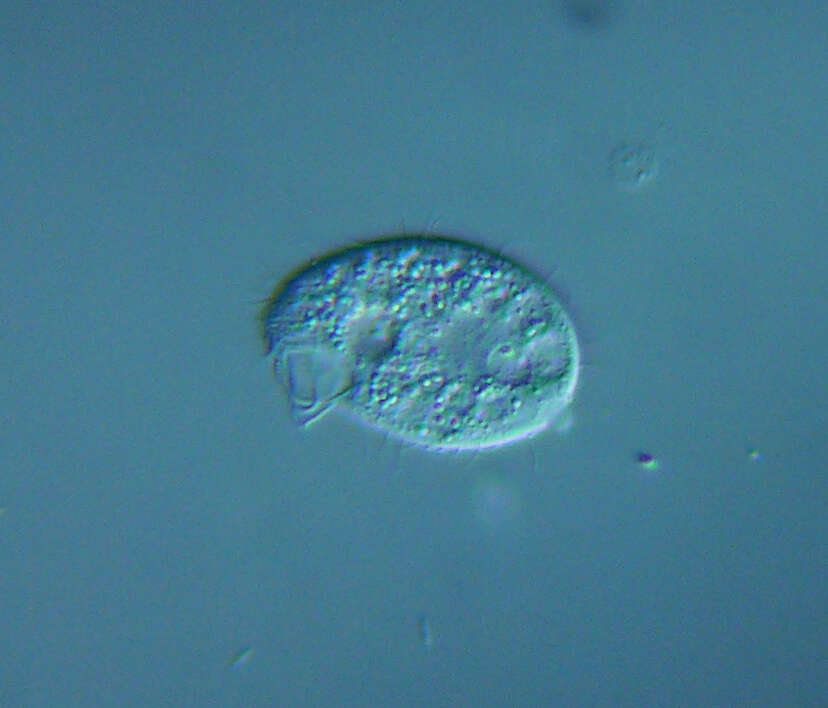
Tetrahymena pyriformis Scale bar indicates 10 µm. From the culture collection of University Duisburg-Essen, AG Boenigk, original culture from University Wuppertal. This image was taken using Zeiss Axioplan with Canon EOS 600D.Image under Creative Commons License V 3.0 (CC BY-NC-SA). Place name: Protist culture of University Duisburg-Essen (Germany) Latitude: 51.463807 Longitude: 7.005321 Multiebenen-Abbildung, manuell gestapelt. Der Messbalken markiert eine Länge von 10 µm. Aus der Kulturensammlung der Uni Duisburg-Essen AG Boenigk; diese Kultur stammt von der Uni Wuppertal. Mikrotechnik: Zeiss Axioplan, Kamera: Canon EOS 600D. Creative Commons License V 3.0 (CC BY-NC-SA). For permission to use of (high-resolution) images please contact postmaster@protisten.de.
-
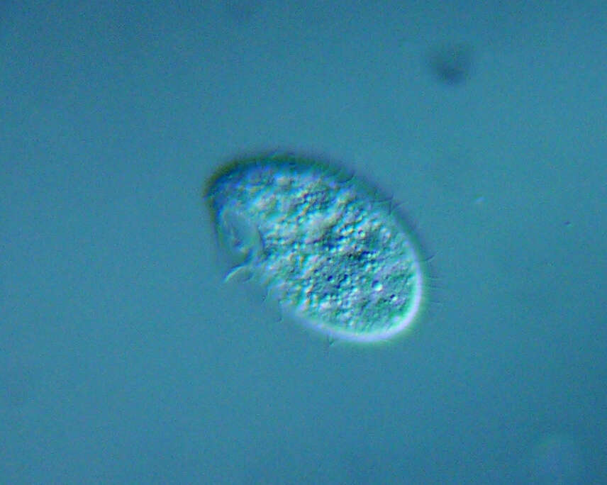
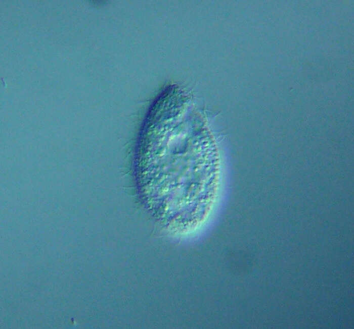
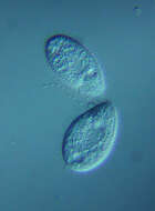
Tetrahymena pyriformis Scale bar indicates 25 µm. From the culture collection of University Duisburg-Essen, AG Boenigk, original culture from University Wuppertal. This image was taken using Zeiss Axioplan with Canon EOS 600D.Image under Creative Commons License V 3.0 (CC BY-NC-SA). Place name: Protist culture of University Duisburg-Essen (Germany) Latitude: 51.463807 Longitude: 7.005321 Multiebenen-Abbildung, manuell gestapelt. Der Messbalken markiert eine Länge von 25 µm. Aus der Kulturensammlung der Uni Duisburg-Essen AG Boenigk; diese Kultur stammt von der Uni Wuppertal. Mikrotechnik: Zeiss Axioplan, Kamera: Canon EOS 600D. Creative Commons License V 3.0 (CC BY-NC-SA). For permission to use of (high-resolution) images please contact postmaster@protisten.de.
-
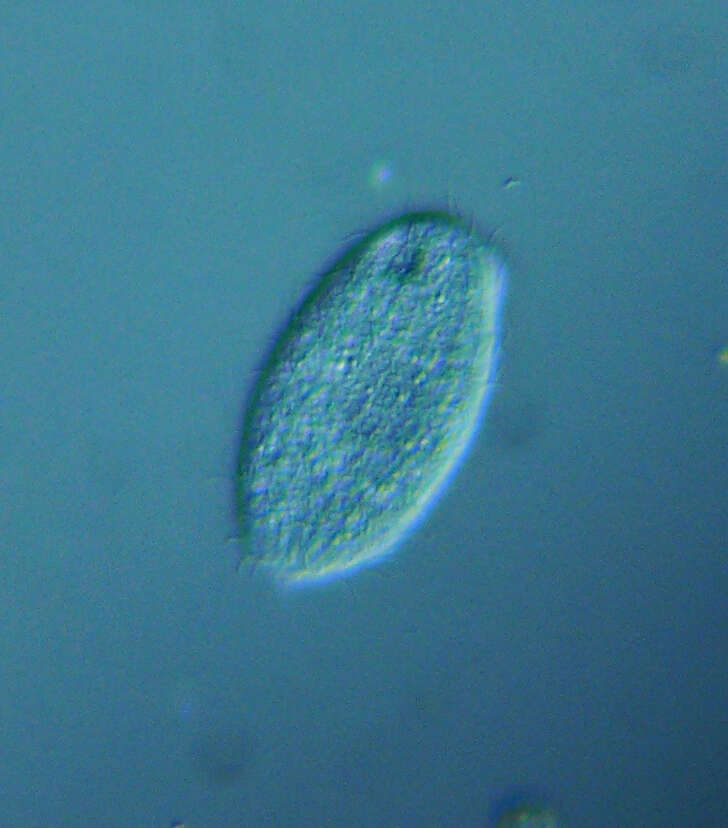
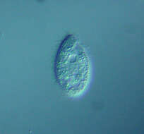
Tetrahymena pyriformis Scale bar indicates 10 µm. From the culture collection of University Duisburg-Essen, AG Boenigk, original culture from University Wuppertal. This image was taken using Zeiss Axioplan with Canon EOS 600D.Image under Creative Commons License V 3.0 (CC BY-NC-SA). Place name: Protist culture of University Duisburg-Essen (Germany) Latitude: 51.463807 Longitude: 7.005321 Multiebenen-Abbildung, manuell gestapelt. Der Messbalken markiert eine Länge von 10 µm. Aus der Kulturensammlung der Uni Duisburg-Essen AG Boenigk; diese Kultur stammt von der Uni Wuppertal. Mikrotechnik: Zeiss Axioplan, Kamera: Canon EOS 600D. Creative Commons License V 3.0 (CC BY-NC-SA). For permission to use of (high-resolution) images please contact postmaster@protisten.de.
-
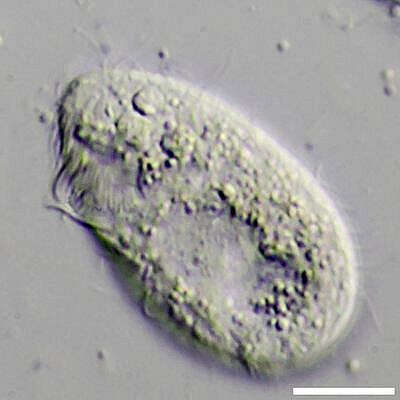
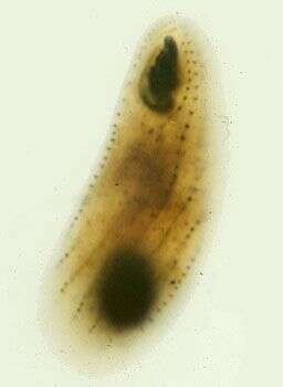
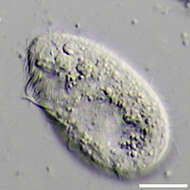
Portrait (ventral view) of the hymenostome ciliate,Tetrahymena pyriformis complex.
-
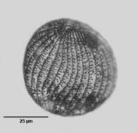
Right ventrolateral view of the silverline system (argyrome) of a ciliate from the Tetrahymena pyriformis complex.The oral aperture is to the viewer's upper right. Stained by the Klein-Foissner dry silver nitrate technic (see Foissner, W. Europ. J. Protistol., 27:313-330;1991). Brightfield, black and white.
-
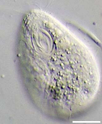
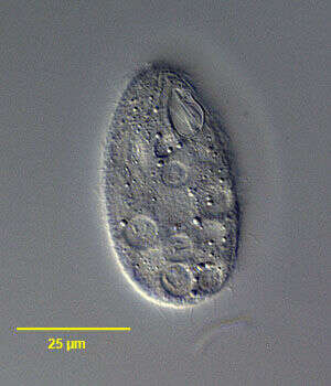
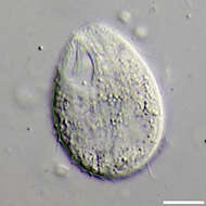
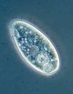
Cell from strain W, oral cilia are near the front of the cell (top of image), large clear area is the macronucleus, there is no micronucleus in this strain. Phase contrast image.
-
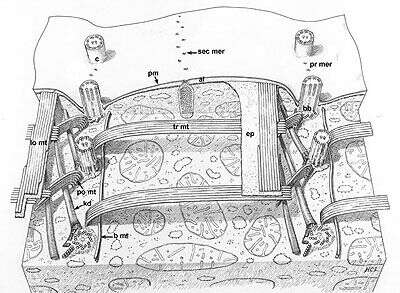
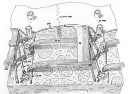
A classic drawing of the organization of this model ciliate. Segments of two kineties or rows of basal body (bb)/cilium (c) complexes. Kinetodesmal fibers (kd) arise from the left side of the proximal end of each basal body and angle up to the cell pellicle. Transverse microtubules (tr mt) extend to the right and postciliary microtubules (po mt) angle posteriorly and to the left of the kinety. One or two basal microtubules (b mt) extend along the right of the kinety at the proximal end of the basal bodies. A band of longitudinal microtubules (lo mt) lies between the epiplasm (ep) and the alveoli (al) to the left of the kinety. Mucocysts (mu) approach the plasma membrane (pm) through the septa separating alveoli along the primary (pr mer) and secondary meridians (sec mer). Drawn by H. C. Lyman.
This image is available in Richard Allen's collection.
-
The protargol stains the bases of the cilia and the nucleus. The image shows the kineties, the three membranelles and the single curved undulating membrane that make up the mouth ciliature, and the macronucleus (this strain lacks a micronucleus).
-
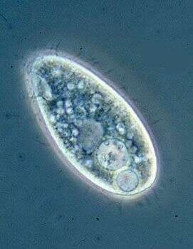
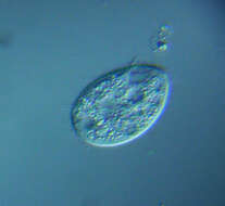
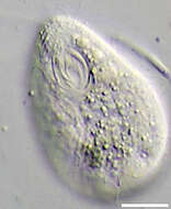
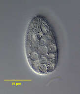
Phase contrast image of living cell. The mouth and some of the associated ciliature can be seen upper left, the surface is coated with somatic cilia that are used for cell locomotion, there is a contractile vacuole near the posterior end of the cell, and a light-coloured macronucleus near the centre of the cell. the other vacuoles are food vacuoles (phagosomes)
-
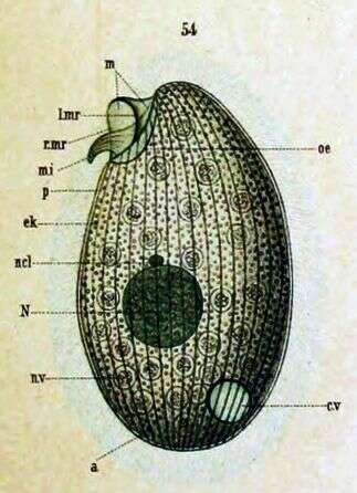
Originally described as Glaucoma pyriformis (Ehrenberg) Key to Schewiakoff's abbreviations: a -- Anus cv -- Contractile vacuole ek -- Ectoplasm l. mr -- Left membrane edge m -- Undulating membrane mi -- Inner undulating membrane N -- Macronucleus ncl -- Micronucleus oe -- Throat r.mr --Right membrane edge p -- Pellicle
-
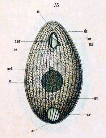
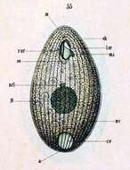
(Originally described under the name Glaucoma pyriformis) Ventral view. Key to Schewiakoff's abbreviations: a -- Anus cv -- Contractile vacuole el -- Ectoplasm l.or -- Left edge of mouth m -- Undulating membrane mi -- Inner Undulating membrane N -- Macronucleus ncl -- Micronucleus nv -- Food vacuole oe -- Throat r.or -- Right edge of mouth
-
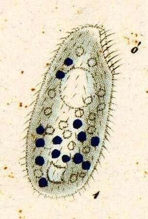
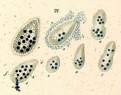
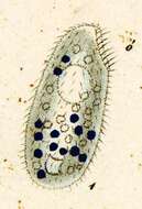
Originally described by Ehrenberg under the name Leucophrys pyriformis.
-
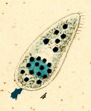
Originally described by Ehrenberg under the name Leucophrys pyriformis.
-
Originally described by Ehrenberg under the name Leucophrys pyriformis.