عن
تعليم
ناقش
بنك الصفات
تسجيل الدخول
أنشئ حسابًا
اللغة:
Deutsch
English
Español
français
italiano
Nederlands
Piemontèis
Português do Brasil
suomi
Türkçe
Čeština
Ελληνικά
македонски
Українська
العربية
简体中文
繁體中文
الأسماء في صفحات التنقل
عامية
علمية
عن
تعليم
ناقش
بنك الصفات
تسجيل الدخول
أنشئ حسابًا
ar
Deutsch
English
Español
français
italiano
Nederlands
Piemontèis
Português do Brasil
suomi
Türkçe
Čeština
Ελληνικά
македонски
Українська
العربية
简体中文
繁體中文
الأسماء في صفحات التنقل
عامية
علمية
اسم غير محسوم
الاسم غير المحسوم:
Biota
»
حقيقيات النوى
»
فطر
»
Dikarya
»
فطريات زقية
»
Pezizomycotina
»
عشوفيات
«
Sordariomycetidae
تجميع
نظرة عامة
وسائط
مقالات
أسماء
الترخيص
أي ترخيص
CC-BY-NC
CC-BY-NC-SA
No copyright
صورة
أي نوع
فيديو
صورة
مقدم المحتوى
أي مقدم المحتوى
CalPhotos
iNaturalist
INBio
Public Health Image Library
CreatureCast
1
2
Last »
cc-publicdomain
موثوق
cc-publicdomain
موثوق
cc-publicdomain
موثوق
cc-by-nc-sa-3.0
موثوق
cc-by-nc-sa-3.0
موثوق
cc-publicdomain
موثوق
cc-publicdomain
موثوق
cc-by-nc-sa-3.0
موثوق
cc-publicdomain
موثوق
cc-publicdomain
موثوق
cc-by-nc-sa-3.0
موثوق
cc-publicdomain
موثوق
cc-publicdomain
موثوق
cc-by-nc-sa-3.0
موثوق
cc-publicdomain
موثوق
cc-publicdomain
موثوق
cc-by-nc-sa-3.0
موثوق
cc-publicdomain
موثوق
cc-publicdomain
موثوق
cc-publicdomain
موثوق
cc-publicdomain
موثوق
cc-publicdomain
موثوق
cc-publicdomain
موثوق
cc-publicdomain
موثوق
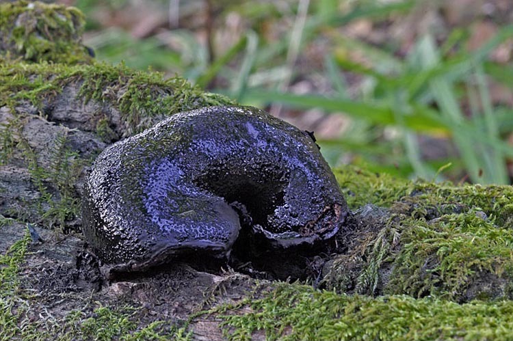
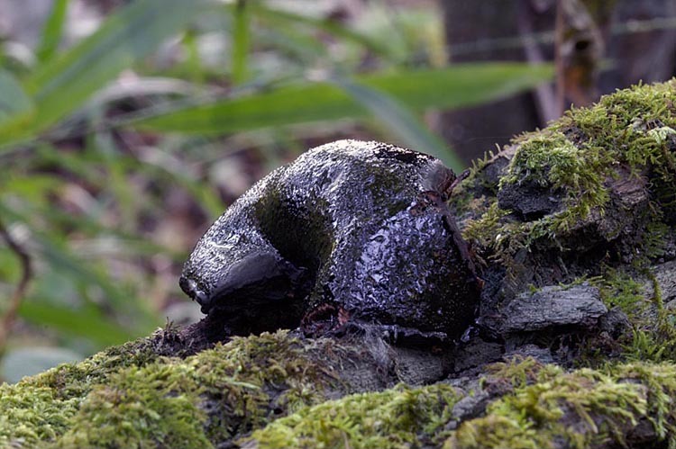
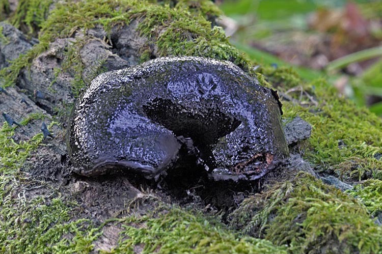
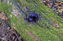
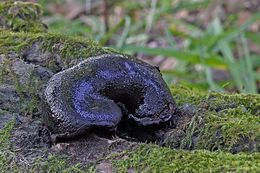
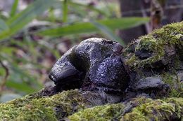
صورة Camarops petersii (Berk. & M. A. Curtis) Nannf. 1972
cc-publicdomain
2005 Nick Kurzenko
CalPhotos
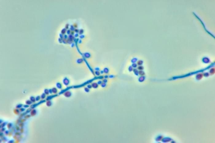
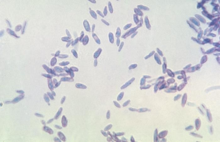
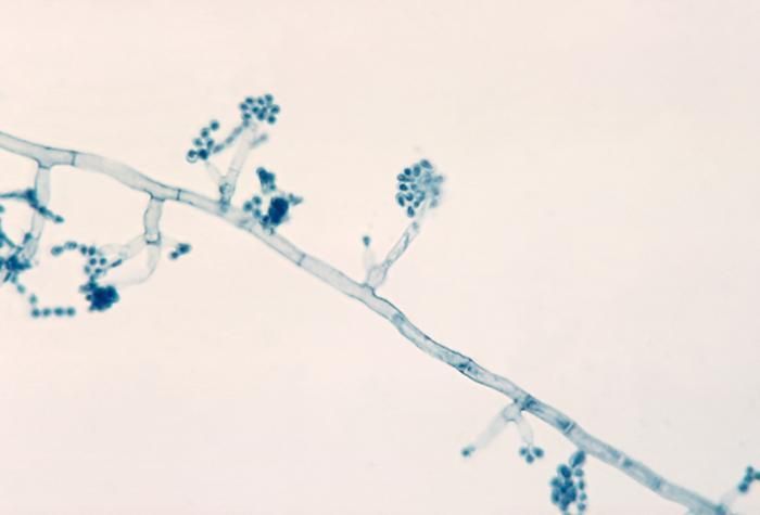
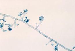
صورة Sporothrix
cc-publicdomain
Public Health Image Library
Magnified 500X, this photomicrograph revealed the presence of
Sporothrix sp.
fungal organisms that were isolated from a peat moss specimen.Created: 1971
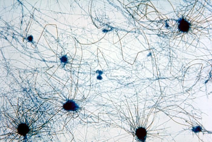
صورة Chaetomiaceae
cc-publicdomain
Public Health Image Library
This photomicrograph reveals multiple perithecia, or fruiting bodies, of a
Chaetomium spp.
fungus.Created: 1955
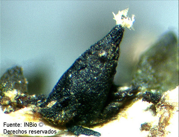
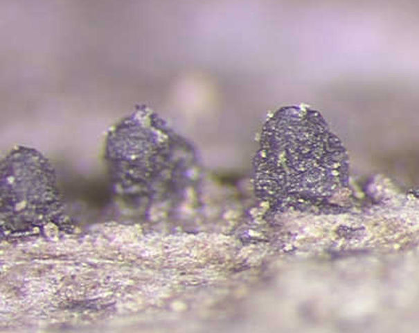
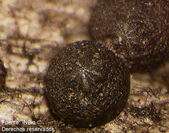
صورة Striatosphaeria
cc-by-nc-sa-3.0
Instituto Nacional de Biodiversidad - INBio, Costa Rica.
INBio
Peritecios de
Striatosphaeria codinaeaphora
Samuels & E. Müller.Foto: Ronald Rodríguez
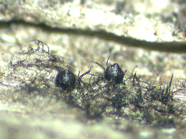
صورة Cercophora atropurpurea A. N. Mill. & Huhndorf 2001
cc-by-nc-sa-3.0
Instituto Nacional de Biodiversidad - INBio, Costa Rica.
INBio
Cuerpo fructífero de
Cercophora atropurpurea
A.N. Miller & Huhndorf. Foto: Fernando A. Fernández
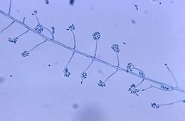
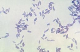
صورة Sporothrix schenckii Hektoen & C. F. Perkins 1900
cc-publicdomain
Public Health Image Library
This photomicrograph reveals the conidiophores and conidia of the fungus
Sporothrix schenckii
.Created: 1972
صورة Chaetomiaceae
cc-publicdomain
Public Health Image Library
This photomicrograph reveals multiple perithecia, or fruiting bodies, of a
Chaetomium spp.
fungus.Created: 1955
صورة Chaetosphaeria cubensis Hol.-Jech. 1982
cc-by-nc-sa-3.0
Fernando A. Fernández.
INBio
Cuerpos fructíferos de
Chaetosphaeria cubensis
V. Holubová-Jechová. Foto: Fernando Fernández.
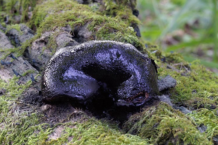
صورة Camarops petersii (Berk. & M. A. Curtis) Nannf. 1972
cc-publicdomain
2005 Nick Kurzenko
CalPhotos
صورة Sporothrix schenckii Hektoen & C. F. Perkins 1900
cc-publicdomain
Public Health Image Library
Shown here is a photomicrograph of the fungus
Sporothrix schenckii
during yeast phase.Created: 1964
صورة Chaetosphaeria cylindrospora F. A. Fernández, Huhndorf, Joanne E. Taylor & K. D. Hyde 2001
cc-by-nc-sa-3.0
Fernando A. Fernández.
INBio
Cuerpos fructíferos de
Chaetosphaeria cylindrospora
F. A. Fernández, S. M. Huhndorf, J. E. Taylor, K. D. Hyde. Foto: Fernando Fernández.
صورة Camarops petersii (Berk. & M. A. Curtis) Nannf. 1972
cc-publicdomain
2005 Nick Kurzenko
CalPhotos
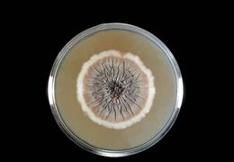
صورة Sporothrix schenckii Hektoen & C. F. Perkins 1900
cc-publicdomain
Public Health Image Library
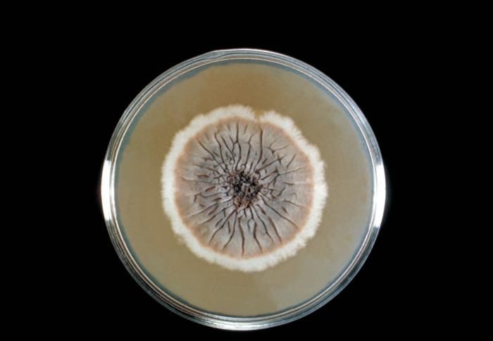
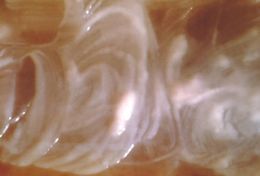
This Sabouraud's dextrose agar plate culture is growing the fungus
Sporothrix schenckii
.Created: 1964
صورة Cercophora costaricensis (G. C. Carroll & Munk) O. Hilber & R. Hilber 1979
cc-by-nc-sa-3.0
Fernando A. Fernández.
INBio
Peritecios de
Cercophora costaricensis
(G.C. Carroll & Munk) O. Hilber & R. Hilber Foto: Fernando Fernández.
صورة Camarops petersii (Berk. & M. A. Curtis) Nannf. 1972
cc-publicdomain
2005 Nick Kurzenko
CalPhotos
صورة Sporothrix schenckii Hektoen & C. F. Perkins 1900
cc-publicdomain
Public Health Image Library
Shown here is a close-up of a
Sporothrix schenckii
culture during yeast phase.Created: 1964
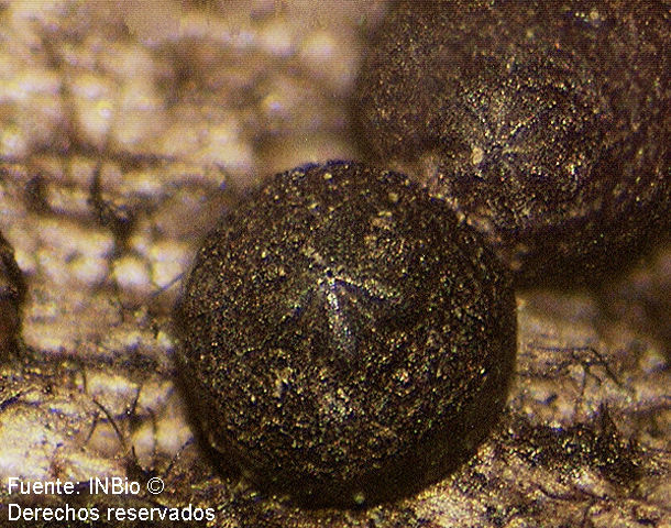
صورة Cercophora macrocarpa (G. C. Carroll & Munk) O. Hilber & R. Hilber 1979
cc-by-nc-sa-3.0
Instituto Nacional de Biodiversidad - INBio, Costa Rica.
INBio
Peritecios de
Cercophora macrocarpa
(G.C. Carroll & Munk) O. Hilber & R. Hilber Foto: Silvia Soto.
صورة Camarops petersii (Berk. & M. A. Curtis) Nannf. 1972
cc-publicdomain
2005 Nick Kurzenko
CalPhotos
صورة Sporothrix schenckii Hektoen & C. F. Perkins 1900
cc-publicdomain
Public Health Image Library
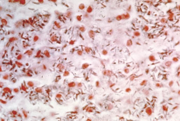
This photomicrograph shows the presence of
Sporothrix schenckii
in a smear obtained from a rat.Created: 1964
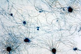
صورة Sporothrix schenckii Hektoen & C. F. Perkins 1900
cc-publicdomain
Public Health Image Library
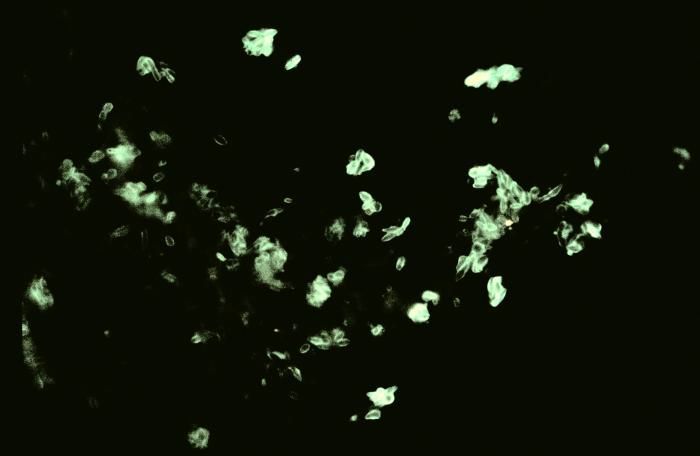
Using a direct FA stain, this slide demonstrates the histopathology of sporotrichosis due to
Sporothrix schenckii
.Created: 1977
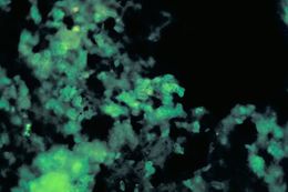
صورة Sporothrix
cc-publicdomain
Public Health Image Library
Using a direct FA stain, this slide demonstrates the histopathology of sporotrichosis due to
Sporothrix schenckii
.Created: 1977
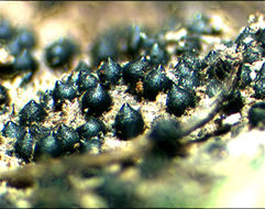
صورة Madurella mycetomatis (Laveran) Brumpt 1905
cc-publicdomain
Public Health Image Library
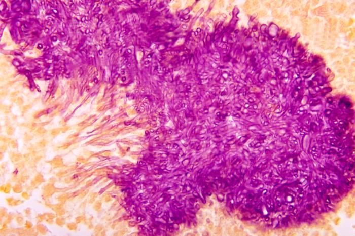
Note the histopathologic appearance of black grain mycetoma due to
Madurella mycetomatis
using a Gridley stain.Created: 1972
صورة Madurella mycetomatis (Laveran) Brumpt 1905
cc-publicdomain
Public Health Image Library
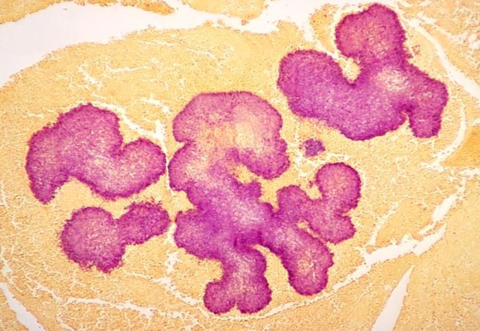
Note the histopathologic appearance of black grain mycetoma due to
Madurella mycetomatis
using a Gridley stain.Created: 1972
صورة Madurella mycetomatis (Laveran) Brumpt 1905
cc-publicdomain
Public Health Image Library
This photomicrograph shows phialides with terminal conidia of the
Madurella mycetomatis
fungus.Created: 1961
1
2
Last »