-
All Biocode files are based on field identifications to the best of the researcher’s ability at the time.
-
Iriomote-jima, Japan
-
Klinteskoven Møn
-
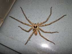
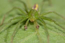
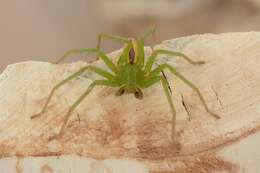
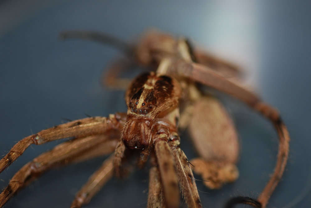
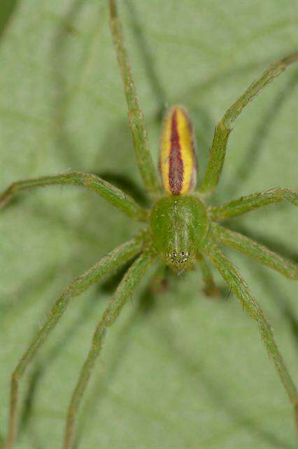
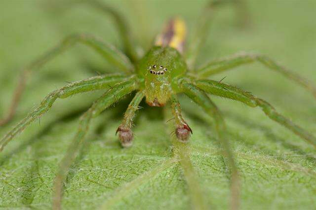
Heteropoda maxima - Giant HuntsmanThe Giant huntsman spider, the largest spider in the world.From
Flicker28 April 2012
-
Dan Quan, Jian Chen, Jie Liu
Zookeys
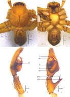
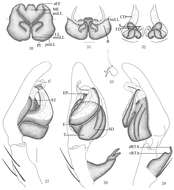
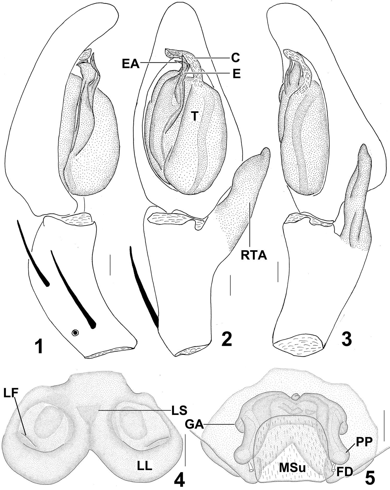
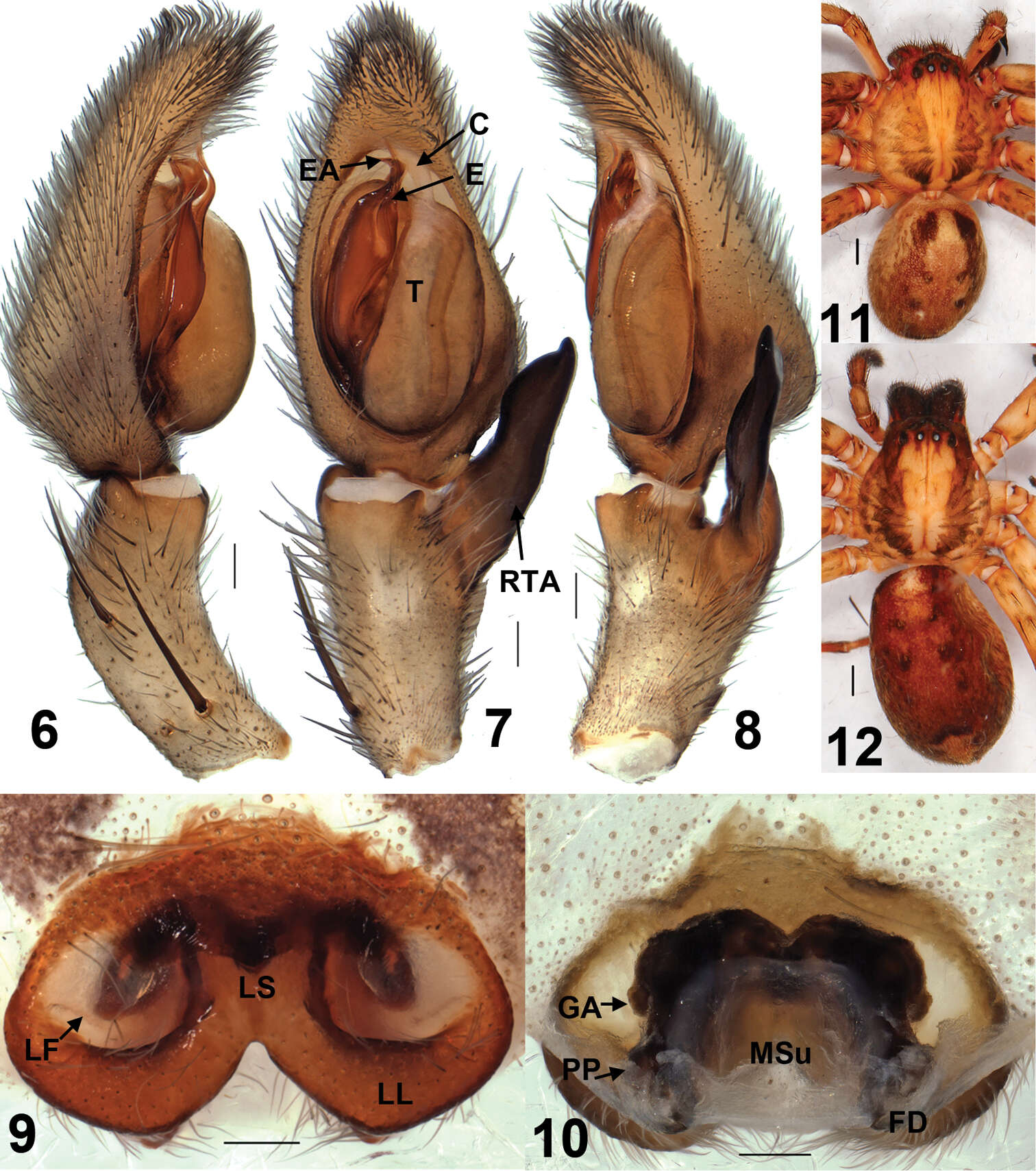
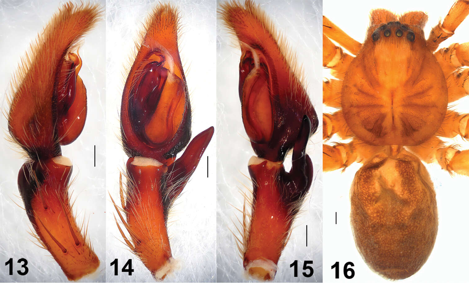
Figures 1–5.Sinopoda serrata (Wang, 1990), from Tiantangzhai National Forest Park (Hubei Province, China). 1 Left male palp, prolateral view 2 Left male palp, ventral view 3 Left male palp, retrolateral view 4 Epigyne, ventral view 5 Vulva, dorsal view. Scales = 0.2 mm. C conductor, E embolus, EA embolic apophysis, FD fertilization duct, GA glandular appendage, LF lateral furrow, LL lateral lobes, LS lobal septum, MSu membranous sac unexpanded, RTA retrolateral tibial apophysis, PP posterior part of spermathecae, T tegulum.
-
Feng Zhang, Bao-Shi Zhang, Zhi-Sheng Zhang
Zookeys
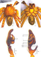
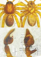
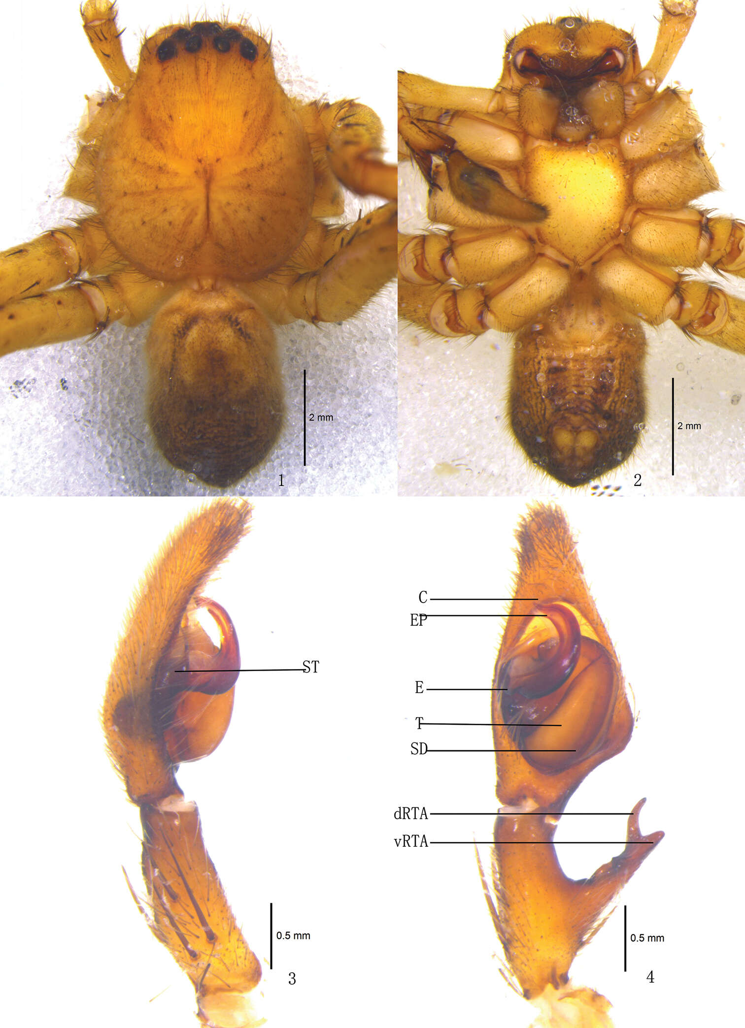
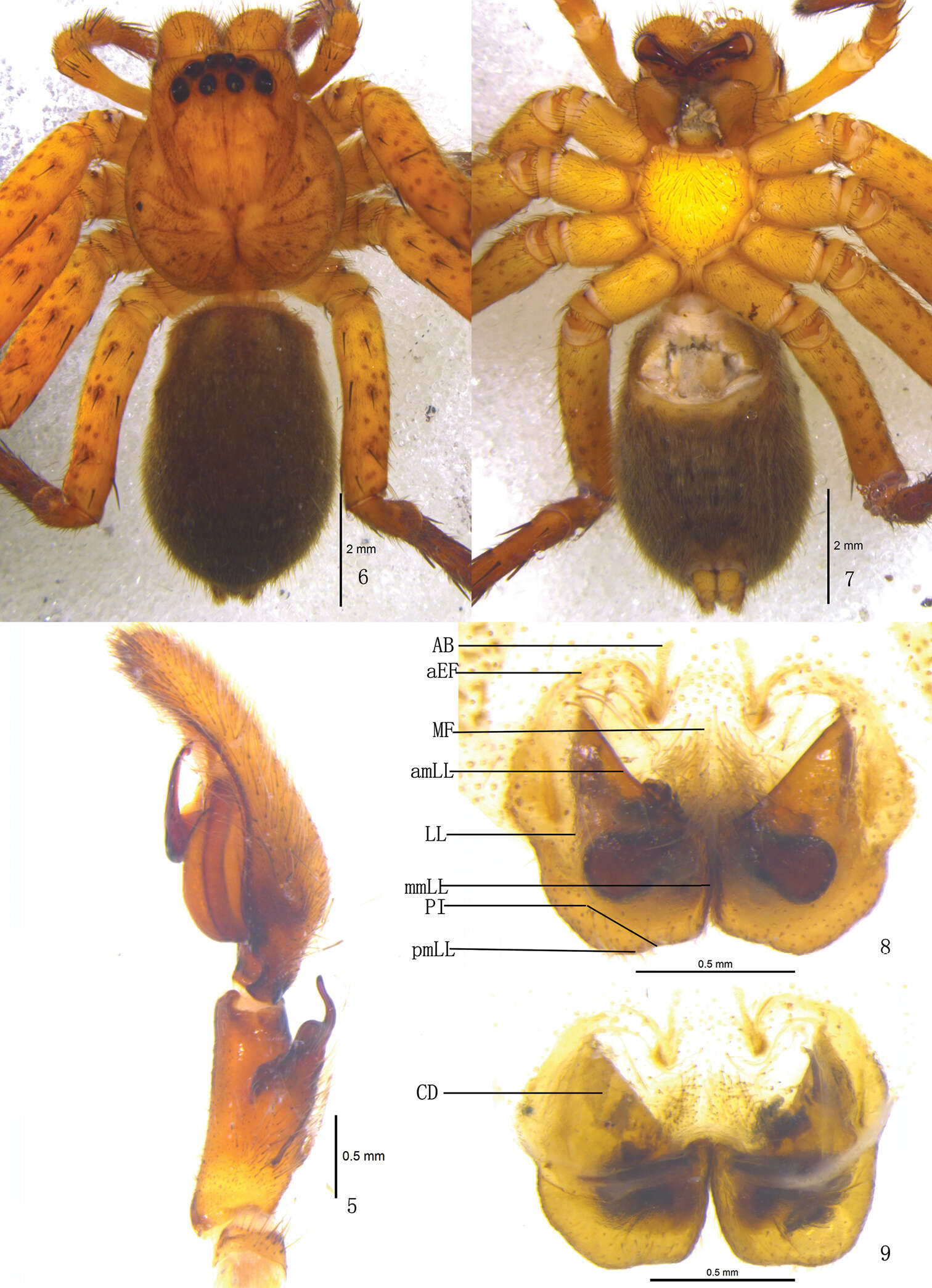
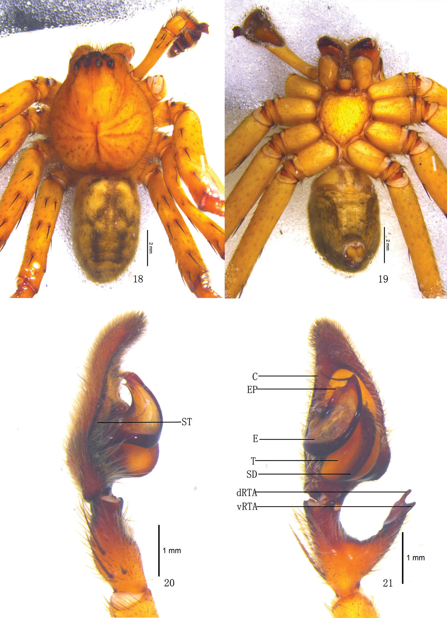
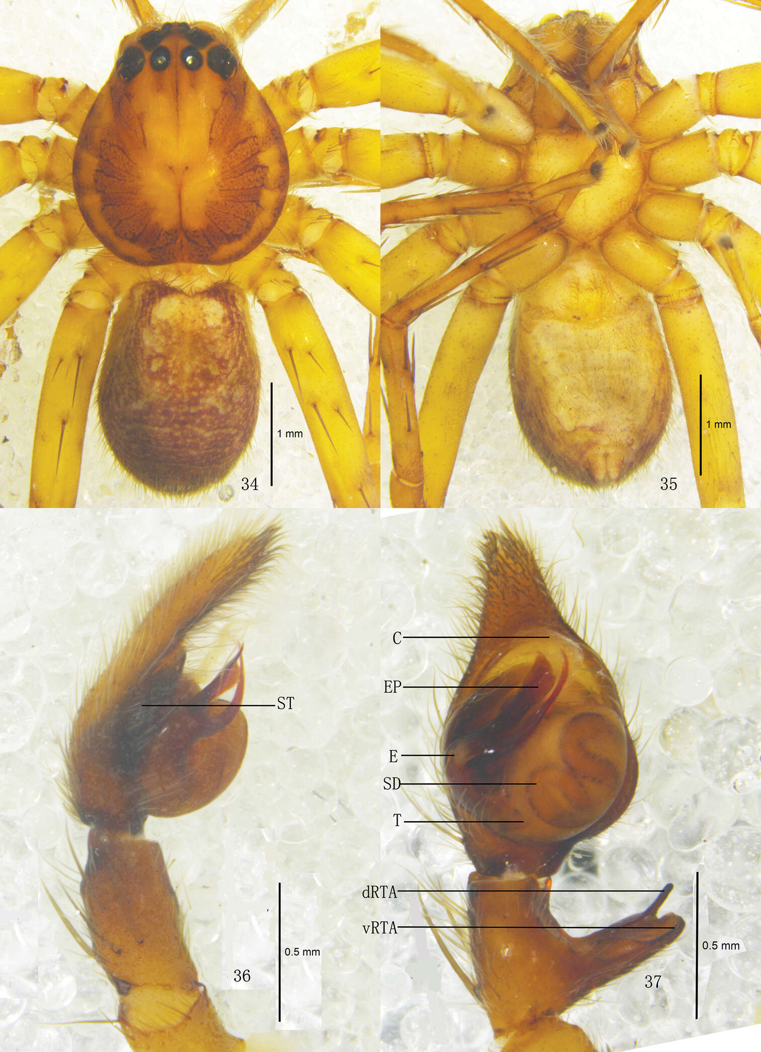
Figures 1–4.Pseudopoda acuminata sp. n., Male (SP–SC–03–0050): 1–2 Body (1 dorsal 2 ventral) 3–4 Left palp (3 prolateral 4 ventral). Abbreviations: C conductor; dRTA dorsal branch of retrolateral tibial apophysis; E embolus; EP embolic projection; SD sperm duct; ST subtegulum; T tegulum; vRTA ventral branch of retrolateral tibial apophysis. Scale bars: 2 mm (1–2); 0.5 mm (3–4).
-
All Biocode files are based on field identifications to the best of the researcher’s ability at the time.
-
Klinteskoven Møn
-
Dan Quan, Jian Chen, Jie Liu
Zookeys
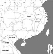
Figure 17.Collection localities of Sinopoda serrata (Wang, 1990) in China.
-
Feng Zhang, Bao-Shi Zhang, Zhi-Sheng Zhang
Zookeys
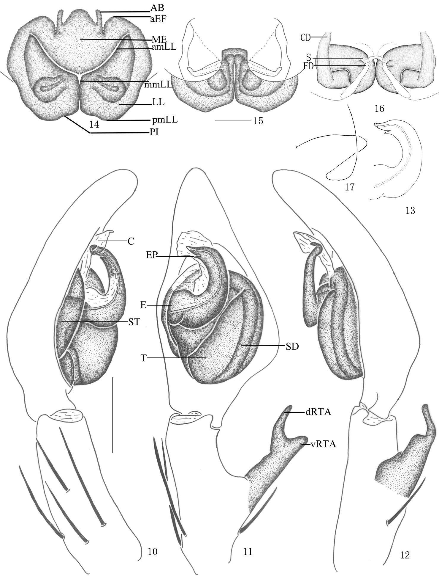
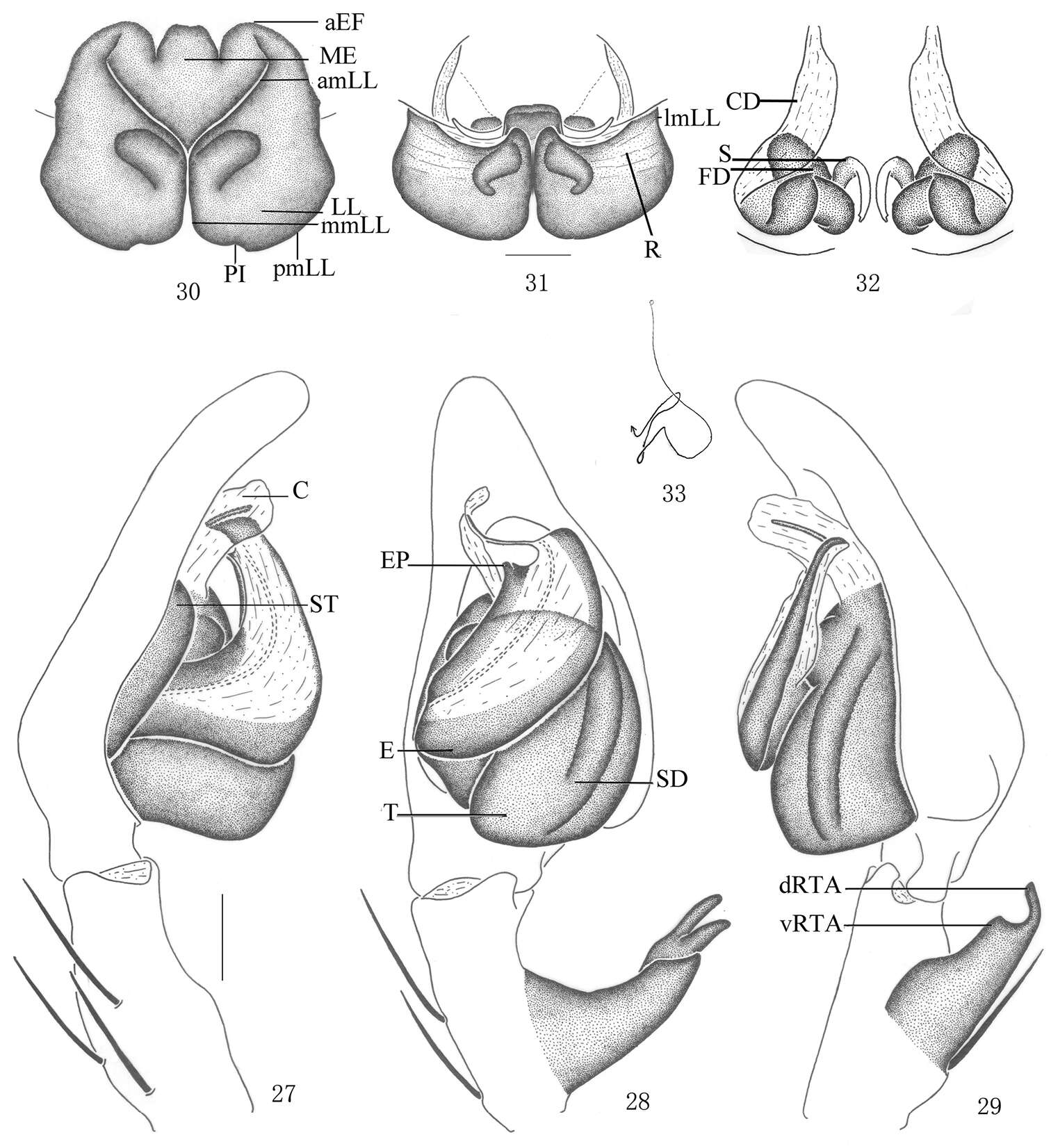
Figures 10–17.Pseudopoda acuminata sp. n., 10–13 Male (SP–SC–03–0050): 10–12 Left palp (10 prolateral 11 ventral 12 retrolateral) 13 embolus (ventral) 14–17 Female (SP–SC–03–0052): 14–16 Epigyne (14 ventral 15 dorsal 16 apical); 17 Schematic course of internal duct system, dorsal. Abbreviations: AB anterior bands; aEF anterior margin of epigynal field; amLL anterior margin of lateral lobes; C conductor; CD copulatory duct; dRTA dorsal branch of retrolateral tibial apophysis; E embolus; EP embolic projection; FD fertilization duct; LL lateral lobes of epigyne; MF median field of epigyne; mmLL median margin of lateral lobes; pmLL posterior margins of lateral lobes; PI posterior incisions; S spermathecae; SD sperm duct; ST subtegulum; T tegulum; vRTA ventral branch of retrolateral tibial apophysis. Scale bars: 0.5 mm.
-
All Biocode files are based on field identifications to the best of the researcher’s ability at the time.
-
Klinteskoven Møn
-
Dan Quan, Jian Chen, Jie Liu
Zookeys
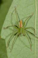
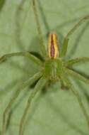
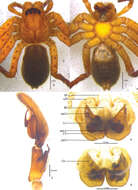
Figures 6–12.Sinopoda serrata (Wang, 1990), from Tiantangzhai National Forest Park (Hubei Province, China). 6 Left male palp, prolateral view 7 Left male palp, ventral view 8 Left male palp, retrolateral view 9 Epigyne, ventral view 10 Vulva, dorsal view11 Male habitus, dorsal view12 Female habitus, dorsal view. Scales = 0.2 mm (6–10), scales = 1 mm (11–12). C conductor, E embolus, EA embolic apophysis, FD fertilization duct, GA glandular appendage, LF lateral furrow, LL lateral lobes, LS lobal septum, MSu membranous sac unexpanded, RTA retrolateral tibial apophysis, PP posterior part of spermathecae, T tegulum.
-
Feng Zhang, Bao-Shi Zhang, Zhi-Sheng Zhang
Zookeys
Figures 5–9.Pseudopoda acuminata sp. n., 5 Left palp of male (retrolateral). 6–9 Female (SP–SC–03–0052): 6–7 Body (6 dorsal 7 ventral) 8–9 Epigyne (8 ventral 9 dorsal). Abbreviations: AB anterior bands; aEF margin of epigynal field; amLL anterior margin of lateral lobes; CD copulatory duct; LL lateral lobes of epigyne; MF median field of epigyne; mmLL median margin of lateral lobes; pmLL posterior margins of lateral lobes; PI posterior incisions. Scale bars: 2 mm (6–7); 1 mm (5, 8–9).
-
Klinteskoven Møn
-
Dan Quan, Jian Chen, Jie Liu
Zookeys
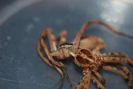
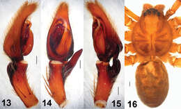
Figures 13–16.Sinopoda serrata (Wang, 1990), holotype, from Mt. Lushan (Jiangxi Province, China). 13 Left male palp, prolateral view 14 Left male palp, ventral view 15 Left male palp, retrolateral view 16 Male habitus, dorsal view. Scales = 0.2 mm (13–15), scale = 1 mm (16).
-
Feng Zhang, Bao-Shi Zhang, Zhi-Sheng Zhang
Zookeys
Figures 18–21.Pseudopoda emei sp. n., Male (SP–SC–03–0050): 18–19 Body (18 dorsal 19 ventral) 20–21 Left palp (20 prolateral 21 ventral). Abbreviations: C conductor; dRTA dorsal branch of retrolateral tibial apophysis; E embolus; EP embolic projection; SD sperm duct; ST subtegulum; T tegulum; vRTA ventral branch of retrolateral tibial apophysis. Scale bars: 2 mm (18–19); 1 mm (20–21).
-
Klinteskoven Møn
-
Feng Zhang, Bao-Shi Zhang, Zhi-Sheng Zhang
Zookeys
Figures 27–33.Pseudopoda emei sp. n., 27–29 Male (SP–SC–03–0050): Left palp (27 prolateral 28 ventral 29 retrolateral). 30–33 Female (SP–SC–03–0052): 30–32 Epigyne (30 ventral 31 dorsal 32 apical) 33 Schematic course of internal duct system, dorsal. Abbreviations: aEF anterior margin of epigynal field; amLL anterior margin of lateral lobes; C conductor; CD copulatory duct; dRTA dorsal branch of retrolateral tibial apophysis; E embolus; EP embolic projection; FD fertilization duct; LL lateral lobes of epigyne; lmLL lateral margin of lateral lobes; MF median field of epigyne; mmLL median margin of lateral lobes; pmLL posterior margins of lateral lobes; PI posterior incisions; R ridges; S spermathecae; SD sperm duct; ST subtegulum; T tegulum; vRTA ventral branch of retrolateral tibial apophysis. Scale bars: 0.5 mm.
-
Klinteskoven Møn
-
Feng Zhang, Bao-Shi Zhang, Zhi-Sheng Zhang
Zookeys
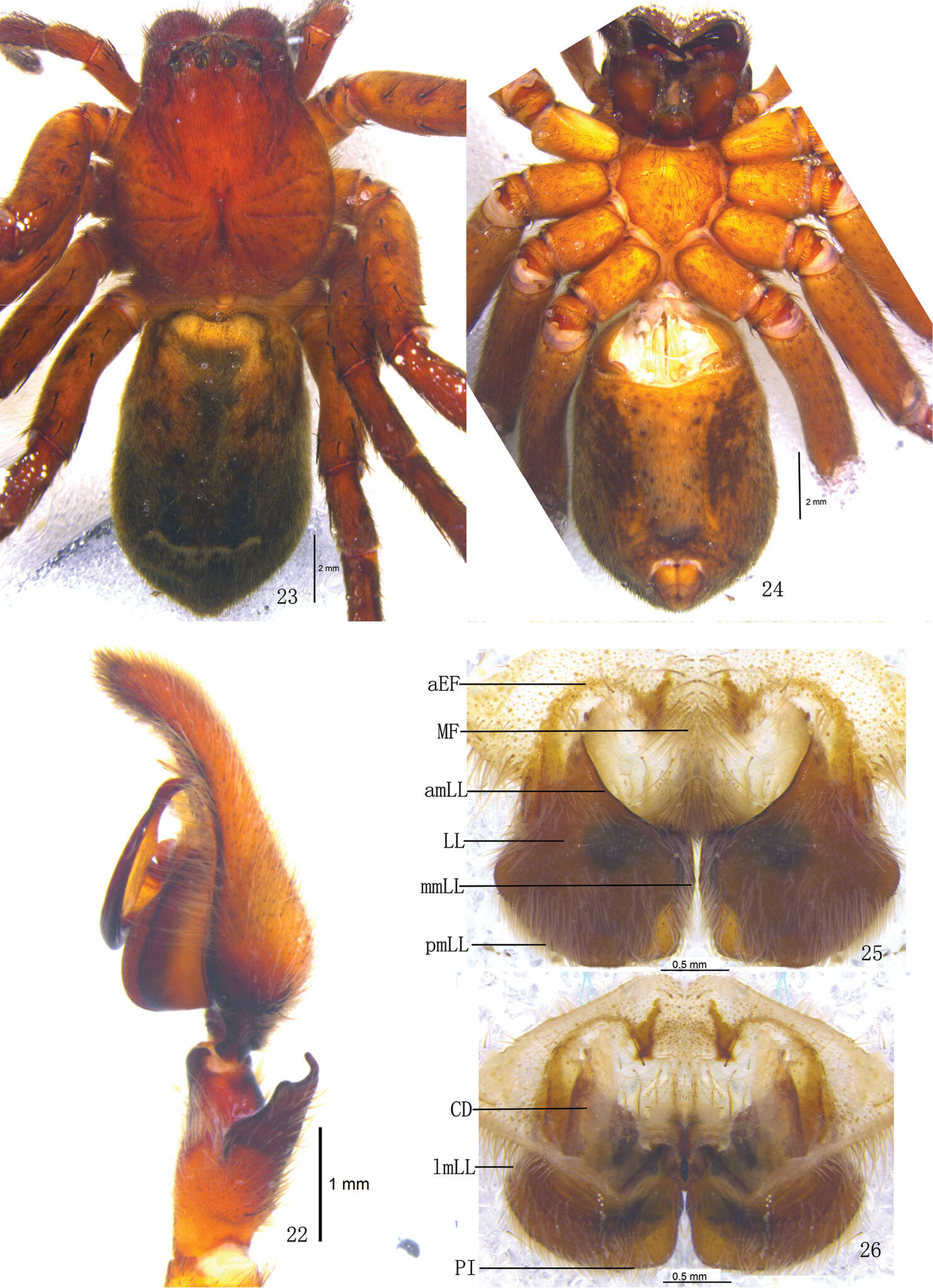
Figures 22–26.Pseudopoda emei sp. n., 22 Left palp of male (retrolateral). 23–26 Female (SP–SC–03–0052): 23–24 Body (23 dorsal 24 ventral) 25–26 Epigyne (25 ventral 26 dorsal). Abbreviations: aEF anterior margin of epigynal field; amLL anterior margin of lateral lobes; CD copulatory duct; LL lateral lobes of epigyne; lmLL lateral margin of lateral lobes; MF median field of epigyne; mmLL median margin of lateral lobes; pmLL posterior margins of lateral lobes; PI posterior incisions. Scale bars: 1 mm (22); 2 mm (23–24); 0.5 mm (25–26).
-
Løvenholm Skov
-
Feng Zhang, Bao-Shi Zhang, Zhi-Sheng Zhang
Zookeys
Figures 34–37.Pseudopoda lacrimosa sp. n., Male (SP–GLGS–11–41): 34–35 Body (17 dorsal 18 ventral) 36–37 Left palp (36 prolateral 37 ventral). Abbreviations: C conductor; dRTA dorsal branch of retrolateral tibial apophysis; E embolus; EP embolic projection; SD sperm duct; ST subtegulum; T tegulum; vRTA ventral branch of retrolateral tibial apophysis. Scale bars: 1 mm (34–35); 0.5 mm (36–37).
-
Løvenholm Skov