الأسماء في صفحات التنقل
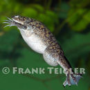
Xenopus laevis has a unique morphology because it lacks a tongue and a visible ear. The body is flattened and head is wedge-shaped and smaller than the body. It has two small eyes found on the top of the head and no eyelids. Its front limbs are small and are not webbed, and its hind legs are large and webbed and the three inside toes on either foot have claws. It has smooth slippery skin which is multicolored on its back with blotches of olive gray or brown and gray, while the underside is creamy white with a yellow tinge. It has lateral lines along its back. Males weigh about 60 grams, are about 5 to 6 centimeters long, and lack a vocal sac, which most male frogs have. Females weigh about 200 grams, are about 10 to 12 centimeters long, and have cloacal extensions at the end of the abdomen.
(Kaplan, 1995; Chang 1998)
Range mass: 60 to 200 g.
Range length: 5 to 12 cm.
Other Physical Features: ectothermic ; heterothermic ; bilateral symmetry
Sexual Dimorphism: female larger
Average basal metabolic rate: 0.012 W.
It is an invasive species all over world because it was used in human pregnancy tests in the 1940's. When more effective means of pregnancy tests were made available, many X. laevis were released all over the world.
IUCN Red List of Threatened Species: least concern
Development - Life Cycle: metamorphosis
Human activities have transplanted this African frog all over the globe, where some claim it is pushing native species out of their niche (Beck 1994). Others argue that there is no documented case of this occurring (Jack Crayon, pers. comm.)
Xenopus laevis has been used extensively as a laboratory research animal, mostly in the field of vertebrate embryology because females are prolific egg layers and embryos are transparent, making it easy to observe the development of the embryo. During the 1940's, female X. laevis were injected with the urine of a woman. If the human was pregnant, then the injected frog would start to produce eggs. Xenopus laevis was the first vertebrate cloned in the laboratory. Magainins are a family of antibiotics found in the skin of X. laevis, which heals wounded skin rapidly. Magainin is an antibiotic, antifungal, antiparasitic, and antiviral, probably useful to the frog because of the stagnant, microbe filled waters in which it lives in. These magainins have been tested as an antibiotic cream, which works just as well as an oral antibiotic, but without the side effects. Xenopus laevis is also used in lab because it is very easy to care for, breed, and observe.
Positive Impacts: source of medicine or drug ; research and education
Xenopus laevis is a scavenger and eats living, dead, or dying arthropods and other pieces of organic waste. It has a voracious appetite and attacks anything that passes in front of it. It uses extremely sensitive fingers, an acute sense of smell, and its lateral line systems to locate food. Lateral line systems, usually found in fish, detect vibrations in the water. It uses a hyobranchial pump to suck food into its mouth. The claws on its hind feet tear apart larger pieces of food. Tadpoles are exclusively filter feeders
(Avila and Frye, 1977; Beck, 1994)
Animal Foods: insects; aquatic or marine worms; aquatic crustaceans
Primary Diet: carnivore (Insectivore , Scavenger )
Xenopus laevis occurs naturally in southern Africa. There are substantial introduced populations in California, Chile, Great Britain, and probably many other locations around the world. (Nieukoop and Faber, 1994)
Biogeographic Regions: nearctic (Introduced ); palearctic (Introduced ); ethiopian (Native )
Xenopus laevis lives in warm, stagnant grassland ponds as well as in streams in arid and semi-arid regions. The ponds are usually devoid of any higher plant vegetation, and covered in green algae. Xenopus laevis can tolerate wide variation in water pH, but the presence of metal ions proves toxic. It thrives in temperatures from 60 to 80 degrees Fahrenheit. It is almost totally aquatic, only leaving the water when forced to migrate.
(Nieuwkoop and Faber, 1994; Beck, 1994; Kaplan, 1995, Jack Crayon, personal communication)
Habitat Regions: temperate ; tropical ; freshwater
Aquatic Biomes: lakes and ponds; rivers and streams
African clawed frogs can reach 15 to 16 years old in wild and feral populations. Captive animals have been known to live as long as 20 years.
Range lifespan
Status: wild: 16 (high) years.
Range lifespan
Status: captivity: 20 (high) years.
Average lifespan
Status: captivity: 8.8 years.
Average lifespan
Status: captivity: 15.0 years.
Xenopus laevis is sexually mature in 10 to 12 months. Mating can take place during any time of the year, but is most common in the spring, and can take place up to four times per year. Males vocalize during the evening to attract females. Although the male lacks a vocal sac, it produces a mating call by rapid contractions of the intrinsic laryngeal muscles. This mating call sounds like alternating long and short trills. After the female hears this, she responds with either an acceptance call (a rapping sound) or a rejection call (slow ticking sound). This is a nearly unique behavior in the animal world; rarely does a female answer the males call. Mating often takes place at night, when there are few disturbances. The male develops mating pads on the underside of his forearms and hands. The mating embrace, amplexus, is pelvic, whereas most frogs have axillary (front limb) amplexus. The female can release hundreds of sticky eggs during the 3 to 4 hour event, which are typically attached to plants or other anchors, one or more at a time. The eggs grow into tadpoles, which filter feed. The tadpole metamorphoses into a small froglet, with the tail being absorbed into the body and sustaining its nutritional requirements during this period, which lasts about 4 to 5 days. The total change from egg to small frog takes about 6 to 8 weeks.
(Kaplan, 1995; Beck, 1994; Chang, 1998; Kelley, 1998, Jack Crayon, personal communication)
Breeding interval: African clawed frogs can breed up to 4 times each year.
Breeding season: Mating can take place during any time of the year, but is most common in the spring.
Range age at sexual or reproductive maturity (female): 10 to 12 months.
Range age at sexual or reproductive maturity (male): 10 to 12 months.
Key Reproductive Features: iteroparous ; year-round breeding ; gonochoric/gonochoristic/dioecious (sexes separate); sexual ; fertilization (External ); oviparous
African clawed frogs were first brought to California decades ago to help doctors figure out whether their patients were pregnant. After new technology made those pregnancy tests obsolete, the creatures were let loose, and thrived for decades in the state's drainage ditches and ponds.
Now there are signs that the proliferation of African clawed frogs may be putting native species in peril. A study published last week in the journal PLOS ONE reveals that they carry a deadly fungus responsible for wiping out vast numbers of amphibians around the world.
The spread of the deadly Batrachochytrium dendrobatidis fungus is contributing to one of the greatest disease-caused losses of biodiversity in recorded history. The fungus causes amphibians' skin to harden, interfering with the regulation of electrolytes and eventually causing cardiac arrest.
By Geoffrey Mohan, Los Angeles Times May 19, 2013, 9:56 p.m.
Die gewone platanna (Xenopus laevis) is 'n padda wat endemies is aan Afrika suid van die Sahara. In Engels staan dit bekend as die African clawed frog. Hierdie padda was gebruik om swangerskap van vroue te bevestig. Dit het tot gevolg gehad dat lewende paddas uitgevoer is na die res van die wêreld waar hulle indringerspesies geword het.
Die padda is plat met die oë bo-op die voorkop en dit het 'n afgeplatte lyf. Die voorpote is kort en dun en die agterpote groot en sterk met swemvliese tussen die tone. Die padda is byna totaal waterlewend en kom slegs tydens reën land toe.
Hulle vreet waterinsekte, paddavissies en vissies.
Die gewone platanna (Xenopus laevis) is 'n padda wat endemies is aan Afrika suid van die Sahara. In Engels staan dit bekend as die African clawed frog. Hierdie padda was gebruik om swangerskap van vroue te bevestig. Dit het tot gevolg gehad dat lewende paddas uitgevoer is na die res van die wêreld waar hulle indringerspesies geword het.
La granota amb ungles africana (Xenopus laevis, també coneguda com a platanna) és una espècie de granota sud-africana. El seu nom comú deriva de les tres ungles curtes de cada pota posterior, que utilitza per arrencar trossos dels seus aliments. El nom del gènere, Xenopus, significa "peu estrany" i l’epítet específic, laevis, significa "llis".
Aquesta granota és un organisme model
Té una llargada de 12 cm. Té el cap i el cos aplanat però no té ni llengua ni orelles externes.
Es troba a la major part de l'Àfrica subsahariana i ha estat introduïda en llocs aïllats de l'Amèrica del Nord, Sud-amèrica i Europa. .[1] Totes les espècies de la família Pipidae no tenen llengua ni dents i són totalment aquàtiques. Les Pipidae són animals carronyers a més d'alimentar-se de moltes menes de preses vives.
És un organisme model important en biologia del desenvolupament. És tetraploide i té un embrió fàcilment manipulable.
Els oòcits de Xenopus proporcionen un sistema d’expressió important per la biologia molecular[2]
El primer vertebrat que es va aconseguir clonar va ser aquesta espècie de granota.
A més diversos exemplars de la granota amb ungles africana van anar a l’espai amb la llançadora espacial Endeavour l’any 1992 per veure si a gravetat zero la reproducció i el desenvolupament eren normals[3][4]
X. laevis també es va fer servir com test de l’embaràs.[5]
Xenopus laevis s’ha usat com animal de companyia des de l’inici de la dècada de 1950. Són animals molt resistents i viuen força temps en captivitat, fins a 30 anys.[6]
Són animals voraços i s’adapten a molts hàbitats.[7] Per això pot ser una espècie invasora.
La granota amb ungles africana (Xenopus laevis, també coneguda com a platanna) és una espècie de granota sud-africana. El seu nom comú deriva de les tres ungles curtes de cada pota posterior, que utilitza per arrencar trossos dels seus aliments. El nom del gènere, Xenopus, significa "peu estrany" i l’epítet específic, laevis, significa "llis".
Aquesta granota és un organisme model
Té una llargada de 12 cm. Té el cap i el cos aplanat però no té ni llengua ni orelles externes.
Es troba a la major part de l'Àfrica subsahariana i ha estat introduïda en llocs aïllats de l'Amèrica del Nord, Sud-amèrica i Europa. . Totes les espècies de la família Pipidae no tenen llengua ni dents i són totalment aquàtiques. Les Pipidae són animals carronyers a més d'alimentar-se de moltes menes de preses vives.
Drápatka vodní (Xenopus laevis) je druh bezjazyčné vodní žáby z Afriky. České jméno vychází z toho, že tato žába má na zadních nohou malé černé drápky.
Samice dorůstají délky 10 až 15 cm, samci bývají asi o třetinu menší. Tvar těla je přizpůsoben výlučně vodnímu životu, zadní nohy jsou posunuty za tělo. Zbarvení se pohybuje v tónech hnědé, šedé či olivově zelené, časté jsou více nebo méně výrazné skvrny na hřbetě. Břicho je světlejší. V chovech existují také albíni této žáby, kteří jsou často prodáváni v obchodech se zvířaty. Drápatka vodní, stejně jako všechny žáby z čeledi pipovitých, nemá jazyk. Oči jsou opatřeny víčky. Malé přední nohy mají 4 prsty bez plovací blány. Zadní nohy, sloužící k plavání, jsou mohutně vyvinuty a je na nich 5 prstů, opatřených plovací blánou. Na 3 vnitřních prstech zadních nohou jsou černé drápky. Na bocích mají tyto žáby zvláštní světlé útvary, připomínající stehy. Obsahují smyslový orgán, který je schopný rozeznávat vibrace ve vodě a umožňuje drápatce objevit kořist nebo nepřítele i v kalné vodě.[2]
Samice jsou obecně mohutnější. Navíc jsou u samičky viditelné kloakální pysky jako malá trubička mezi zadníma nohama. Samečci během období páření mají na prstech předních nohou tmavší pářicí mozoly.[3]
Přirozený areál rozšíření je v Africe okolo rovníku (nejseverněji patrně ve Středoafrické republice) a ve zbytku Afriky na jih od rovníku (nejjižněji v JAR). Tato žába se ale mimo to nachází i na jiných místech světa, kam byla uměle zavlečena. Vyskytuje se v USA, Mexiku, Chile, Indonésii, na jihu Walesu a byla také spatřena ve Francii a na Jávě.[4]
Vypadá to, že drápatky stojí za úbytkem početnosti některých druhů žab. Na místa, kam byly drápatky zavlečeny, s sebou přinesly jisté houbové onemocnění. Samotné drápatky jsou proti němu odolné, ale ostatní druhy žab v zasažených ekosystémech toto onemocnění postihlo.[4][5] V roce 2007 drápatky vodní pronikly do vodní nádrže v San Francisku. Panuje teď velká debata o tom, jak se těchto žab zbavit a zamezit jim v šíření.[6][7]
Drápatky jsou výlučně vodní žáby. Obývají stojaté i tekoucí vody. Pokud vodní nádrž nebo tok vyschne, přežívají ve stavu strnulosti.[8] Dokáží plavat velice rychle. Živí se různými vodními živočichy - vodním hmyzem, malými korýši nebo rybkami. Pravidelně svlékají kůži a tu pak požírají.
Ve své domovině se drápatky páří během deštivých měsíců.[3] Přestože nemají zvukový vak (na rozdíl od většiny žab), samci při námluvách skřehotají. Střídají dlouhé a krátké tóny stahováním vnitřních hrtanových svalů. Zřídka se stává, že samice také odpovídají zvukem. Ochotu k páření signalizují rychlým kuňkáním, odmítnutí signalizují pomalým kuňkáním.[9]
Samice během páření vyloučí do vody 500 až 2000 vajíček a samec je průběžně oplodňuje. Pulci se líhnou po 2 až 3 dnech. Proměna pulců v malé žabky nastává po 5 až 7 týdnech, pohlavní dospělosti žáby dosáhnou přibližně po jednom roce. Samice rostou rychleji.[3]
Obvyklá délka života se pohybuje mezi 5 a 15 lety, ale jsou známy i případy, kdy se tyto žáby dožily v zajetí 30 let.[10]
Drápatka vodní nemá tak krátký životní cyklus a genetickou jednoduchost, což je obecně požadováno u modelových organismů v genetickém výzkumu. Přesto je to důležitý modelový organismus ve vývojové biologii. Drápatka vodní dosahuje pohlavní dospělosti po jednom roce a (jako většina zástupců rodu Xenopus) je tetraploidní. Na druhou stranu má velká a snadno manipulovatelná embrya. Roger Wolcott Sperry použil drápatky vodní pro své slavné experimenty popisující vývoj zrakového systému. Vajíčka drápatek poskytují dobrý prostředek k vyjádření účelu dané DNA, což se využívá v molekulární biologii. Vložením DNA nebo mRNA do vajíčka nebo vyvíjejícího se embrya mohou vědci studovat tvorbu proteinů v uzavřené kontrolované soustavě. To umožňuje rychlé vyjádření manipulované DNA (nebo mRNA).
Předtím, než byly vynalezeny moderní těhotenské testy, byly k prokázání těhotenství využívány drápatky vodní. Byl to rozšířený způsob zejména ve 30. až 60. letech 20. století. Test se prováděl tak, že moč ženy se vstříkla do žáby. Pokud je žena těhotná, hormony v její moči podnítí ovulaci a žába začne během několika hodin klást vajíčka. Je to reakce na hormon gonadotropin, který se ve velkém množství nachází v krvi a moči těhotných žen.[5][11]
Tyto žáby jsou na chov nenáročné. K chovu páru žab postačí akvárium o objemu 40 litrů. Nemusí mít žádné zařízení, takovéto „holé“ akvárium je pak snadněji udržovatelné. Souš také není potřeba, drápatky ji vůbec nevyužívají. Teplota vody by se měla pohybovat okolo 20 až 25 °C, ale žáby mohou žít v chladnější i v teplejší vodě. Drápatky není vhodné chovat společně s akvarijními rybami, protože dříve nebo později by se ryby pravděpodobně staly žabí potravou. Ze stejného důvodu by společně chované žáby měly mít přibližně stejnou velikost. Jako krmivo se používají komáří larvy, rybky, žížaly a podobně - podle velikosti žab. Občas se dá se přikrmovat i syrovým libovým masem. Nejuniverzálnějším krmivem jsou mražené larvy pakomárů, dostupné v obchodech se zvířaty.
Páření v akváriu lze stimulovat čerstvou teplou vodou po několika chladných týdnech. Vajíčka je třeba od rodičů oddělit, žáby je totiž rády požírají. Pulci se živí jemnou potravou (kopřivový prášek, kvasnice).[3][12]
V tomto článku byl použit překlad textu z článku African clawed frog na anglické Wikipedii.
Drápatka vodní (Xenopus laevis) je druh bezjazyčné vodní žáby z Afriky. České jméno vychází z toho, že tato žába má na zadních nohou malé černé drápky.
Xenopus laevis er en afrikansk sporefrø.
I 1940'erne og 1950'erne blev Xenopus laevis brugt til at lave nogle af de første pålidelige graviditetstest. I 1930'erne opdagede forskere at urin fra gravide kvinder kunne få hunsporefrøer til at lægge æg. Det skyldes at urin (og blod) fra den gravide indeholder hormonet gonadotropin. I den gravide kvindes krop er gonadotropins funktion at forhindre ægløsning og afstødelse af fosteret. Graviditetstestene foregik ved, at man injicerede urin fra en kvinde i Xenopus laevis, og hvis den så inden for 24 timer begyndte at lægge æg, var kvinden med stor sandsynlighed gravid. Man skulle naturligvis være omhyggelig med at sikre sig at æglægningen ikke skete af andre årsager. I 1960'erne blev frøerne afløst at kemiske analyser.
Der Glatte Krallenfrosch (Xenopus laevis), auch Afrikanischer Krallenfrosch, Apothekerfrosch, Glatter Spornfrosch oder oft einfach nur Krallenfrosch genannt, ist eine der Arten aus der Gattung der Krallenfrösche (Xenopus) innerhalb der Zungenlosen Frösche (Familie Pipidae). Wissenschaftlich nicht mehr gütige Synonyme: Pipa laevis, Pipa bufonia, Engystoma laevis, Xenopus boiei.[1] Die Krallenfrösche stellen das afrikanische Gegenstück zu den südamerikanischen Wabenkröten (Gattung Pipa) aus derselben taxonomischen Familie dar.
Krallenfrösche sind an vielen Universitäten seit langem bevorzugte Modellorganismen für Lehre und Forschung.
Die natürliche Verbreitung beschränkt sich auf Afrika südlich der Sahara. Schwerpunkte liegen in Angola, Namibia, Eswatini, Malawi und Simbabwe. Aufgrund einer jahrzehntelangen, massenhaften Verwendung als Labortier (früher als Indikator-Organismus für Schwangerschaftstests, heute in der Entwicklungsbiologie; siehe unten) sowie für den Zoohandel haben sich die Tiere durch Unachtsamkeit des Menschen auch in großen Teilen des Südens der USA und teilweise sogar in Europa (Frankreich, Italien und Portugal) im Freiland angesiedelt.[2][3] Hier halten sich die anpassungsfähigen Krallenfrösche bevorzugt in Tümpeln und anderen warmen, meist stehenden Gewässern auf.
Als Aquarientier in menschlicher Obhut kann man diesen Lurch oft finden, da er aufgrund seiner aquatilen Lebensform und seiner einfachen Haltung sehr beliebt ist.
Der Glatte Krallenfrosch hat einen flachen Kopf und einen stromlinienförmigen Körper, der bei weiblichen Exemplaren etwa 10 bis 13 Zentimeter lang wird – Männchen bleiben deutlich kleiner. Die Rückenfarbe ist vorwiegend oliv-braun und der Bauch sowie die Innenseiten der Schenkel meistens hell-beige mit zahlreichen unregelmäßig verteilten, kleinen Pigmentierungen. Die wenig entwickelten Vorderbeine haben je vier lange Finger ohne Schwimmhäute (im Gegensatz zu den Zwergkrallenfröschen). Die auffallend muskulösen Hinterbeine tragen je fünf Zehen, wobei die drei inneren mit kräftigen, schwarzen Hornkrallen versehen sind, die der Gattung ihren Namen gaben. Zwischen den Zehen befinden sich große Schwimmhäute, die bis an die Zehenspitzen reichen. Die Frösche sind hervorragende Schwimmer, die sich unter Wasser durch Rudern und Stoßen mit den Hinterbeinen sehr schnell fortbewegen. Die kleinen, runden Augen sind nach oben gerichtet und erfassen sofort jede Bewegung, die sich über der Wasseroberfläche abspielt. Trotz der rein aquatilen Lebensweise handelt es sich um ein lungenatmendes Tier; nur die Kaulquappen besitzen Kiemen. Die Haut des Krallenfrosches ist derart glatt und schlüpfrig, dass man ihn mit bloßen Händen kaum ergreifen kann. Farbliche Varianten, wie bei vielen Amphibien, sind auch bei dieser Art vorhanden. Albinismus tritt vor allem bei gezüchteten Tieren häufig auf.
Larve mit "Barteln" (Sinnesorgane)
Larve mit bereits entwickelten Beinen
Zwei Jungtiere nach der Metamorphose
Adulter Albino mit roten Augen
Der Frosch, der rund 15 bis etwa 25 Jahre alt werden kann, lebt ständig in ruhigen Gewässern, die er nur notgedrungen wie bei Austrocknung oder Nahrungsmangel verlässt. Er ist vorwiegend dämmerungs- und nachtaktiv. Die Männchen rufen unter Wasser mit dunkler Stimme („gra-gra-gra“ bzw. „kreik-kreik“). Die Weibchen wachsen schneller heran und sind entsprechend früher geschlechtsreif als die Männchen. Man erkennt sie daran, dass sie bei gleichem Alter gut ein Viertel größer sind als die männlichen Exemplare. Außerdem haben nur sie drei lappige Fortsätze an der Kloake. Während der Laichzeit tragen die Männchen deutlich sichtbare, dunkle Brunstschwielen an den Innenseiten der Arme. Sie umklammern, wie alle Arten der Mesobatrachia (und auch der Archaeobatrachia), die Weibchen in der Leistengegend – man spricht von einem inguinalen Amplexus. In der freien Wildbahn laichen Krallenfrösche bei guten Umweltbedingungen mehrmals im Jahr ab.
Während des Laichaktes, der sich auf bis zu fünf Tage hinziehen kann, heftet das Weibchen die Eier an Wasserpflanzen oder anderes. Nach knapp einer Woche schlüpfen etwa vier Millimeter große Larven, die in ihren ersten Lebenstagen von ihrem Dottersack zehren. Im Anschluss daran stellen die Larven ihre Ernährung auf Filtration von Feinstpartikeln um. Die Kaulquappen von Xenopus fallen dadurch auf, dass sie sich mit nach unten gesenktem Kopf und ondulierenden Schwanzschlägen langsam durchs Wasser bewegen. Am Mund weisen sie zwei lange Barteln auf, die an den Seiten der Mundspalte entspringen und nach vorne gerichtet sind. Mit ihnen ertasten die Tiere sowohl die Umgebung als auch kleinste Nahrungsorganismen am Boden. Ständig öffnet und schließt sich ihr Maul – sie nehmen so Wasser auf, um dieses mit Hilfe der Kiemenbögen nach planktischen Nahrungspartikeln (z. B. Grünalgen, Kieselalgen) zu filtern. Durch die paarigen Atemlöcher (Spiracula) wird das Wasser wieder abgegeben. Nach etwa vier Wochen stellen die nun schon sehr den Alttieren ähnelnden Jungtiere ihre Ernährung auf feste tierische Kost um. Die Barteln werden in dieser Zeit zurückgebildet.
Seine Nahrung sucht der Krallenfrosch in freier Wildbahn in den oberen Sedimentschichten des Gewässergrundes, indem er mit seinen Vorderarmen das Substrat nach Kleinorganismen durchwühlt. Da X. laevis weder Zunge noch Zähne besitzt, werden die aufgewirbelten Beutetiere über ein spezielles Verhalten, das „Saugschnappen“, erfasst und geschluckt. Man weiß, dass Krallenfrösche am ganzen Körper ungefähr 200 Sinneszellen besitzen, die wie bei den Fischen als Seitenlinienorgan fungieren. Mit Hilfe dieses Organs registriert der Frosch in seinem näheren Umfeld sowohl Wasserbewegungen als auch wasserchemische Veränderungen. Dank dieser Sinnesleistung sind die Tiere in der Lage, sich ein genaues Bild über Art und Position der Beute zu machen. Man erkennt das Seitenlinienorgan recht gut anhand erhabener, „nahtähnlicher“ Linien aus kleinen Wulsten auf der Hautoberfläche des Frosches.
Normalerweise ernähren sich Krallenfrösche von wasserlebenden Insektenlarven und Würmern; aber auch kleinere Fische, Krebse sowie Amphibienlarven werden gerne verzehrt. Bei hohen Besatzdichten in Aquarien oder Zuchtanlagen kann unter den Fröschen Kannibalismus auftreten. Dabei werden die eigenen Gelege oder Kaulquappen erbeutet. Vor potentiellen Fressfeinden (Wasservögel, Raubfische) schützt sich der Krallenfrosch mittels seiner glatten Hautoberfläche und eines Drüsensekrets, das giftig ist. Beim direkten Hautkontakt kann es beim Menschen Allergien auslösen.
Krallenfrösche sind an vielen Universitäten seit langem bevorzugte Modellorganismen für die zellbiologische und entwicklungsphysiologische Lehre und Forschung.[4][5] Ihre Embryonen in den zahlreich ins Wasser abgegebenen Eiern (10.000 bis 15.000 Stück pro Weibchen und Jahr) sind leicht zugänglich, und auch unbefruchteter Laich kann durch die Gabe des menschlichen Hormons Choriongonadotropin erhalten und später jederzeit befruchtet werden. Die Eier sind relativ groß und resistent gegen Infektionen nach Eingriffen, wie etwa Transplantationen. Unter normalen Lebensbedingungen wird X. laevis bereits mit zwei Jahren geschlechtsreif. Generell sind die Tiere im Labor leicht zu halten. Größere Plastikwannen mit beheiztem Wasser um die 22 °C sind völlig ausreichend. Für die Zucht empfehlen sich höhere Temperaturen (24–26 °C). Wichtig sind feste Abdeckungen über den Bassins, da die Frösche sehr springfreudig sind.
Bis in die 1960er-Jahre wurden mit dem Krallenfrosch auch in deutschen Apotheken noch Schwangerschaftstests durchgeführt (der sogenannte Froschtest) – daher der Name „Apothekerfrosch“.[6] Frauen, bei denen möglicherweise eine Schwangerschaft vorlag, brachten dem Apotheker ihren Morgenurin, von dem dann einem jungen Krallenfroschweibchen etwas unter die Haut in den dorsalen Lymphsack gespritzt wurde. Produzierte das Tier innerhalb von 12 Stunden Eier (manchmal bis zu 2000 Stück), galt dies als ein positiver Schwangerschaftsbefund.[7]
Der Frosch reagiert bei diesem auch „Hogben-Test“ genannten Ablauf, den der englische Forscher Lancelot Hogben (1895–1975) Anfang der dreißiger Jahre in Kapstadt entdeckte,[8] auf das gleiche Hormon (Humanes Choriongonadotropin oder hCG), auf das auch bei heute üblichen Verfahren zur Schwangerschaftsindikation geprüft wird. Theoretisch wären andere Froschlurche, beispielsweise die heimischen Erdkröte (Bufo bufo), für diesen Test ebenfalls geeignet, aber die Haltungsbedingungen von adulten Erdkröten sind um ein Vielfaches aufwändiger. Bis zum Zweiten Weltkrieg herrschte ein schwunghafter Handel mit Wildfängen von afrikanischen Krallenfröschen. In den 1940er-Jahren gelang dann erstmals die erfolgreiche Nachzucht. Man hatte entdeckt, dass auch die Männchen mit den wirksamen Faktoren des Schwangerenurins, dem so genannten „Prolan“, behandelt werden müssen, um die Fortpflanzung in Gefangenschaft besonders zu fördern.
Die zum Froschtest eingeführten afrikanischen Krallenfrösche waren häufig mit dem Chytridpilz (Batrachochytrium dendrobatidis) infiziert, der dadurch weltweit verbreitet wurde und heute als eine der Ursachen für das globale Amphibiensterben angesehen wird.[9][10] Wie in einem im März 2019 veröffentlichten Science-Artikel festgestellt wurde, ist B. dendrobatidis (Bd) derzeit für erhebliche Bestandsrückgänge bei mehr als 500 Amphibienarten verantwortlich.[11] In Deutschland ist nachweislich die stark gefährdete Wechselkröte davon betroffen.[12]
Rechtliches
Seit dem 1. Januar 1989 besteht in Deutschland durch die Versuchstier-Meldeverordnung eine gesetzliche Verpflichtung zur Erfassung der für wissenschaftliche Versuche verwendeten Tiere. Das deutsche Bundesministerium für Ernährung und Landwirtschaft veröffentlicht dazu jedes Jahr entsprechende Statistiken. Jede Einrichtung, die Tierversuche durchführen möchte, muss einen Tierschutzbeauftragten benennen. Diese sind entweder Tierärzte, Ärzte oder Biologen der Fachrichtung Zoologie sein. Sie sind dafür verantwortlich, im Betrieb auf die Einhaltung der tierschutzrechtlichen Regelungen zu achten sowie die Personen, die mit den Tieren umgehen, zu beraten. Im Mai 2002 wurde der Tierschutz auch in das Grundgesetz aufgenommen, um ihm mehr Gewicht zu verleihen. Eine Novellierung des Tierschutzgesetzes trat am 13. Juli 2013 in Kraft unter anderem mit Bestimmungen zu den Versuchstierrichtlinien. Diese gelten auch für den Krallenfrosch.
Bereits in der bekannten und Anfang des 20. Jh. weit verbreiteten Enzyklopädie Brehms Tierleben (1920) findet sich eine ausführliche und gut bebilderte Beschreibung von Xenopus laevis, damals noch als Glatter Spornfrosch bezeichnet.[13] Schon zu Lebzeiten von Alfred Brehm wurden Exemplare nach Europa gebracht und in Aquarien zur genaueren Beobachtung gehalten. Eine weitere aussagekräftige Illustration findet sich in dem Grundlagenwerk von Hans Friedrich Gadow: Amphibia and Reptiles. London, New York, Macmillan and Co., auf Seite 147 (Fig. 29), aus dem Jahr 1901. Die Abbildung zeigt X. laevis mit Larven im natürlichem Umfeld.
Xenopus laevis, Brehms Tierleben, Bd. 4 (1) Lurche und Kriechtiere (1920)
Xenopus laevis, Clawed Toad, adult and larvae (Amphibia and Reptiles 1901)
Der Glatte Krallenfrosch (Xenopus laevis), auch Afrikanischer Krallenfrosch, Apothekerfrosch, Glatter Spornfrosch oder oft einfach nur Krallenfrosch genannt, ist eine der Arten aus der Gattung der Krallenfrösche (Xenopus) innerhalb der Zungenlosen Frösche (Familie Pipidae). Wissenschaftlich nicht mehr gütige Synonyme: Pipa laevis, Pipa bufonia, Engystoma laevis, Xenopus boiei. Die Krallenfrösche stellen das afrikanische Gegenstück zu den südamerikanischen Wabenkröten (Gattung Pipa) aus derselben taxonomischen Familie dar.
Krallenfrösche sind an vielen Universitäten seit langem bevorzugte Modellorganismen für Lehre und Forschung.
The African clawed frog (Xenopus laevis), also known as the xenopus, African clawed toad, African claw-toed frog or the platanna) is a species of African aquatic frog of the family Pipidae. Its name is derived from the three short claws on each hind foot, which it uses to tear apart its food. The word Xenopus means 'strange foot' and laevis means 'smooth'.
The species is found throughout much of Sub-Saharan Africa (Nigeria and Sudan to South Africa),[2] and in isolated, introduced populations in North America, South America, Europe, and Asia.[1] All species of the family Pipidae are tongueless, toothless and completely aquatic. They use their hands to shove food in their mouths and down their throats and a hyobranchial pump to draw or suck things in their mouth. Pipidae have powerful legs for swimming and lunging after food. They also use the claws on their feet to tear pieces of large food. They have no external eardrums, but instead subcutaneous cartilaginous disks that serve the same function.[3] They use their sensitive fingers and sense of smell to find food. Pipidae are scavengers and will eat almost anything living, dying, or dead and any type of organic waste.
It is a pest in many countries, including across Europe.[4]
These frogs are plentiful in ponds and rivers within the south-eastern portion of Sub-Saharan Africa. They are aquatic and are often greenish-grey in color. African clawed frogs have been frequently sold as pets, and sometimes incorrectly misidentified as African dwarf frogs. Albino clawed frogs are common and sold as animals for laboratories.
They reproduce by fertilizing eggs outside of the female's body (see frog reproduction). Of the seven amplexus modes (positions in which frogs mate), these frogs are found breeding in inguinal amplexus, where the male clasps the female in front of the female's back legs and squeezes until eggs come out. The male then sprays sperm over the eggs to fertilize them.
African clawed frogs are highly adaptable and will lay their eggs whenever conditions allow it. During wet rainy seasons they will travel to other ponds or puddles of water to search for food.[5] During times of drought, the clawed frogs can burrow themselves into the mud, becoming dormant for up to a year.[6]
Xenopus laevis have been known to survive 15 or more years in the wild and 25–30 years in captivity.[7] They shed their skin every season, and eat their own shed skin.
Although lacking a vocal sac, the males make a mating call of alternating long and short trills, by contracting the intrinsic laryngeal muscles. Females also answer vocally, signaling either acceptance (a rapping sound) or rejection (slow ticking) of the male.[8][9] This frog has smooth slippery skin which is multicolored on its back with blotches of olive gray or brown. The underside is creamy white with a yellow tinge.
Male and female frogs can be easily distinguished through the following differences. Male frogs are small and slim, while females are larger and more rotund. Males have black patches on their hands and arms which aid in grabbing onto females during amplexus. Females have a more pronounced cloaca and have hip-like bulges above their rear legs where their eggs are internally located.

Both males and females have a cloaca, which is a chamber through which digestive and urinary wastes pass and through which the reproductive systems also empty. The cloaca empties by way of the vent which in reptiles and amphibians is a single opening for all three systems.[10]
African clawed frogs are fully aquatic and will rarely leave the water except to migrate to new water bodies during droughts or other disturbances. Clawed frogs have powerful legs that help them move quickly both underwater and on land. Feral clawed frogs in South Wales have been found to travel up to 2 kilometres (1.2 mi) between locations.[11] The feet of Xenopus species have three black claws on the last three digits. These claws are used to rip apart food and scratch predators.
Clawed frogs are carnivores and will eat both living and dead prey including fish, tadpoles, crustaceans, annelids, arthropods, and more. Clawed frogs will try to consume anything that is able to fit into their mouths. Being aquatic, clawed frogs use their sense of smell and their lateral line to detect prey rather than eyesight like other frogs. However, clawed frogs can still see using their eyes and will stalk prey or watch predators by sticking their heads out of the water.[12] Clawed frogs will dig through substrate to unearth worms and other food. Their tongue is unable to extend like other frogs, so clawed frogs use their hands to grab food and shovel it into their mouths.
These frogs are particularly cannibalistic; the stomach contents of feral clawed frogs in California have revealed large amounts of the frog's larvae.[13] Clawed frog larvae are filter feeders and collect nutrients from plankton, allowing adult frogs that consume the tadpoles to have access to these nutrients. This allows clawed frogs to survive in areas that have little to no other food sources.
Clawed frogs are nocturnal and most reproductive activity and feeding occurs after dark. Male clawed frogs are very promiscuous and will grab onto other males and even other species of frogs.[14][15] Male frogs that are grasped will make release calls and attempt to break free.
If not feeding, clawed frogs will just sit motionless on top of the substrate or floating at the top with their heads sticking out.
The X. laevis liver responds to low temperatures by increasing production of type II iodothyronine deiodinase through increased food intake. This in turn spurs the thyroid to increase T3 to increase body temperature. (This T3 increase also induces germ cell apoptosis, mediated through genes left over from tadpole metamorphosis.)[16]
The effects of provocation of T hormone release are broadly differentiated by where it starts: If centrally, within the mediobasal hypothalamus, then it stimulates seasonal testicular growth; if peripherally, then testicular regression and cold-season thermogenesis.[16]
These observations are regarded as widely applicable across vertebrate thyroid systems.[16]
The lipidomics of Xenopus oocytes has been studied by Tian et al 2014 and Phan et al 2015.[17]

In the wild, X. laevis are native to wetlands, ponds, and lakes across arid/semiarid regions of Sub-Saharan Africa.[2][19] X. laevis and X. muelleri occur along the western boundary of the Great African Rift. The people of the sub-Saharan are generally very familiar with this frog, and some cultures use it as a source of protein, an aphrodisiac, or as fertility medicine. Two historic outbreaks of priapism have been linked to consumption of frog legs from frogs that ate insects containing cantharidin.[20]
X. laevis in the wild are commonly infected by various parasites,[18] including monogeneans in the urinary bladder.
Xenopus embryos and eggs are a popular model system for a wide variety of biological studies, in part because they have the potential to lay eggs throughout the year.[21][22][23] This animal is widely used because of its powerful combination of experimental tractability and close evolutionary relationship with humans, at least compared to many model organisms.[21][22] For a more comprehensive discussion of the use of these frogs in biomedical research, see Xenopus.
Xenopus laevis is also notable for its use in the first widely used method of pregnancy testing. In the 1930s, two South African researchers, Hillel Shapiro and Harry Zwarenstein,[24] students of Lancelot Hogben at the University of Cape Town, discovered that the urine from pregnant women would induce oocyte production in X. laevis within 8–12 hours of injection.[25] This was used as a simple and reliable test up through to the 1960s.[26] In the late 1940s, Carlos Galli Mainini[27] found in separate studies that male specimens of Xenopus and Bufo could be used to indicate pregnancy[28] Today, commercially available hCG is injected into Xenopus males and females to induce mating behavior and to breed these frogs in captivity at any time of the year.[29]
Xenopus has long been an important tool for in vivo studies in molecular, cell, and developmental biology of vertebrate animals. However, the wide breadth of Xenopus research stems from the additional fact that cell-free extracts made from Xenopus are a premier in vitro system for studies of fundamental aspects of cell and molecular biology. Thus, Xenopus is the only vertebrate model system that allows for high-throughput in vivo analyses of gene function and high-throughput biochemistry.[21]
Xenopus oocytes are a leading system in their own right for studies of various systems, including ion transport and channel physiology.[21] Xanthos et al 2001 uses oocytes to uncover T-box expression earlier than previously found in vertebrates.[30]
Although X. laevis does not have the short generation time and genetic simplicity generally desired in genetic model organisms, it is an important model organism in developmental biology, cell biology, toxicology and neurobiology. X. laevis takes 1 to 2 years to reach sexual maturity and, like most of its genus, it is tetraploid. It does have a large and easily manipulated embryo, however. The ease of manipulation in amphibian embryos has given them an important place in historical and modern developmental biology. A related species, Xenopus tropicalis, is now being promoted as a more viable model for genetics.
Roger Wolcott Sperry used X. laevis for his famous experiments describing the development of the visual system. These experiments led to the formulation of the chemoaffinity hypothesis.
Xenopus oocytes provide an important expression system for molecular biology. By injecting DNA or mRNA into the oocyte or developing embryo, scientists can study the protein products in a controlled system. This allows rapid functional expression of manipulated DNAs (or mRNA). This is particularly useful in electrophysiology, where the ease of recording from the oocyte makes expression of membrane channels attractive. One challenge of oocyte work is eliminating native proteins that might confound results, such as membrane channels native to the oocyte. Translation of proteins can be blocked or splicing of pre-mRNA can be modified by injection of Morpholino antisense oligos into the oocyte (for distribution throughout the embryo) or early embryo (for distribution only into daughter cells of the injected cell).[31]
Extracts from the eggs of X. laevis frogs are also commonly used for biochemical studies of DNA replication and repair, as these extracts fully support DNA replication and other related processes in a cell-free environment which allows easier manipulation.[32]
The first vertebrate ever to be cloned was an African clawed frog in 1962,[33] an experiment for which Sir John Gurdon was awarded the Nobel Prize in Physiology or Medicine in 2012 "for the discovery that mature cells can be reprogrammed to become pluripotent".[34]
Additionally, several African clawed frogs were present on the Space Shuttle Endeavour (which was launched into space on September 12, 1992) so that scientists could test whether reproduction and development could occur normally in zero gravity.[35][36]
Xenopus laevis also serves as an ideal model system for the study of the mechanisms of apoptosis. In fact, iodine and thyroxine stimulate the spectacular apoptosis of the cells of the larval gills, tail and fins in amphibians metamorphosis, and stimulate the evolution of their nervous system transforming the aquatic, vegetarian tadpole into the terrestrial, carnivorous frog.[37][38][39][40]
Stem cells of this frog were used to create xenobots.
Early work on sequencing of the X. laevis genome was started when the Wallingford and Marcotte labs obtained funding from the Texas Institute for Drug and Diagnostic Development (TI3D), in conjunction with projects funded by the National Institutes of Health. The work rapidly expanded to include de novo reconstruction of X. laevis transcripts, in collaboration with groups around the world donating Illumina Hi-Seq RNA sequencing datasets. Genome sequencing by the Rokhsar and Harland groups (UC Berkeley) and by Taira and collaborators (University of Tokyo, Japan) gave a major boost to the project, which, with additional contributions from investigators in the Netherlands, Korea, Canada and Australia, led to publication of the genome sequence and its characterization in 2016.[41]
X. laevis oocytes are often used as an easy model for the artificially induced expression of transgenes. For example, they are commonly used when studying chloroquine resistance produced by specialized transporter mutants.[42] Even so the foreign expression tissue may itself confer some alterations to the expression, and so findings may or may not be entirely identical to native expression: For example, iron has been found by Bakouh et al 2017 to be an important substrate for one such transporter in X. l. oocytes, but as of 2020 iron is merely presumptively involved in native expression of the same gene.[42]
Xenbase[43] is the Model Organism Database (MOD) for both Xenopus laevis and Xenopus tropicalis.[44] Xenbase hosts the full details and release information regarding the current Xenopus laevis genome (9.1).
Xenopus laevis have been kept as pets and research subjects since as early as the 1950s. They are extremely hardy and long lived, having been known to live up to 20 or even 30 years in captivity.[45]
African clawed frogs are frequently mislabeled as African dwarf frogs in pet stores. Identifiable differences are:
African clawed frogs are voracious predators and easily adapt to many habitats.[46] For this reason, they can easily become a harmful invasive species. They can travel short distances to other bodies of water, and some have even been documented to survive mild freezes. They have been shown to devastate native populations of frogs and other creatures by eating their young.
In 2003, Xenopus laevis frogs were discovered in a pond at San Francisco's Golden Gate Park. Much debate now exists in the area on how to exterminate these creatures and keep them from spreading.[47][48] It is unknown if these frogs entered the San Francisco ecosystem through intentional release or escape into the wild. San Francisco officials drained Lily Pond and fenced off the area to prevent the frogs from escaping to other ponds in the hopes they starve to death.
Due to incidents in which these frogs were released and allowed to escape into the wild, African clawed frogs are illegal to own, transport or sell without a permit in the following US states: Arizona, California, Kentucky, Louisiana, New Jersey, North Carolina, Oregon, Vermont, Virginia, Hawaii,[49] Nevada, and Washington state. However, it is legal to own Xenopus laevis in New Brunswick (Canada) and Ohio.[50][51]
Feral colonies of Xenopus laevis exist in South Wales, United Kingdom.[52] In Yunnan, China there is a population of albino clawed frogs in Lake Kunming, along with another invasive: the American bullfrog. Because this population is albino, it suggests that the clawed frogs originated from the pet trade or a laboratory.[53]
The African clawed frog may be an important vector and the initial source of Batrachochytrium dendrobatidis, a chytrid fungus that has been implicated in the drastic decline in amphibian populations in many parts of the world.[2] Unlike in many other amphibian species (including the closely related western clawed frog) where this chytrid fungus causes the disease Chytridiomycosis, it does not appear to affect the African clawed frog, making it an effective carrier.[2]
{{cite journal}}: CS1 maint: multiple names: authors list (link) The African clawed frog (Xenopus laevis), also known as the xenopus, African clawed toad, African claw-toed frog or the platanna) is a species of African aquatic frog of the family Pipidae. Its name is derived from the three short claws on each hind foot, which it uses to tear apart its food. The word Xenopus means 'strange foot' and laevis means 'smooth'.
The species is found throughout much of Sub-Saharan Africa (Nigeria and Sudan to South Africa), and in isolated, introduced populations in North America, South America, Europe, and Asia. All species of the family Pipidae are tongueless, toothless and completely aquatic. They use their hands to shove food in their mouths and down their throats and a hyobranchial pump to draw or suck things in their mouth. Pipidae have powerful legs for swimming and lunging after food. They also use the claws on their feet to tear pieces of large food. They have no external eardrums, but instead subcutaneous cartilaginous disks that serve the same function. They use their sensitive fingers and sense of smell to find food. Pipidae are scavengers and will eat almost anything living, dying, or dead and any type of organic waste.
It is , including across Europe.
La rana de uñas africana (Xenopus laevis) es una especie acuática de anuro sudafricano de la familia Pipidae. Llega a medir 12 cm de largo con cabeza y cuerpo aplanados pero sin lengua. Su nombre proviene de las tres uñas de las patas traseras, cuya función es remover el fango para ocultarse de los depredadores. Introducida en Europa y América,[1] se ha convertido en algunos países en una plaga que amenaza la fauna local.[2]
Aunque el X. laevis no tenga un tiempo generacional corto ni la simplicidad genética deseadas para ser un modelo experimental, es muy usado como tal en biología del desarrollo, más específicamente para estudios de segmentación embrionaria. El X. laevis tarda entre 1 y 2 años en alcanzar la madurez sexual y, al igual que la mayoría de especies de su género, es tetraploide. A pesar de todo, la facilidad para manipular sus embriones y ovocitos (de gran tamaño) han colocado al X. laevis en un puesto de privilegio en la biología del desarrollo, tanto pasada como actual. Roger Wolcott Sperry usó X. laevis en su conocido experimento en el que describía el desarrollo del sistema visual.
Su condición de tetraploide ha permitido que su uso en la investigación genética vaya más allá del estudio de la biología del desarrollo, postulándose como un buen modelo de poliploidía en vertebrados.[3]
Xenopus laevis es considerado un organismo modelo en estudios de desarrollo embriológico de anfibios. En estos organismos se da una segmentación holoblástica (completa) de tipo mesolecítica, debido a la concentración de vitelo en el polo vegetal, que genera impedimentos en la segmentación. Por esta razón, la primera división comienza en el polo animal y se va moviendo lentamente hacia el vegetal. A partir de esta división se ensancha el polo vegetal y se forma una cavidad que posteriormente se expande para dar lugar al blastocele. La segunda segmentación comienza en el polo animal, antes de que la primera división haya dividido el citoplasma vegetal. Esta segunda división se da en forma perpendicular a la primera y al igual que esta es meridional. La tercera segmentación se da de manera ecuatorial, pero no exactamente en el ecuador sino un poco desplazada hacia el polo animal, debido al vitelo. De esta manera el embrión queda dividido en cuatro micrómeras en el polo animal y cuatro macrómeras en el polo vegetal. Las micrómeras se dividirán rápidamente mientras que las macrómeras lo harán de forma lenta.[4]
El ciclo celular de estas blastómeras está determinado por el promotor de la mitosis (FPM). Este promotor actúa como inductor de la mitosis y para el mantenimiento e iniciación de la profase. FPM es un complejo proteínico muy conservado evolutivamente, ya que se encuentra en formas similares desde levaduras hasta humanos.[5] El FPM consta de dos subunidades, una subunidad mayor denominada ciclina B que regula la subunidad menor, la cinasa dependiente de la ciclina (cdc2). La subunidad de la ciclina es la reguladora y la subunidad de la cinasa es la encargada de transferir los grupos fosfatos del ATP a residuos específicos.[6] En Xenopus no hay fase celular G en las primeras 12 divisiones, porque cuando FPM hace que las células entren en mitosis y cuando se degrada las células entran en fase S. Cuando el embrión tiene de 16 a 64 células se conoce como mórula. Luego de este estadio, cuando tiene 128 células, el embrión es considerado una blástula ya que comienza a verse el blastoporo.
Las células que forman la blástula tendrán diferentes destinos dependiendo de su posición en el embrión. Las capas que se encuentran al interior darán origen al mesodermo mientras que el endodermo y el ectodermo provienen de las capas superficiales. Por otro lado, los precursores de la notocorda y de los tejidos mesodérmicos se encuentran en la región ecuatorial bajo la capa superficial.
X. laevis presenta una dotación cromosómica 2n=36, casi el doble que otras especies cercanas de ranas, lo que llevó a pensar que su genoma surgió mediante un fenómeno de poliploidía.
Existen diversas hipótesis capaces de explicar la aparición de un organismo poliploide, en el caso de X. laevis se propuso que su alotetraploidia fue consecuencia de la hibridación interespecífica de dos progenitores diploides seguida de la duplicación espontánea del genoma; este fenómeno se sitúa hace 17-18 Ma.
Los subgenomas han evolucionado de forma asimétrica: uno ha preservado la organización cromosómica ancestral mientras que el otro ha sufrido reordenamientos intracromosómicos (inversiones principalmente), pérdida de genes por deleción, procesos de pseudogenización y cambios en los patrones de metilación del ADN y de las histonas. La relativa estabilidad del cariotipo de los anfibios ha favorecido la ausencia de reordenamientos intercromosómicos entre los subgenomas.
Tras el proceso de alotetraploidización, se conservaron más del 56% de los genes codificantes de proteínas. Sin embargo, dependiendo de la función de las proteínas codificadas, los genes presentaban distintas tendencias de pérdida y de conservación; por ejemplo, la mayoría de genes que intervenían en rutas de regulación del ciclo celular se conservaron mientras que la mayoría de genes involucrados en la reparación del ADN se perdieron.
Estas ranas están aumentando en los acuarios hogareños debido al hecho de que son relativamente fáciles de cuidar como animales domésticos. Se crían generalmente conviviendo con peces o ranas acuáticas más grandes que ellas. Se alimentan de gusanos e insectos, peces pequeños y camarón de salmuera vivo o desecado.
Según ASW:[7]
Gástrula de Xenopus laevis
Embrión de Xenopus laevis de 3 días de desarrollo
Debido a su potencial colonizador y constituir una amenaza grave para las especies autóctonas, los hábitats o los ecosistemas, esta especie ha sido incluida en el Catálogo Español de Especies Exóticas Invasoras, regulado por el Real Decreto 630/2013, de 2 de agosto, estando prohibida en España su introducción en el medio natural, posesión, transporte, tráfico y comercio.[9]
La especie ha sido introducida accidentalmente al país en los años 1970, esto hizo que se infectara los ríos como el Copiapó y Limarí, ocupando actualmente la zona central de Chile en un área de más de 20.000 km² aproximadamente. Una de las consecuencias más graves de esta invasión biológica, es el desplazamiento de la especie nativa de rana chilena, Calyptocephalella gayi, la cual está en peligro de extinción. Esta incluida en el catálogo de las especies exóticas asilvestradas/naturalizadas en Chile.[10]
Esta especie ha sido utilizada para la detección de embarazos. En 1930 se desarrolló un test (prueba de la rana) según el cual se inyectaba orina humana a una hembra de la especie Xenopus laevis. La ovulación de la misma se veía estimulada si la orina inyectada contenía hCG (gonadotropina coriónica humana). En ese caso, la prueba de embarazo era considerada positiva.[11]
La rana de uñas africana (Xenopus laevis) es una especie acuática de anuro sudafricano de la familia Pipidae. Llega a medir 12 cm de largo con cabeza y cuerpo aplanados pero sin lengua. Su nombre proviene de las tres uñas de las patas traseras, cuya función es remover el fango para ocultarse de los depredadores. Introducida en Europa y América, se ha convertido en algunos países en una plaga que amenaza la fauna local.
Xenopus laevis Xenopus generoko animalia da. Anfibioen barruko Pipidae familian sailkatuta dago, Anura ordenan.
Afrikankynsisammakko eli kynsisammakko (Xenopus laevis) on vedessä lähes koko elämänsä viettävä sammakko. Sadekautena se nousee maalle ja kuivan kauden koittaessa kynsisammakko kaivautuu mutaan odottamaan sateita. Kynsisammakkonaaras kasvaa jopa 13 cm:n mittaiseksi. Kynsisammakoiden selkä on ruskea tai harmaa ja niiden vatsa on vaalea. Albiinot kynsisammakot ovat vaaleanpunaisia. Sen tunnusmerkkinä on ommelta muistuttavat valkoiset juovat kummassakin kyljessä. Se muodostuu tuntoaistielimistä, jotka tuntevat veden värähtelyn. Siten sammakko havaitsee mutaisessa vedessä vaanivat viholliset ja paikantaa mahdollisen ravinnon. Pienissä eturaajoissa on kolme kynnellistä varvasta. Vahvoissa takaraajoissa on pitkät räpylävarpaat, minkä ansiosta afrikankynsisammakko on taitava uimari. Pienet silmät osoittavat ylöspäin mahdollistaen havaitsemaan sitä uhkaavat saalistajat. Vartalo on litteä ja virtaviivainen.[3]
Afrikankynsisammakkoja elää Angolassa, Itä- ja Etelä-Afrikassa. Niitä käytetään koe-eläiminä ja pidetään myös lemmikkeinä, ja ihmisen mukana laji on levinnyt Chileen, Isoon-Britanniaan ja Yhdysvaltoihin.[4]
Afrikankynsisammakko on lihansyöjä, jolle kelpaa ravinnoksi kaikki mikä mahtuu sen suuhun, hyönteiset, kalat ja pienemmät sammakot, myös lajikumppanit. Vesikilpikonnan ruoka on hyvää ravintoa akvaariossa kasvatettaville sammakoille, samoin katkaravut ja pakastetut hyttysentoukat.
Afrikankynsisammakko tarvitsee vähintään 80 litraisen akvaarion jossa vettä on noin 20 cm. Pariskunnalle tulee olla vähintään 150 litrainen akvaario jossa on 30 cm vettä. Akvaarioon tarvitaan ulkosuodatin, koska sammakon iho on erittäin herkkä infektioille joita tulee jos vesi on likaista. Veden tulee olla noin 20-27-asteista ja sen virtausnopeus säädetään hiljaiseksi ja tasaiseksi. Terraarioon tulee laittaa kasvillisuutta, kunhan pitää huolen että kaikki sinne laitettavat kasvit ovat myrkyttömiä. Pohjamateriaaliksi käy esim. muovimatto ja kasvillisuus tuetaan pohjaan purkkeineen. Kelluvat kasvit on suositeltavia.
Kynsisammakot saattavat lisääntyä jopa neljä kertaa vuodessa. Kutuaika on yleisintä keväällä, mutta voi tapahtua mihin vuodenaikaan tahansa.[4]
Kynsisammakot voivat lisääntyä myös vankeudessa jolloin munitut ja hedelmöitetyt noin 100-200 sammakonmunaa siirretään omaan akvaarioonsa. Toukkien alkaessa aukoa suutaan aloitetaan niiden ruokinta, niille sopii akvaariokalanpoikasten ruoka ja ne ruokitaan noin 2-3 kertaa päivässä.
Afrikankynsisammakko eli kynsisammakko (Xenopus laevis) on vedessä lähes koko elämänsä viettävä sammakko. Sadekautena se nousee maalle ja kuivan kauden koittaessa kynsisammakko kaivautuu mutaan odottamaan sateita. Kynsisammakkonaaras kasvaa jopa 13 cm:n mittaiseksi. Kynsisammakoiden selkä on ruskea tai harmaa ja niiden vatsa on vaalea. Albiinot kynsisammakot ovat vaaleanpunaisia. Sen tunnusmerkkinä on ommelta muistuttavat valkoiset juovat kummassakin kyljessä. Se muodostuu tuntoaistielimistä, jotka tuntevat veden värähtelyn. Siten sammakko havaitsee mutaisessa vedessä vaanivat viholliset ja paikantaa mahdollisen ravinnon. Pienissä eturaajoissa on kolme kynnellistä varvasta. Vahvoissa takaraajoissa on pitkät räpylävarpaat, minkä ansiosta afrikankynsisammakko on taitava uimari. Pienet silmät osoittavat ylöspäin mahdollistaen havaitsemaan sitä uhkaavat saalistajat. Vartalo on litteä ja virtaviivainen.
Xenopus laevis est une espèce d'amphibiens de la famille des Pipidae[1]. En français, elle est nommée Xénope lisse, Xénope du Cap ou Dactylère du Cap.
Cette espèce se rencontre à l'origine en Afrique australe de l'Afrique du Sud au Malawi[2]. Les populations plus nordiques, de la Zambie au Nigeria, ont été attribuées à Xenopus poweri.
Elle a été introduite sur l'île de l'Ascension, en France[3], en Italie en Sicile, au Chili, au Mexique en Basse-Californie, aux États-Unis en Californie et en Arizona et en Indonésie à Java[1].
Les mâles mesurent de 75,6 à 187,5 mm et les femelles de 57 à 147 mm pour un poids allant de 60 g a 85 g[4].
Xenopus laevis a un nombre de chromosomes 2n = 36 qui est environ le double de celui plus habituel chez les autres Xenopus. Par exemple pour Xenopus tropicalis, qui est aussi utilisé dans les laboratoires, notamment pour faire de la transgenèse, 2n = 20. Concernant les origines de Xenopus laevis, on suppose la survenue d’une fécondation croisée exceptionnelle entre deux espèces à 2n = 18 générant des allotétraploïdes qui auraient été fertiles. Cet événement aurait eu lieu il y a 17 millions d'années[5].
Cette espèce fut utilisée pour des tests de grossesse dans le test de Hogben, une méthode développée par Lancelot Hogben dans les années 1940-1950. Ce test consistait à injecter l'urine de la femme testée dans l'ovaire de la grenouille, si cette dernière pondait dans les 24 heures suivantes, cela signifiait que le test était positif[6]. Ce test constituait un progrès car l'animal n’avait pas besoin d'être tué et pouvait être réutilisé.
La fin de ce type de test a eu pour effet la dissémination hors des élevages de cette grenouille porteuse saine d'un parasite, le Chytridiomycète Batrachochytrium dendrobatidis, un des responsables du déclin des populations d'amphibiens dans différentes régions du monde[7].
Cette espèce est très utilisée dans les laboratoires comme organisme modèle en biologie du développement. Après stimulation hormonale par le hCG, une femelle xénope pond plusieurs centaines d'ovocytes qui sont fécondés in vitro avec des broyats de testicules de xénope mâle. On peut ensuite suivre le développement simultané de plusieurs centaines d'embryons. On peut étudier le rôle des gènes impliqués dans le développement en injectant dans les premières cellules de l'embryon des ARN messagers (surexpression d'un gène) ou des morpholinos (inhibition de l'expression d'un gène).
La première description connue de chytridiomycose chez les amphibiens fut faite sur cette grenouille. Et comme ces xénopes sont vendus en animaleries et utilisés dans des laboratoires du monde entier, il est possible que le champignon se soit transmis depuis l'Afrique jusqu’aux Amériques et à l'Australie par ce biais.
Les têtards de Xenopus sont également utilisés comme organisme modèle, notamment pour leur capacité de régénérer leur queue quand celle-ci a été amputée lors de leur développement. Il existe des têtards de Xenopus compétents et incompétents pour la régénération de la queue, ce qui en fait un organisme modèle idéal pour une étude comparative. Par exemple, en comparant par séquençage d'ARN les têtards capables de régénération avec ceux incapables de régénérer un nouveau membre, les cellules appelées ROC sont mises en évidence. Elles font partie de l'épiderme recouvrant la plaie après amputation de la queue et servent de centre d'organisation et de signalisation pour la prolifération des différents types cellulaires pour la reconstitution d'une nouvelle queue[8].
Le développement de la larve de cette grenouille et de celle de Xenopus laevis en impesanteur simulée a montré que son embryogenèse était sensible au champ de gravité terrestre. En impesanteur simulée, l'embryon se développe différemment[9].
Xenopus laevis est une espèce d'amphibiens de la famille des Pipidae. En français, elle est nommée Xénope lisse, Xénope du Cap ou Dactylère du Cap.
Xenopus laevis é unha especie de anfibio acuático africano da familia Pipidae moi utilizado en investigación como organismo modelo. Presenta tres curtas uñas características nos pés das patas traseiras, que utiliza para desgarrar a súa comida. A palabra Xenopus significa "pé estraño" e laevis significa "liso". Esta especie pode medrar ata os 13 cm. Ten un corpo e cabeza aplanados, e carece de lingua.
A especie encóntrase en gran parte da África subsahariana (Nixeria e Sudán ata Suráfrica),[2] e en poboacións illadas, como especie introducida, en Norteamérica, Suramérica e Europa.[1] Todas as especies da familia Pipidae carecen de lingua e de dentes e son completamente acuáticas. Utilizan as súas mans para meter a comida na boca e introducila na gorxa, e un bombeo hipobranquial para arrastrar ou succionar o alimentos que están na boca. Os Pipidae teñen patas potentes para nadar e botarse sobre as presas. Tamén utilizan as uñas das patas traseiras para desgarrar cachos dos alimentos que son grandes. Presentan oído interno e medio.[3][4] Teñen liña lateral que percorre todo o seu corpo e parte inferior, coa cal poden percibir os movementos e vibracións na auga. Utilizan para procurar o seu alimento os seus dedos sensibles, o seu sentido do olfacto e o sistema da liña lateral. Son preeiros e comen case todo tipo de lixo orgánico ou ser vivo morto, moribundo ou vivo que poidan atrapar.
Estes anfibios abundan en lagoas, pozas e ríos agás na parte sueste da África subsahariana. Son de vida acuática e xeralmente de cor verdosa cincenta. As variedades albinas comercialízanse como mascotas e tamén as de “tipo silvestre". Ás veces confúndese co Hymenochirus pero poden distinguirse ben porque o X. laevis ten pés palmados só nas patas de atrás, e a outra especie nas catro patas. Caracterízanse entre os anfibios por ter verdadeiras uñas que usan para trepar e desgarrar os peixes e cágados que comen. Poñen ovos desde o inverno á primavera. Durante a estación de chuvias viaxan a outras pozas de auga para procurar comida.[5]
A duración media da súa vida está entre 5 e 15 anos, e hai rexistros dalgúns individuos que viviron de 20 a 25 anos.[6] Mudan a pel todas as estacións, e comen a súa pel desprendida.
Aínda que carecen de saco vocal, os machos fan unha chamada de apareamento consistente en vibracións curtas e longas, ao contraer os músculos larínxeos intrínsecos. As femias tamén responden vocalmente, indicando a súa aceptación (mediante un son de golpes secos) ou rexeitamento (sucesión lenta de clics).[7][8] Teñen unha pel lisa escorregadiza que está multicoloreada no dorso con manchas oliváceas cincentas ou marróns. A parte ventral é cor crema clara con tons amarelos.
O macho e a femia poden distinguirse doadamente polas seguintes diferenzas: Os machos son xeralmente un 20% máis pequenos, con corpos e pernas delgadas. Os machos fan chamadas de apareamento para atraer as femias, soando como un grilo que estive baixo a auga. As femias son meirandes, cunha aparencia máis repoluda e con protuberancias parecidas a cadeiras sobre as súas patas traseiras, nas cales almacenan os ovos internamente. A femia non canta ou chama coma o macho, pero si responde ás súas chamadas (algo extremadamente raro entre os anfibios).
Tanto os machos coma as femias teñen cloaca, a través da cal eliminan os residuos dixestivos e urinarios e pola que saen os ovos (nas femias).[9]
Na natureza Xenopus laevis vive en zonas húmidas, lagoas, e lagos de rexións áridas ou semiáridas da África subsahariana.[2][11] Xenopus laevis e Xenopus muelleri viven ao longo do límite occidental do Gran Val Rift. A xente da zona subsahariana está xeralmente moi familiarizada con esta especie, e algunhas culturas úsana como unha fonte de proteínas, un afrodisíaco, ou unha medicina para a fertilidade, xa que con frecuencia estes anfibios comen insectos que conteñen cantaridina.[12]
Na natureza os Xenopus laevis son frecuentemente infectados por varios parasitos,[10] como os platihelmintos Monogenea que infectan a vexiga urinaria.
Segundo Anphibian Species of the World existen tres subespecies:[13]
Os embrións e ovos de Xenopus son un sistema modelo moi utilizado para unha ampla variedade de estudos biolóxicos.[14][15] Este animal é amplamente utilizado porque combina unha alta tratabilidade experimental e polas súas maiores similitudes co ser humano, en comparación con moitos outros organismos modelo.[14][15].
Xenopus foi desde hai tempo un importante sistema para facer estudos en bioloxía molecular, celular e do desenvolvemento de animais vertebrados. Os extractos libres de célula de Xenopus son un importante sistema in vitro para estudos de aspectos fundamentais da bioloxías molecular e celular. É o único sistema modelo de vertebrados que permite análises in vivo de alto rendemento da función xénica e de bioquímica. Finalmente, os ovocitos de Xenopus son un sistema moi axeitado para estudos sobre transporte de ións e fisioloxia de canais iónicos.[14]
Aínda que X. laevis non ten un tempo curto de xeración nin a simplicidabioloxía do desenvolvemento, bioloxía celular, toxicoloxía e neurobioloxía. X. laevis tarda de 1 a 2 anos en chegar á madurez sexual e é tetraploide. Porén o seu embrión é grande e fácil de manipular, o que fixo que se usase moito en estudos de bioloxía do desenvolvemento. Outra especie relacionada, Xenopus tropicalis, está agora sendo promovida como un modelo máis viable para estudos xenéticos. Roger Wolcott Sperry utilizou X. laevis nos seus famosos experimentos para describir o desenvolvemento do sistema visual, que levaron á formulación da hipótese da cromoafinidade.
Os ovocitos de Xenopus son un interesante sistema de expresión en bioloxía molecular. Ao inxectaren ADN ou ARNm no ovocito ou no embrión en desenvolvemento, os científicos poden estudar os produtos proteicos nun sistema controlado. Isto permite a expresión funcional rápida de ADNs manipulados (ou ARNm). Isto é especialmente útil en electrofisioloxía, onde a facilidade de rexistro no ovocito fai que a expresión de canais de membrana sexa moi útil. Un problema no traballo con ovocitos é eliminar as proteínas nativas que poderían crear confusión nos resultados, como canais e membrana nativos. A tradución de proteínas pode ser bloqueada ou o empalme do pre-ARNm pode ser modificado por inxección de oligos antisentido Morpholino no ovocito (para que se distribúan por todo o embrión) ou nas fases temperás do embrión (para que se distribúan só nas células fillas da célula do embrión inxectada).[16]
Os extractos dos ovos de X. laevis son tamén utilizados comunmente para estudos bioquímicos da replicación e reparación do ADN, xa que estes extractos soportan unha completa replicación do ADN e outros procesos relacionados nun ambiente libre de células que permite unha doada manipulación.[17]
O primeiro vertebrado que foi clonado foi X. laevis, un experimento polo cal Sir John Gurdon foi galardoado co Premio Nobel de Medicina e Fisioloxía en 2012 "polo seu descubrimento de que as células maduras poden ser reprogramadas para facerse pluripotentes".[18]
Adicionalmente, varios individuos de X. laevis foron enviados ao espazo na lanzadeira espacial Endeavour en 1992 para que os científicos puideses comprobar se a reprodución e o desenvolvemento podían ocorrer con normalidade en gravidade cero.[19][20]
X. laevis é tamén notable polo seu uso no primeiro método ben documentado de proba de embarazo ao descubrirse que a urina das mulleres preñadas inducía a produción de ovocitos de X. laevis. Na urina das mulleres preñadas hai cantidades substanciais da hormona gonadotropina coriónica humana (HCG), e pode inxectarse HCG da que se dispón comercialmente en machos e femias de Xenopus para inducir o comportamento reprodutor destes anfibios en catividade en calquera época do ano.[21]
Xenbase é a base de datos de organismo modelo para Xenopus laevis e Xenopus tropicalis.[22]
Xenopus laevis utilízase como mascota e como animal de laboratorio desde a década de 1950. Son moi resistentes e de vida longa, e en catividade chegaron algúns a vivir ata 20 ou 30 anos.[23]
Nas tendas de mascota os X. laevis son con frecuencia confundidos con Hymenochirus, que soin moi similares. Porén poden distinguirse doadamente polas seguintes características:
Este anfibio é un voraz predador e adáptase doadamente a moitos hábitats.[24] Por esta razón, pode facilmente converterse nunha especie invasora daniña. Poden viaxar a curtas distancias ata outros corpos de auga, e documentáronse casos de que algúns poden sobrevivir a conxelacións suaves. Poden devastar as poboacións nativas de ras e outras criaturas ao comérenlles as súas crías.
En Estados Unidos descubríronse en 2003 poboacións de Xenopus laevis nunha lagoa de San Francisco,[25][26] e a súa tenencia, transporte ou venda está prohibida en varios estados de EUA.[27][28][29] En Europa existen colonias silvestres no sur de País de Gales.[30]
X. laevis pode ser un importante vector epidemiolóxico e a fonte inicial do fungo Chytridiomycota Batrachochytrium dendrobatidis, o cal foi implicado no drástico declive das pobaoacións de anfibios en moitas partes do mundo ao causar quitridiomicose, que non afecta a X. laevis, o cal fai del un portador moi eficaz.[2]
Xenopus laevis é unha especie de anfibio acuático africano da familia Pipidae moi utilizado en investigación como organismo modelo. Presenta tres curtas uñas características nos pés das patas traseiras, que utiliza para desgarrar a súa comida. A palabra Xenopus significa "pé estraño" e laevis significa "liso". Esta especie pode medrar ata os 13 cm. Ten un corpo e cabeza aplanados, e carece de lingua.
A especie encóntrase en gran parte da África subsahariana (Nixeria e Sudán ata Suráfrica), e en poboacións illadas, como especie introducida, en Norteamérica, Suramérica e Europa. Todas as especies da familia Pipidae carecen de lingua e de dentes e son completamente acuáticas. Utilizan as súas mans para meter a comida na boca e introducila na gorxa, e un bombeo hipobranquial para arrastrar ou succionar o alimentos que están na boca. Os Pipidae teñen patas potentes para nadar e botarse sobre as presas. Tamén utilizan as uñas das patas traseiras para desgarrar cachos dos alimentos que son grandes. Presentan oído interno e medio. Teñen liña lateral que percorre todo o seu corpo e parte inferior, coa cal poden percibir os movementos e vibracións na auga. Utilizan para procurar o seu alimento os seus dedos sensibles, o seu sentido do olfacto e o sistema da liña lateral. Son preeiros e comen case todo tipo de lixo orgánico ou ser vivo morto, moribundo ou vivo que poidan atrapar.
Lo xenopo liscio o platanna[2] (Xenopus laevis Daudin, 1802) è una rana acquatica appartenente alla famiglia Pipidae, endemica dell'Africa australe[3]. È un importante organismo modello negli studi di biologia evolutiva dello sviluppo.
I maschi di X. laevis sono lunghi 5–6 cm e raggiungono un peso di circa 60 g, mentre le femmine raggiungono i 10–12 cm e i 200 g.
La pelle è liscia e presenta una colorazione variegata dal grigio al verde oliva sul dorso, mentre il ventre è biancastro con sfumature giallastre. Sono state descritte forme albine, prive di pigmentazione.
Il corpo è appiattito e la testa di forma triangolare. Sono privi di lingua e di dentatura, e le orecchie esterne sono anch'esse assenti. Nei maschi manca il sacco vocale. Gli occhi, piccoli e situati nella parte anteriore del capo, sono privi di palpebre. Gli arti anteriori sono piccoli e non palmati, mentre le zampe posteriori sono grandi e palmate e sulle falangi distali delle prime tre dita sono presenti dei piccoli artigli.[4]
Queste rane passano la maggior parte del loro ciclo vitale in acqua, che abbandonano solo nei periodi di siccità e sono notturne[2]. Quando si verifica un periodo di siccità, possono ricorrere a due possibili strategie: alle volte si scavano una tana nel fango di essiccazione, ove possono sopravvivere fino ad un anno senza cibo; in altre occasioni possono migrare alla ricerca di nuove pozze stagionali.[5].
La loro dieta si basa quasi completamente sulla predazione di organismi acquatici, in prevalenza crostacei (copepodi e cladoceri), insetti nectonici (efemerotteri, zigotteri e anisotteri) e bentonici (chironomidi e ceratopogonidi). Una quota inferiore della dieta deriva da animali terrestri caduti accidentalmente in acqua e da cannibalismo nei confronti di uova e larve di altre specie di anfibi.[6]
L'accoppiamento può avvenire durante tutto l'anno, più comunemente in primavera; sono stati documentati sino a quattro cicli riproduttivi per anno.
I maschi emettono il loro richiamo di accoppiamento prevalentemente durante le ore serali. La mancanza del sacco vocale è sopperita da rapide contrazioni dei muscoli laringei, che producono un richiamo caratterizzato dalla alternanza di trilli lunghi e corti[7][8]. Le femmine possono rispondere al richiamo sia con un canto di consenso sia con un canto di rifiuto, comportamento assolutamente inusuale tra gli anfibi. Ottenuto il consenso della femmina il maschio la cinge per la vita (amplesso pelvico) e dà inizio all'accoppiamento.
Dopo l'accoppiamento la femmina depone centinaia di uova, attaccandole, singolarmente o in piccoli gruppi, a piante acquatiche, rocce o altre strutture bentoniche. Dalle uova si sviluppano i girini che completano il loro sviluppo in 6-8 settimane.[4]
L'areale della specie va dall'Angola meridionale alla provincia del Capo in Sudafrica, comprendendo Zambia, Malawi, Mozambico, Zimbabwe, Botswana, Namibia, Lesotho e Swaziland. Esiste inoltre una sottospecie X. laevis sudanensis (da alcuni considerata al rango di specie a sé stante) con un areale disgiunto che comprende Nigeria, Camerun, Repubblica Centrafricana e Repubblica Democratica del Congo.[1]
A causa del grande utilizzo come animali da laboratorio e da acquario, si sono diffuse in alcuni stati degli Stati Uniti d'America meridionali (California, Arizona), in Messico, in Cile, in Galles, in Francia e, recentemente, in Italia (Sicilia)[9]. La popolazione siciliana risulta avere un areale particolarmente ampio, ben più ampio di quello della popolazione francese (considerata fino a qualche anno fa la più estesa in Europa): i censimenti condotti negli ultimi anni hanno evidenziato un'estensione di circa 225 km² che include le valli del fiume Jato e del Belice; la specie è rinvenibile inoltre in un grande invaso artificiale e in numerosi stagni agricoli del territorio limitrofo. Ha dimostrato di avere notevole capacità invasiva e influenze negative sulla riproduzione di specie locali di anfibi tra cui Hyla intermedia, Discoglossus pictus e Pelophylax hispanicus.[10]
X. laevis è un importante organismo modello in biologia evolutiva dello sviluppo. È stato uno dei primi vertebrati ad essere clonato, nel 1958 da John Bertrand Gurdon (premio Nobel per la Medicina e la Fisiologia nel 2012 insieme al giapponese Shinya Yamanaka[11]) e collaboratori[12][13].
Raggiunge la maturità sessuale in 1-2 anni e, come la maggior parte delle specie del suo genere, ha un corredo cromosomico tetraploide (numero cromosomico 2n=36 ) e ha embrioni di grandi dimensioni, in grado di svilupparsi "in vitro" in una semplice soluzione salina.
X. laevis è stato utilizzato per uno dei primi test di gravidanza, attualmente obsoleto: la gonadotropina corionica (HCG) contenuta nell'urina delle donne gravide è infatti in grado di indurre la deposizione di uova nella femmina adulta di X. laevis. Oggigiorno tale proprietà viene utilizzata a fini di ricerca, per stimolare l'accoppiamento negli animali da laboratorio.[14]
Gli oociti di X. laevis sono spesso usati in biologia molecolare per studiare l'espressione del DNA o del mRNA in un sistema controllato.[15][16].
Alcuni esemplari di X. laevis erano presenti sullo Space Shuttle Endeavour lanciato in orbita il 12 settembre del 1992, al fine di studiare le possibilità di accoppiamento e gestazione in assenza di gravità.[17][18]
Studi condotti sui girini di X. laevis hanno consentito di dimostrare che gli endocannabinoidi, e in particolare, l'endocannabinoide 2-arachidonoilglicerolo (2-AG), modulano l'attività dei neuroni sensoriali dell'epitelio olfattivo. Il 2-AG, sintetizzato sia dalle cellule neuronali che da quelle della glia, controlla la soglia olfattiva tramite l'attivazione del recettore cannabinoide CB(1), influenzando i comportamenti di ricerca del cibo.[19]
Gli esemplari adulti di X. laevis sono infine utilizzati come modello per gli studi sulla chitridiomicosi, una malattia della pelle letale, causata dal fungo chitride Batrachochytrium dendrobatidis, che affligge le popolazioni di anfibi in molte parti del mondo.[20]
La rana acquatica Xenopus laevis serve anche come modello ideale per lo studio del meccanismo della apoptosi. Infatti lo iodio e la tiroxina stimolano la spettacolare apoptosi delle cellule larvali delle branchie, della coda e delle pinne dei girini durante la metamorfosi degli anfibi, e inoltre stimolano anche la evoluzione del loro sistema nervoso trasformando il girino acquatico e vegetariano in rana carnivora.[21][22][23][24]
Lo xenopo liscio o platanna (Xenopus laevis Daudin, 1802) è una rana acquatica appartenente alla famiglia Pipidae, endemica dell'Africa australe. È un importante organismo modello negli studi di biologia evolutiva dello sviluppo.
De klauwkikker[2] (Xenopus laevis) is een kikker uit de familie tongloze kikkers (Pipidae).[3]
Van alle soorten klauwkikkers uit het geslacht Xenopus is dit de bekendste soort. De klauwkikker wordt veel in aquaria gehouden, er worden medische proeven mee gedaan en de ontwikkeling van de eieren wordt bestudeerd door medici en genetici. De kikker werd vroeger zelfs als een primitieve zwangerschapstest gebruikt. Om verschillende redenen is de klauwkikker terechtgekomen in andere werelddelen en wordt soms als een plaag gezien.
De klauwkikker is eenvoudig te herkennen aan het afgeplatte plompe lichaam, de grote zwemvliezen aan de tenen en met name de ogen die boven op de kop gepositioneerd zijn.De kikker wordt ongeveer tien tot twaalf centimeter lang en heeft een grijze tot bruine kleur, albino's komen ook voor.[4]
De klauwkikker leeft van nature in delen van centraal- en zuidelijk Afrika. De habitat bestaat uit moerassen en andere vochtige omgevingen die af en toe droogvallen. De kikker begraaft zich dan in de modder tot het weer regent. De larven of kikkervisjes filteren voedseldeeltjes uit het water. Ze hebben twee opvallende voelsprieten aan de voorzijde van de kop. De volwassen kikkers leven van kleine dieren en aas maar eten ook kleinere soortgenoten.
De klauwkikker wordt ook wel beschreven onder andere Nederlandstalige namen zoals gewone klauwkikker,[5] of reuzenklauwkikker.[6] Daarnaast wordt ook de naam Afrikaanse klauwkikker wel gebruikt.[7] Dit is een vertaling van de Engelse naam African clawed frog. In de Engelse taal wordt de kikker daarnaast ook wel African clawed toad (Afrikaanse geklauwde pad) en 'African claw-toed frog' (Afrikaanse klauwteenkikker) genoemd.
De Nederlandstalige naam klauwkikker slaat op de kleine nageltjes aan de achterpoten. Ook in andere talen wordt een dergelijke naam gebruikt zoals het Duitse krallenfrosch, het Zweedse klo(-)groda en het Deense sporefrø. In andere talen wordt verwezen naar andere lichaamskenmerken zoals de grote achterpoten, bijvoorbeeld het Zuid-Afrikaanse plathander of platanna, dat afgeleid is van 'platte handen'.[5] In de Franse taal wordt de naam 'Xénope lisse' gebruikt, wat zoiets betekent als 'gladde klauwkikker'.
De wetenschappelijke geslachtsnaam Xenopus betekent vrij vertaald 'vreemde voeten' en slaat op de geklauwde achterpoten, klauwtjes komen bij andere kikkers weinig voor. Xenopus is een samenstelling van Oudgrieks ξένος (xenos), 'vreemd' en πούς (pous), 'voet'.[8]
De soortnaam laevis komt uit Latijn en betekent 'glad', de soortnaam slaat op de relatief gladde huid in vergelijking met andere klauwkikkers.
Het natuurlijke verspreidingsgebied beperkt zich tot delen van zuidelijk Afrika. De kikker komt voor in de landen Angola, Botswana, Centraal-Afrikaanse Republiek, Congo-Brazzaville, Congo-Kinshasa, Gabon, Kameroen, Kenia, Lesotho, Malawi, Mozambique, Namibië, Nigeria, Swaziland, Tanzania, Zambia, Zimbabwe en Zuid-Afrika.[4]
De habitat bestaat bij voorkeur uit heldere tot modderige stilstaande tot langzaam stromende wateren. De waterbodem bestaat vaak uit modder. In snelstromend water komt de klauwkikker niet voor.[9] De kikker is geheel aquatisch en komt in principe nooit uit het water, omdat de huid anders uitdroogt en de lichaamsbouw niet geschikt is om te lopen.
In de zomer drogen in delen van het verspreidingsgebied de poelen op en begraaft de klauwkikker zich in de modder.[2] De huid vormt dan een soort slijmlaag die niet snel uitdroogt. De klauwkikker brengt de droge tijd in een soort sluimertoestand door. Alleen als de bodem gortdroog wordt komt de kikker naar de oppervlakte op zoek naar water en kan dan springend op het land worden aangetroffen. De dieren komen meestal pas tevoorschijn na de eerste regenbuien in de lente waarna ze zich voortplanten.
De klauwkikker is in verschillende delen van de wereld aangetroffen als exoot in delen van Azië, Europa, Noord-Amerika en Zuid-Amerika. In veel landen waar de kikker van nature niet voorkomt wordt de soort beschouwd als een invasieve soort. Uit waarnemingen is bekend dat de klauwkikker andere amfibieën opeet of beconcurreert. In sommige landen zijn intensieve uitroeiingsprogramma's opgezet om van de klauwkikker af te komen.
De klauwkikker komt van nature niet voor in noordelijke delen zoals Europa. Binnen Europa is de kikker echter aangetroffen in Groot-Brittannië in het noorden tot op Sicilië in het zuiden. In België en Nederland is de kikker nog nooit in het wild aangetroffen.
In Frankrijk is de kikker gevonden in de departementen Deux-Sèvres en Maine-et-Loire.
In Groot-Brittannië zijn de dieren uitgezet en hebben zich gehandhaafd, ondanks het relatief koude klimaat. De eerste populaties werden in 1962 ontdekt op het eiland Wight. Later werden twee grote populaties ontdekt in Zuid-Wales in 1979. Tegenwoordig is de klauwkikker op Wight uitgestorven en zijn de populaties in Zuid-Wales veel kleiner dan voorheen.[10]
In Italië komt de klauwkikker alleen voor op het zuidelijke eiland Sicilië. De kikker is in de jaren negentig geïntroduceerd, waarschijnlijk stammen de populaties af van ontsnapte laboratoriumdieren. Uit een onderzoek uit 2007 bleek dat de klauwkikker heeft verspreid tot een gebied van driehonderd vierkante kilometer.[11] In Portugal werden in 2006 populaties aangetroffen in het stadje Oeiras, ongeveer 20 kilometer ten westen van de hoofdstad Lissabon. In 2007 en 2008 werden expedities opgezet om de soort in kaart te brengen. De klauwkikker bleek aanwezig in twee riviertjes, er werden geen hoogontwikkelde larven gevonden.[12]
In Mexico komt de klauwkikker alleen voor in het extreme noordwesten in de staat staat Baja California. De kikker wordt gevonden van de Tijuanarivier in het zuiden tot noordwaarts tegen de grens van Californië.
In de Verenigde Staten werd de klauwkikker in de jaren 40 ingevoerd als zwangerschapstest, zie ook onder het kopje #Zwangerschapstest. De kikker werd echter ook populair als huisdier en ontsnapte in de natuur. Tegenwoordig zijn er alleen stabiele populaties bekend in de staten Arizona en Californië. In verschillende Amerikaanse staten is de klauwkikker bewust uitgezet maar heeft zich nog niet verspreid. In de jaren tachtig werd in de staat Virginia bijvoorbeeld een vijver ontdaan van alle exemplaren om te voorkomen dat zij zich zouden verspreiden. Ook in de staat Texas zijn weleens exemplaren aangetroffen, maar hier zijn geen zichzelf in stand houdende populaties bekend.[13] In veel ander staten van de VS zijn er wel vermoedens van de aanwezigheid van de klauwkikker, maar er zijn geen officiële waarnemingen bekend. Voorbeelden van dergelijke staten zijn Nevada, New Mexico, Utah en Wyoming.[13]
De kikker wordt in de Verenigde Staten in verband gebracht met de achteruitgang van de noordelijke pad (Anaxyrus boreas) binnen het areaal. Ook is waargenomen dat de klauwkikker zeldzame vissen opeet, zoals de grondel Eucyclogobius newberryi en de driedoornige stekelbaars (Gasterosteus aculeatus).[14]
In Arizona komt de klauwkikker voor in het zuiden rond de steden Phoenix en Tucson. In Tucson is al sinds de jaren 60 een populatie bekend bij de golfbaan Arthur Pack Golf Course.[15]
In de Amerikaanse staat Californië richt de klauwkikker veel schade aan door vissen en amfibieën op te eten en te beconcurreren. In Californië werd de soort voor het eerst in het wild waargenomen in 1968. De kikker wordt als een plaag gezien omdat het dier andere kikkers eet die van nature voorkomen maar zeldzaam zijn. Het is tegenwoordig verboden om de klauwkikker in te voeren of zelfs te bezitten in Californië. Verschillende pogingen om de kikker uit te roeien zijn mislukt, zoals het gebruik van gif, het verlagen van de grondwaterspiegel en het verzamelen en verwijderen van exemplaren uit het wild.[16]
In Chili werden exemplaren van de klauwkikker in een meertje gedumpt nabij Santiago's internationale vliegveld. Sindsdien heeft de kikker zich verspreid naar andere wateren zoals meren, vijvers en afgedamde rivieren.[17] Het verspreidingsgebied van de klauwkikker in Chili beperkt zich tot de omgeving van het vliegveld en is is relatief klein.
In Indonesië zou de klauwkikker voorkomen op het eiland Java. Waarschijnlijk berust dit op een misverstand waarbij een wetenschapper het woord geintroduceerd heeft gebruikt in plaats van het correcte geïmporteerd.[18]
In verschillende landen verspreid over de wereld is de klauwkikker ook wel aangetroffen, maar dit betreft plaatselijk ontsnapte dieren. Hier zijn geen stabiele populaties bekend die zich langere tijd kunnen handhaven. Een voorbeeld is Israël, waar in 1996 een exemplaar werd aangetroffen in Emek Hefer. In Japan werden exemplaren gevonden in 2009 en 2012 op verschillende locaties.[13] Dergelijke vondsten kunnen als incidenteel worden beschouwd en worden hier verder niet besproken.
De klauwkikker is een opvallende soort vanwege het platte lichaam en de afwijkende kenmerken ten opzichte van andere groepen van kikkers.
De lichaamskleur is olijfgroen tot grijsbruin met op de rug vale groene vlekken en een lichtere, vaak witte buik. De voor- en achterpoten hebben een sterk verschillende configuratie. De ogen zijn duidelijk zichtbaar op de kop, de achterpoten zijn gespierd en hebben kleine klauwtjes aan de tenen.
De ogen van de klauwkikker zijn boven op de kop gepositioneerd, in tegenstelling tot de meeste kikkers waarbij de ogen meer aan de zijkant gelegen zijn. De ogen zijn ook veel kleiner in vergelijking met de ogen van andere kikkers. De klauwkikker heeft geen oogleden en het oog heeft net als veel andere tongloze kikkers ook geen knipvlies, dit is een soort derde ooglid waarmee het oog gesloten kan worden. De pupil van de kikker is klein en rond.
Klauwkikkers hebben net zoals alle tongloze kikkers niet de beschikking over een tong. Andere kikkers hebben juist een lange, kleverige tong die snel naar buiten schiet en de prooi vangt. In de bek van de klauwkikker zijn vele zintuiglijke cellen aanwezig die voedsel proeven. Deze ontbreken juist bij andere kikkers. De smaakzintuigen spelen een belangrijkere rol bij het zoeken naar voedsel dan de ogen, terwijl de meeste kikkers juist echte zichtjagers zijn.
De mannetjes van de klauwkikker hebben geen kwaakblaas zoals bij de meeste kikkers het geval is. De klauwkikker heeft geen uitwendig trommelvlies ontbreekt, het trommelvlies of tympanum is bij veel andere kikkers juist prominent aanwezig. De klauwkikker is wel in staat om geluiden waar te nemen.
De klauwkikker heeft een afgeplat, peer-vormig lichaam. De huid is glad en draagt geen kleine wratjes zoals sommige verwante tongloze kikkers. De lichaamskleur is groenachtig tot bruin of grijs, op de rugzijde is een niet altijd even duidelijke, netachtige tekening te zien. De klauwkikker is net als veel andere kikkers in staat om van kleur te veranderen. Als een exemplaar in een aquarium met zwart gekleurd glas wordt geplaatst kleurt het zeer donker. Als het dier in een aquarium met wit gekleurd glas wordt geplaatst verandert de lichaamskleur naar geelwit en als het glas van een vlekkenmotief wordt voorzien vertoont de kikker een duidelijke lichaamstekening.[19]
De meeste klauwkikkers blijven klein en worden nooit langer dan 10 centimeter. De mannetjes van de klauwkikker worden gemiddeld 4,5 tot 9,7 centimeter lang en de grotere vrouwtjes kunnen 5,7 tot 14,7 centimeter lang worden. De mannetjes bereiken een lichaamsgewicht van ongeveer 60 gram, de vrouwtjes worden ongeveer 200 gram zwaar.[20]
De klauwkikker heeft op iedere flank en aan de onderzijde een rij opvallende structuren die nog het meest lijken op een flinke hechting van witte "stikseldraadjes". Deze organen zijn vergelijkbaar met die van een zijlijn van vissen, dit een rij tastzintuigjes op de zijkant van een vis. Deze zintuigjes bevatten zintuiglijke cellen waarmee de kikker trillingen in het water kan waarnemen. Hierdoor kan de klauwkikker ook in modderig en troebel water prooien waarnemen en vijanden ontwijken.
Vrouwtjes zijn van de mannetjes te onderscheiden doordat ze gemiddeld groter worden. Daarnaast hebben vrouwtjes duidelijk zichtbare cloacalippen, vooral bij de grotere vrouwtjes zijn deze goed te zien als een soort buisje aan de achterzijde.
De klauwkikker is een zeer goede zwemmer dankzij de grote zwemvliezen aan de achterpoten. Ook de voorpoten worden gebruikt om mee te zwemmen, ze worden gebruikt als roer. Dit is niet gebruikelijk bij kikkers want meestal worden de voorpoten tegen het lichaam gedrukt tijdens het snel zwemmen, ook bij de waterminnende groene kikkers die ook in België en Nederland voorkomen. De klauwkikker kan zelfs achteruit zwemmen door de achterpoten snel naar voren te bewegen. Dit doet het dier alleen bij bedreiging om aan een belager te ontsnappen.
De achterpoten dragen ieder vijf tenen, aan het uiteinde van de binnenste drie tenen zijn kleine nageltjes aanwezig die gebruikt worden om zich vast te ankeren aan onderwaterobjecten. De buitenste twee tenen dragen nooit van dergelijke klauwtjes.[9]
De achterpoten worden ook gebruikt op de bodem te beroeren bij het zoeken naar voedsel. Soms worden de achterpoten gebruikt om het voedsel in de bek te duwen en met de nagels kan het voedsel aan kleinere stukken worden gescheurd.[16] De nagels worden waarschijnlijk ook gebruikt ter verdediging, als een klauwkikker wordt opgepakt kan deze de huid beschadigen.[5] Ten slotte worden de achterpoten gebruikt om een wolk van modder op de bodem in het water te brengen zodat de kikker beter kan vluchten.[19]
De voorpoten zien er heel anders uit dan de achterpoten. De voorpoten dragen beide vier zeer spitse vingers die niet voorzien zijn van een zwemvlies en ook geen klauwtjes dragen.[5] De vingers zijn zeer tastgevoelig en worden gebruikt om prooien te vinden. De vingers kunnen ook voedsel oppakken zoals regenwormen en in de mond brengen.[19] Omdat een klauwkikker geen tong heeft moet de prooi met de voorpoten in de keel worden geduwd, en een kikker die met de handen eet is een opmerkelijk gezicht. Het mannetje ontwikkelt in de paartijd klein paarkussentjes op de eerste drie vingers van de voorpoten. Deze zijn donker van kleur en dienen om een vrouwtje beter vast te houden tijdens de amplexus.[9] Ook op de voorarmen verschijnen dergelijke kussentjes.
Van de vier soorten uit het geslacht van de dwergklauwkikkers (Hymenochirus) is de klauwkikker makkelijk te onderscheiden doordat dwergklauwkikkers aanmerkelijk kleiner blijven.
Daarnaast hebben dwergklauwkikkers een huid die bedekt is met kleine korrelige insluitsels, de klauwkikker heeft een gladde huid. De dwergklauwkikkers hebben ten slotte zwemvliezen aan zowel de tenen als de vingers en niet alleen aan de tenen zoals de klauwkikker.
Met andere soorten klauwkikkers is het onderscheid lastiger. De Keniaanse klauwkikker (Xenopus borealis) is meestal duidelijk donker gevlekt.
Veel soorten zijn te herkennen aan de vindplaats. De soort Xenopus lenduensis komt endemisch voor in Congo, Xenopus longipes is alleen te vinden in Kameroen. De soort Xenopus gilli komt uitsluitend voor in Zuid-Afrika en de Keniaanse klauwkikker is alleen te vinden in Tanzania en Kenia. De combinatie van de onderling verschillende verspreidingsgebieden en
De klauwkikker bevindt zich meestal op de bodem van het water als er gerust wordt. De klauwkikker is sterk aangepast aan verschillende omstandigheden, zoals een woestijnachtig klimaat of juist in bevroren water. Ook water dat voor 40 procent uit zeewater bestaat wordt getolereerd.[16] De klauwkikker kan tot acht maanden zonder voedsel als de omstandigheden tegenzitten, zoals een hete zomer of een droge winter. De kikker houdt gedurende deze periode een aevestatie onder de grond. De temperatuurtolerantie is groot en loopt uiteen van 15 tot 35 graden Celsius. De ideale temperatuur is ongeveer 22 tot 25 graden.[9] De klauwkikker heeft tevens een hoge tolerantie betreft de zuurgraad van het water. De aanwezigheid van metaalionen is echter schadelijk voor de kikker.[20]
Net als alle reptielen en amfibieën moet de klauwkikker af en toe vervellen. Hierbij wordt de oude huid afgeworpen waarbij reeds een nieuwe huid aanwezig.De klauwkikker eet de oude huid in de regel op, wat ook veel andere kikkers doen. In de natuur schuurt de kikker zijn oude huid af door het lichaam langs rietstengels te wrijven. Ook e klauwtjes aan de eerste drie tenen worden gebruikt bij het afstropen van de huid. Van in gevangenschap gehouden exemplaren is bekend dat de vervelling niet geheel vlekkeloos verloopt en de dieren kunnen hierdoor beschadigd raken.
Op het menu staan vooral kleine dieren zoals insecten en de larven, kleine kreeftjes, slakjes, kleine tweekleppigen en kleine dieren. Een belangrijk deel van het menu bestaat uit aas, zoals dode vogels, vissen en kleine zoogdieren.[4]
Grotere prooien worden met de vingers vastgehouden en in de mond geduwd. Kleinere dieren zoals insectenlarven worden door de vingers als het ware de bek in gewaaierd.[5] De klauwkikker staat bekend als een onverzadigbaar dier dat vele prooidieren eet.[21]
Een groot deel van het menu bestaat uit de larven en de eieren van muggen. De klauwkikker kan beschouwd worden als een belangrijke verdelger van deze insecten die jaarlijks miljoenen mensen besmetten met ziektes.[5] Met name in het natte seizoen, als de kikkers uit hun zomerslaap ontwaken, zijn ze erg hongerig omdat de lichaamsreserves zijn uitgeput. Het natte seizoen is ook het jaargetijde waarin muggen massaal hun eieren afzetten. De klauwkikker is in deze tijd verantwoordelijk voor een sterke reductie van het aantal muggen door de eieren en larven te eten.
Ook andere soorten kikkers worden in één keer doorgeslikt, dit maakt de klauwkikker in veel streken waar het dier is uitgezet tot een schadelijke soort. Bij de klauwkikker is ook sterke drang tot kannibalisme bekend, zowel kleinere soortgenoten als de eigen eieren en larven worden regelmatig gegeten.[16] Van in gevangenschap gehouden exemplaren is bekend dat een koppeltje klauwkikkers na de afzet van de eieren direct begint met het opeten ervan.[9] Bij de klauwkikker is het zelfs een overlevingsstrategie om de eigen larven als voedsel te nuttigen.[22] Het eten van de eigen larven heeft als voordeel dat een periode van voedselschaarste kan worden overbrugd. Daarnaast kunnen nieuw ontstane wateren snel worden gekoloniseerd doordat de klauwkikker wel voedsel kan vinden (de eigen eieren en larven) terwijl andere kikkers -die geen eieren en larven eten- door het gebrek aan geschikte prooien moeten wegtrekken.
De klauwkikker heeft verschillende vijanden, zoals roofvogels, rovende vissen, krokodilachtigen en schildpadden. De kikkervisjes worden buitgemaakt door een breed scala aan predatoren en zelfs waterinsecten eten de larven van de klauwkikker.
Naast vijanden heeft de klauwkikker ook te lijden onder verschillende parasieten. Omdat de kikker zelf zo gemakkelijk te bestuderen is, zijn de parasieten die op het amfibie leven eveneens goed onderzocht. Een bekende endoparasiet van de klauwkikker -dus een parasiet die in het lichaam leeft- is de platworm Protopolystoma xenopodis. Deze worm dringt in een juveniel stadium de cloaca-opening van de vrouwelijke kikkers binnen en vestigt zich in de urineblaas.
De volwassen kikker zal proberen te vluchten als het dier in het water wordt aangevallen. De klauwkikker kan ook achteruit zwemmen om te ontkomen aan vijanden en gebruikt de achterpoten om de modder op de bodem te laten opdwarrelen zodat een sterk gezichtsbelemmerende modderwolk in het water ontstaat waarin de kikker tracht te ontsnappen.


De regentijd luidt de voortplantingstijd in, de mannetjes gaan dan in de ochtend en de avond kwaken om vrouwtjes te lokken. Mannetjes hebben geen kwaakblaas maar gebruiken spieren rond het strottenhoofd om geluiden te maken.[20]
De lokroep van mannetjeskikkers verschilt per soort. De lokroep van de klauwkikker bestaat uit een tweedelig trillend geluid dat ongeveer een halve seconde duurt en tot 100 keer per minuut herhaald kan worden. Het mannetje van de klauwkikker heeft geen kwaakblaas zoals veel andere kikker. Een ander verschil is dat de mannetjes niet boven water kwaken, maar onderwater. Op het land is het kwaakgeluid nauwelijks te horen. Het vrouwtje zal als ze geen interesse heeft een zacht tikkend geluid laten horen en als ze wel geïnteresseerd is maakt ze een wat harder, bonzend geluid.
Als een mannetje een vrouwtje heeft gevonden springt hij op haar en houdt haar stevig vast. Deze zogenaamde paargreep wordt wel amplexus genoemd en het mannetje wacht af tot zij haar eieren afzet zodat hij ze kan bevruchten. Bij de meeste kikkers omklemmen de mannetjes altijd de oksels van de voorpoten van de vrouwtjes, dit wordt wel een axillaire amplexus genoemd. Bij de klauwkikker is sprake van een lumbaire amplexus, het mannetje grijpt het vrouwtje altijd vast bij haar liezen.[19] De klauwkikker kent geen paringsdans zoals van andere tongloze kikkers bekend is. Tijdens de paring zwemen de dieren vlak boven de waterbodem.
De vrouwtjes zetten uiteindelijk 500 tot 2000 eieren af, de eieren zijn relatief klein en zijn ongeveer 1 millimeter in doorsnede. Het oppervlak is kleverig en de eitjes blijven aan de waterplanten plakken. De klauwkikker kent geen enkele vorm van broedzorg zoals verwante soorten als de pipa. Na ongeveer een week komen de eieren uit en komen de kikkervisjes tevoorschijn. Bij gunstige omstandigheden kunnen de larven al na twee tot drie dagen tevoorschijn komen.[16] De larven leven de eerste week nog van hun dooiervoorraad en bewegen zich niet. Ze hebben nog geen functionele mond en drijven aan het wateroppervlak.[19]
De kikkervisjes worden tot enkele centimeters lang en hebben een grotendeels doorzichtig, glasachtig lichaam. De kop is opvallend breed en de larven hebben lange voeldraden op de kop wat ze een meervalachtig uiterlijk geeft.[21] Deze duidelijk zichtbare voelsprieten worden vermoedelijk gebruikt om de bodem van het water te kunnen voelen.[5] De larven zwemmen met hun kop iets naar beneden gericht boven het bodemoppervlak. Terwijl ze zwemmen wordt het uiteinde van de staart zo heen en weer gezwiept dat deeltjes op de bodem omhoog worden gewerveld. De staart is lang en dun, het uiteinde van de staart is draad-achtig.[16] De larven schrapen geen algen af zoals de meeste ander kikkervisjes, maar filteren hun voedsel uit het water. Ze zuigen water naar binnen in de mond en de voedseldeeltjes blijven achter terwijl het water door spleetachtige openingen achter de kop.[19] Ze eten zeer kleine deeltjes tot ter grootte van een virus.[20]
De larven hebben ongeveer twee maanden nodig voor hun ontwikkeling, aan het einde van het larvestadium vindt de metamorfose plaats. Dan zijn de pootjes volgroeid en wordt het weefsel van de staart door het lichaam opgenomen. Het duurt nog tien tot twaalf maanden eer de kleine kikkertjes volwassen zijn.[5] In het wild kan de klauwkikker een leeftijd van 15 tot 16 jaar bereiken. Van in gevangenschap gehouden dieren is bekend dat ze tot 20 jaar oud kunnen worden.[20]
De klauwkikker wordt door mens onderzocht op verschillende eigenschappen zoals de embryonale ontwikkeling en verbindingen in de huid. De kikker werd vroeger als zwangerschapstest gebruikt. Daarnaast is de kikker een erg populair exotisch huisdier en wordt over de gehele wereld aangeboden.
De klauwkikker leeft in vaak door microben vervuilde omgevingen en de huid maakt verbindingen aan die pathogenen weren. Uit de huid van de klauwkikker zijn diverse stoffen gewonnen die getest worden als medicijn. Een voorbeeld zijn de zogenaamde magaininen, deze verbindingen zijn schimmel- virus- en bacteriewerend en doden ook parasieten.[20]
In 1928 werd in een laboratorium een zwangerschapstest ontwikkeld die zekerheid kon geven over een vroeg stadium van zwangerschap bij de mens. Hierbij werd urine van een vrouw ingespoten bij een vrouwelijke muis. Deze werden na enige tijd gedood waarna de eierstokken werden bestudeerd. Als de urine van een zwangere vrouw afkomstig was werden er specifieke veranderingen waargenomen in de eierstokken van de muis. Hoewel de test betrouwbaar was duurde het minstens vijf dagen voordat de muizen konden worden gedood. Later werden konijnen in eenzelfde test gebruikt, deze vertoonden na twee dagen al veranderingen in de eierstokken waardoor ze eerder konden worden gedood en bestudeerd.
Wetenschappers uit Zuid-Afrika waren de eersten die eenzelfde test ontwikkelden maar dan bij de klauwkikker. Deze reageert op urine van een zwangere vrouw door na vijf tot achttien uur eieren af te zetten. Dit is niet alleen twee keer zo snel maar bovendien hoeft de kikker niet te worden afgemaakt en kan telkens opnieuw worden gebruikt. Sinds deze ontdekking is de klauwkikker de gehele wereld over geëxporteerd om als zwangerschapstest te worden gebruikt.[5] Deze 'kikkertest' was nooit honderd procent betrouwbaar. Er is een geval bekend van een vrouw uit Chicago die in een restaurant pittig had gegeten en waarvan de volgende dag de urinetest werd afgenomen. De kikkers die werden geïnjecteerd bleken korte tijd later allemaal te zijn overleden.[21]
Later werd ook van andere kikkersoorten bekend dat ze deze fysieke reactie vertoonden als ze werden ingespoten met de urine van een zwangere vrouw.[2] Tegenwoordig is de klauwkikker in onbruik geraakt als zwangerschapstest door verbeterde technieken.
De soort is populair in aquaria, maar omdat de enorme benodigde leefruimte per dier vaak wordt onderschat worden veel in gevangenschap gehouden exemplaren niet erg oud. Bovendien is het een nachtactieve soort die alleen tevoorschijn komt als het geheel donker is. Vaak worden albino's aangeboden vanwege hun opvallende kleur. In het wild komen overigens ook regelmatig albino's voor.[21]
De dieren kunnen het best in een aquarium worden gehouden met een waterdiepte van ongeveer 13 tot 15 centimeter. De grootte van de bak moet minimaal ongeveer 75 bij 30 en 40 centimeter hoog zijn voor een koppeltje. De bak moet ook een deksel hebben aangezien de kikker anders gemakkelijk ontsnapt. De ideale watertemperatuur is 20 tot 25,5 graden Celsius. Er zijn weinig geschikte waterplanten om het aquarium mee in te richten aangezien de kikkers deze omwoelen. Drijvende planten zijn meer geschikt, of planten op een landgedeelte waar de kikkers niet bij komen.[23]
De kikker heeft veel voedsel nodig en moet dagelijks gevoerd worden. Voedsel dat niet wordt opgegeten dient te worden weggehaald anders raakt het water vervuild. Omdat de klauwkikker permanent in het water leeft is het bepoederen van het voedsel met supplementen zoals kalk lastig omdat dit van het voer loskomt en in het water terechtkomt. Aangeraden wordt rauw vlees te 'marineren' met supplementen en dit te voeren. Geschikt basisvoedsel bestaat uit regenwormen, Tubifex, rode muggenlarven en andere waterinsecten en watervlooien.[6] Van de klauwkikker is bekend dat het dier onthoud wanneer het gevoerd wordt en zit na enige tijd te wachten op voedsel.[19] De klauwkikker kan beter niet met andere dieren zoals vissen worden gehouden aangezien deze niet lang overleven.[23] Zelfs kleinere kikkers zullen worden opgegeten. Als de vrouwtjes eieren hebben afgezet dienen deze zo snel mogelijk te worden verwijderd want ook het eigen nageslacht wordt als voedsel beschouwd.
De larven zijn ook lastig te voeden aangezien ze kleine plantjes en diertjes eten die in het water zweven. Ze kunnen gevoerd worden met kleine eencellige diertjes, fijn gemalen visvoer of eierdooier in poedervorm. Als de aangeboden voedseldeeltjes te grof zijn kunnen de larven hierin stikken.[6]
Om de voortplanting te stimuleren kan de water temperatuur worden verlaagd tot ongeveer 20 graden gedurende enkele weken. Vervolgens wordt de watertemperatuur verhoogd tot 28 graden en de waterstand wordt verlaagd tot ongeveer tien cm gedurende ongeveer een week. Vervolgens wordt de waterstand weer verhoogd naar de oorspronkelijke hoogte door relatief koud water toe te voegen van ongeveer tien graden.
De klauwkikker is een veel gebruikt modelorganisme binnen verschillende takken van de biologie. De kikker was het eerste gewervelde dier dat ooit werd gekloond, deze gebeurtenis vond plaats in 1958 en werd uitgevoerd door de biologen John Gurdon, Tom Elsdale en Michael Fischberg.[24]
Een expert op het gebied van het gedrag van de klauwkikker in het wild is Elizabeth Deuchar. Naast de klauwkikker wordt ook de Nigeriaanse klauwkikker (Xenopus tropicalis) veel gebruikt. De klauwkikker wordt met een lichaamslengte van ongeveer tien centimeter langer dan de Nigeriaanse klauwkikker die ongeveer vier tot vijf cm lang wordt. Verder lijken deze soorten uiterlijk veel op elkaar.
De klauwkikker kan na ongeveer 2 jaar eitjes afzetten, en zet 300-1000 eitjes af per keer. De Nigeriaanse klauwkikker is na ongeveer 4 maanden volwassen en produceert 1000-3000 eitjes per keer. De klauwkikker heeft een allotetraploïd genoom, terwijl de Nigeriaanse klauwkikker een diploïd genoom heeft, waarvan de sequentie bekend is. Deze verschillende eigenschappen maken dat de Nigeriaanse klauwkikker geschikter is voor veel genetische studies.
De klauwkikker wordt vooral veel gebruikt vanwege zijn oöcyten, die tijdens de maturatie (rijping) uitgroeien tot de eitjes. De oöcyten van de Xenopus laevis worden tijdens de maturatie 1000-1200 micrometer in doorsnede en zijn dus duidelijk zichtbaar voor het oog. Daarmee is de oöcyt van de klauwkikker een van de grootste celtypen die bekend zijn. Ter vergelijking: een menselijke oöcyt heeft een doorsnede van ongeveer 100 micrometer, en een normale dierlijke cel is ongeveer 15 micrometer in doorsnede. De oöcyt van de Nigeriaanse klauwkikker wordt ongeveer 500 micrometer in doorsnede.
Tijdens de maturatie worden de oöcyten gepolariseerd. Deze polarisatie is met het oog zichtbaar: de oöcyt heeft een duidelijk gescheiden wit en zwart gedeelte. In het zwarte gedeelte bevindt zich onder andere het genetisch materiaal. Het witte gedeelte is gevuld met eiwit.
De oöcyten zijn zeer robuust en daardoor geschikt voor invasieve technieken zoals microinjectie en patch-clamping.
Bij microinjectie worden met behulp van een zeer dunne naald stoffen in de oöcyt geïnjecteerd. Deze techniek wordt bijvoorbeeld gebruikt om RNA te injecteren. Xenopus oöcyten zijn namelijk, in tegenstelling tot andere celtypen, in staat om vreemd RNA efficiënt tot expressie te brengen, of in te bouwen in het DNA. Nadat de oöcyten geactiveerd zijn, kan micro-injectie ook gebruikt worden voor fertilisatie. Ook na de bevruchting, tijdens de eerste embryonale stadia, wordt microinjectie gebruikt, vaak met het doel om een gen te inactiveren in een specifiek gedeelte van de embryo. Zo kunnen de functies van bepaalde genen worden bepaald.
Bij patch-clamping worden elektroden over de celmembraan van de oöcyt geplaatst. Zo kunnen elektrofysiologische experimenten worden uitgevoerd. De zogenaamde calcium-waves zijn op deze wijze uitgebreid bestudeerd.
Ten slotte worden oöcyten van klauwkikkers gebruikt voor de bereiding van celvrije extracten door middel van centrifugatie. In deze celvrije extracten kunnen zeer veel cellulaire processen worden bestudeerd, zonder dat de structuur van een cel aanwezig is. Eiwit-expressie, de opbouw en afbraak van de celkern, de werking van het centrosoom en de vervaardiging van het endoplasmatisch reticulum zijn enkele processen die op deze wijze zijn bestudeerd.
De klauwkikker werd voor het eerst wetenschappelijk beschreven door Daudin in 1802.[3] Oorspronkelijk werd de wetenschappelijke naam Bufo laevis gebruikt. Tegenwoordig wordt het geslacht Bufo tot de familie padden (Bufonidae) gerekend. Later werd de soort beschreven onder andere namen zoals Pipa laevis, Pipa bufonia, Engystoma laevis en Dactylethra laevis. Dergelijke namen worden beschouwd als verouderd en worden synoniemen genoemd.
Klauwkikkers uit het geslacht Xenopus komen allemaal voor endemisch voor in delen van Afrika, vooral in de zuidelijke delen. De verwante dwergklauwkikkers komen noordelijker voor, van grofweg Nigeria tot Congo-Kinshasa. De pipa's zijn eveneens sterk verwant maar komen voornamelijk voor in noordelijk Zuid-Amerika.[3]
Klauwkikkers werden samen met alle andere kikkers die geen tong hebben lange tijd tot een aparte familie genaamd Aglosa gerekend. De naam Aglossa komt uit het Grieks en betekent letterlijk zonder (a) tong (glōssa). De Aglossa hebben ook afwijkende kenmerken aan de heup en werden beschouwd als de primitiefste groep van kikkers. Later drong het tot de wetenschap door dat de vertegenwoordigers van de Aglossa wel unieke kenmerken vertoonden, maar dat deze niet per se primitief hoeven te zijn.[25] Later werd de groep Aglossa veranderd naar de huidige familienaam Pipidae, de tongloze kikkers. In veel indelingen hebben de klauwkikkers een eigen (onder)familie, genaamd Xenopodinae.
De klauwkikker werd vroeger verdeeld in vijf ondersoorten, die allemaal een iets ander uiterlijk en verspreidingsgebied hebben. Tegenwoordig worden deze ondersoorten als volwaardige soorten gezien. Voorbeelden zijn Xenopus poweri en Xenopus petersii. In de literatuur worden deze soorten soms nog als ondersoort van de klauwkikker vermeld.[3]
Referenties
Bronnen
De klauwkikker (Xenopus laevis) is een kikker uit de familie tongloze kikkers (Pipidae).
Van alle soorten klauwkikkers uit het geslacht Xenopus is dit de bekendste soort. De klauwkikker wordt veel in aquaria gehouden, er worden medische proeven mee gedaan en de ontwikkeling van de eieren wordt bestudeerd door medici en genetici. De kikker werd vroeger zelfs als een primitieve zwangerschapstest gebruikt. Om verschillende redenen is de klauwkikker terechtgekomen in andere werelddelen en wordt soms als een plaag gezien.
De klauwkikker is eenvoudig te herkennen aan het afgeplatte plompe lichaam, de grote zwemvliezen aan de tenen en met name de ogen die boven op de kop gepositioneerd zijn.De kikker wordt ongeveer tien tot twaalf centimeter lang en heeft een grijze tot bruine kleur, albino's komen ook voor.
De klauwkikker leeft van nature in delen van centraal- en zuidelijk Afrika. De habitat bestaat uit moerassen en andere vochtige omgevingen die af en toe droogvallen. De kikker begraaft zich dan in de modder tot het weer regent. De larven of kikkervisjes filteren voedseldeeltjes uit het water. Ze hebben twee opvallende voelsprieten aan de voorzijde van de kop. De volwassen kikkers leven van kleine dieren en aas maar eten ook kleinere soortgenoten.
Platana szponiasta, żaba szponiasta (Xenopus laevis) – gatunek płaza bezogonowego z rodziny grzbietorodowatych zamieszkujący wody stojące i płynące w środkowej i południowej Afryce. Prowadzi wodny tryb życia, należy do gatunków synantropijnych, może przetrwać porę suchą w wilgotnym mule na dnie zbiorników wodnych.
Gatunek inwazyjny[4].
Osiąga od 8 cm (samce) do 15 cm (samice). Nazwa rodzajowa Xenopus znaczy „dziwna stopa”[5].
Jak wszystkie gatunki rodzaju Xenopus (z wyjątkiem Xenopus epitropicalis) ma po trzy pazurki na palcach tylnych stóp [6].
Po odkryciu żab szponiastych okazało się, że samice są szczególnie wrażliwe na ludzki hormon – gonadotropinę kosmówkową. Z tego względu wykorzystywano je początkowo, zwłaszcza w USA, jako naturalny test ciążowy. Młodej samicy platany szponiastej wstrzykiwano odrobinę kobiecego moczu. Jeżeli kobieta była w ciąży, samica platany w ciągu doby (najczęściej po 8–10 godzinach) zaczynała produkcję własnych jajeczek. Metodę wyparło wejście do użycia testów ciążowych[5].
Żaby te są łatwe w hodowli, ich samice znoszą duże jaja, swobodnie można manipulować zarodkiem, a kijanki są przeźroczyste. Dzięki temu można łatwo śledzić kolejne etapy rozwoju zarodka[5].
Gdy powstawał projekt opisania pierwszego pełnego genomu płaza spodziewano się, że to właśnie platana szponiasta zostanie poddana badaniom. Po konsultacjach naukowych okazało się jednak, że lepszym kandydatem będzie Xenopus tropisalis, gdyż pozwoli to m.in. zaoszczędzić pieniądze[5].
Platana szponiasta, żaba szponiasta (Xenopus laevis) – gatunek płaza bezogonowego z rodziny grzbietorodowatych zamieszkujący wody stojące i płynące w środkowej i południowej Afryce. Prowadzi wodny tryb życia, należy do gatunków synantropijnych, może przetrwać porę suchą w wilgotnym mule na dnie zbiorników wodnych.
Gatunek inwazyjny.
Osiąga od 8 cm (samce) do 15 cm (samice). Nazwa rodzajowa Xenopus znaczy „dziwna stopa”.
Jak wszystkie gatunki rodzaju Xenopus (z wyjątkiem Xenopus epitropicalis) ma po trzy pazurki na palcach tylnych stóp .
Po odkryciu żab szponiastych okazało się, że samice są szczególnie wrażliwe na ludzki hormon – gonadotropinę kosmówkową. Z tego względu wykorzystywano je początkowo, zwłaszcza w USA, jako naturalny test ciążowy. Młodej samicy platany szponiastej wstrzykiwano odrobinę kobiecego moczu. Jeżeli kobieta była w ciąży, samica platany w ciągu doby (najczęściej po 8–10 godzinach) zaczynała produkcję własnych jajeczek. Metodę wyparło wejście do użycia testów ciążowych.
Żaby te są łatwe w hodowli, ich samice znoszą duże jaja, swobodnie można manipulować zarodkiem, a kijanki są przeźroczyste. Dzięki temu można łatwo śledzić kolejne etapy rozwoju zarodka.
Gdy powstawał projekt opisania pierwszego pełnego genomu płaza spodziewano się, że to właśnie platana szponiasta zostanie poddana badaniom. Po konsultacjach naukowych okazało się jednak, że lepszym kandydatem będzie Xenopus tropisalis, gdyż pozwoli to m.in. zaoszczędzić pieniądze.
Xenopus laevis, conhecida pelo nome comum de rã-de-unhas-africana,[1] é uma espécie de anfíbio anuro de origem africana, estritamente aquático. Os Xenobotes, microrrobôs de autocura, foram nomeados em homenagem a essa espécie de rã.[2]
As fêmeas podem atingir cerca de 15 cm de comprimento, chegando os machos a cerca de metade. Possuem cabeça e corpo achatados. Corpo hidrodinâmico, possuindo patas posteriores bem adaptadas ao nado. Não possui língua, tal como os restantes membros da família Pipidae.
Alimenta-se basicamente de invertebrados aquáticos, peixes, e qualquer outro animal que caiba em sua boca, tendo um apetite voraz.
Como todo animal albino, a visão não é seu forte. Por isso costuma atacar o que passe por perto para tentar se alimentar.
Produz sons, embora não utilizando saco vocal.
Originária da África subsariana, tornou-se espécie invasora no Chile, Califórnia, Florida, Sicília e na região ocidental de França. Em 2006 foi encontrada em Portugal, na Ribeira da Lage, no concelho de Oeiras. Mais tarde, também chegou à Ribeira de Barcarena.
Alimenta-se de ovos, larvas e adultos de outros anfíbios, lagostins, peixes de água doce, vermes e moluscos. Na ribeira da Lage, esta rã está a alimentar-se da única outra espécie de anfíbios aí identificada, a rã-verde (Rana perezi).
Foi introduzido acidentalmente no estado da Flórida, EUA, devido à libertação de exemplares em lagos e outras massas de água por indivíduos que os teriam posteriormente adquirido em lojas de animais, sendo agora proibida a sua manutenção em cativeiro em muitos estados deste país, pois trata-se de uma espécie prolífica, capaz de dizimar espécies autóctones com relativa facilidade.
Devido ao alto índice de fertilidade, facilidade de manutenção em cativeiro e peculiaridade, tem sido utilizado em inúmeras experiências laboratoriais, tendo sido o primeiro animal a ser clonado.
Xenopus laevis, conhecida pelo nome comum de rã-de-unhas-africana, é uma espécie de anfíbio anuro de origem africana, estritamente aquático. Os Xenobotes, microrrobôs de autocura, foram nomeados em homenagem a essa espécie de rã.
Afrikansk klogroda (Xenopus laevis) är en vattenlevande art i familjen pipagrodor.
Den säljs ofta i zooaffärer under namnet guldklogroda, eftersom det finns albinovarianter varav vissa har en gulaktig ton. Den vilda varianten är brun. Den blir cirka 12 centimeter lång och bör inte hållas med små fiskar. Den används ofta som laboratoriedjur. Trots att den är vattenlevande kan den uppskatta en landdel, där de gärna gräver ner sig i gruset och ligger och jäser.
Afrika pençeli kurbağası (Xenopus laevis), Pipidae familyasından Güney Afrika'da yaşayan bir kurbağa türü.
Yassı bir kafaya sahip olan bu türün büyüklüğü 12 cm'ye kadar olabilir. İsmini, arka ayaklarında bulunan üç kısa tırnak-pençe görünümlü yapıdan almaktadır. Bu tırnakları büyük olasılıkla, avcı diğer canlılardan saklanmak için kendisini toprağa gömmek için kullanmaktadır.
Her ne kadar X. laevis, model organizmalarda olması istenen özelliklerden biri olan kısa nesil süresine (generation time) ve basit genetik yapıya sahip olmasa da, gelişim biyolojisi için önemli bir model organizmadır. X. laevisin cinsel erginliğe ulaşması 1-2 yıl sürer ve aynı cinsten diğer türlerin birçoğu gibi tetraploittir. Xenopus'un tetraploid olması, knock-out (gen susturumu, gen şutlaması) gibi genetik bir takım tekniklerin bu canlı üzerinde uygulanmasını güç ya da imkânsız kılmaktadır. Yine de tüm dezavantajlarına rağmen, bu kadar yaygın kullanılmasına sebep olan bir takım avantajları da vardır. Örneğin, büyük ve işlemesi kolay yumurta ve embryoları vardır. Bu işleme kolaylığı, Amfibi (İki yaşayışlı) embriyolarına gelişim biyolojisi tarihi boyunca olduğu kadar günümüzde de önemli bir yer sağlamaktadır. Roger Wolcott Sperry X. laevisi, görme sisteminin gelişimini açıklayan ünlü deneylerinde kullanmıştır.
Aşağıda, 4 günlük bir kurbağa embriyosunun, hematoksilin ve eosin boyaması sonucu renklendirilmiş histolojik bir kesiti görülüyor.
Загальна довжина досягає 10—13 см. Спостерігається статевий диморфізм: самиці дещо більші за самців. Має маленьку сплощену голову, коротку круглу морду з повернутими догори ніздрями й маленькими очима, що мають круглі зіниці. В заглибені за ніздрями є 2 мішечки — голвоний та бічний. завдяки ним здатна нюхати повітря. Головний мішечек використовується на суходолі, а бічний — у воді. Верхня повіка у неї атрофувалась до невеликої шкірної складки, що, так само як і редукція язика, пов'язана з водним способом життя. Під оком розташовується коротка дотикова нитка, що нагадує дотикальну нитку безногих земноводних. Пальці передніх лап тонкі, порівняно довгі, втім не з'єднані плавальної перетинкою. На задніх же лапах розвинена широка плавальна перетинка і три перші пальці наділені гострими чорними кігтями. На тілі є органи бічної лінії.
Зверху жаба темно-бурого або оливково-зеленого кольору, часто з великими темними плямами неправильної форми, знизу — білувата, одноколірна або з бурими плямами. Самиця відрізняється тим, що задній прохід закривається у неї трьома шкірними клапанами.
Полюбляє стоячі водойми. Живе виключно у воді і живиться лише під водою, допомагаючи собі засовувати їжу в рот передніми лапами. Полює на водних безхребетних, різних личинок, як особистий захист використовує отруйні секреції, що містяться у шкірі.
Тривалість життя 15—25 років.
Статева зрілість настає у 2 роки. Парування і розмноження відбувається протягом усього літа. У цей час у самця розвиваються так звані «шлюбні щітки» у вигляді чорної смуги з боків пальців і уздовж кінцівки до пахвових западин. Самець обхоплює самицю перед задніми кінцівками. Парування відбувається вночі. У цей час самці голосно квакають, і голос їх нагадує голос райки. При паруванні самиця тримається в цей час задніми ногами за яку-небудь водну рослину. Потім клоака її наближається до переднього кінця поздовжньої борозни, яка тягнеться по череву самця до його клоаки і обмежена двома шкірними складками. Яйце швидко ковзає по цій борозні в кінці, прослизає під клоачним отвором самця, запліднюється і повисає на рослині, яке притримує самиця. Наступна ікринка відкладається вже в іншому місці.
Зазвичай на ранок після парування обидві тварини линяють, при цьому шкіра відстає спочатку на кінцівках, потім просувається до рота і нарешті проковтується.
Відкладання яєць починається приблизно через годину після початку парування. Яйця відкладаються здебільшого по одному, рідше по 3-4, швидко одне за іншим. Одна самиця може відкласти до 15 000 ікринок. Процес ікрометання своєрідний і виявляє цікаві пристосування, що збільшують можливість запліднення. Відкладені яйця спочатку тримаються між трьома шкірними клаптями, що оточуюють клоаку самиці. Яйця мають у поперечнику близько 3 мм і оточені прозорою, надзвичайно липкою студенистой оболонкою, зовнішній шар якої протягом небагатьох годин відкладання ікри міцніє і стає схожим на ріг.
При температурі 22°C личинки з'являються через 2 доби. За будовою рота личинка цієї жаби швидше нагадує личинок хвостатих. У неї немає воронки, навколишнього ротового отвора, рогових щелеп і зубів. Живиться вона фільтруючи їжу з води. Цідильний апарат міститься у пуголовки на зябрових дугах замість внутрішніх зябер, які не розвиваються. Пуголовка ковтає воду ротом і випускає її через зяброві отвори, яких у неї 2 з обох боків тіла. Протягом короткого часу личинки можуть зробити воду, на початку зелену, абсолютно прозорою.
До моменту появи личинки вже втрачають зовнішні зябра. Через дві години, після того як починається самостійне полювання, включається легеневе дихання. У перші дні життя на нижній стороні голови у личинки є цементна залоза, за допомогою якої тварина тримається на місці. Незабаром цей орган зникає. На 3—4 день після вилуплення морда пуголовки стає клиноподібною, у кутах рота з'являються довгі дотикові нитки, аналогічні органам рівноваги у личинок тритонів. Зазвичай пуголовка тримає вусики, витягнутими вперед, лише при швидкому підйомі на поверхню води вони направляються назад.
Від запліднення яйця до повного перетворення при температурі 22°C проходить 2,5—3 місяці. Власне метаморфоз займає з цього часу 15—20 днів. Перетворення починається з того, що з'являються передні кінцівки. Вони виходять з обох сторін від мішечка над зябровим отвором. В результаті спосіб життя личинки не змінюється, тоді як у пуголовків інших безхвостих прорив передніх кінцівок припиняє дихання внутрішніми зябрами. Три дні потому після появи передніх кінцівок задні лапи починають проробляти плавальні рухи, займаючи характерне для дорослої амфібії положення. Тепер вони швидко ростуть, і на трьох внутрішніх пальцях з'являються чорні кігті. Тіло пуголовки набуває забарвлення, схоже на забарвлення дорослої жаби. Через 14 днів після появи передніх кінцівок щупальця починають зникати, протягом 4—6 годин рот набуває свою остаточну форму, зяброві отвори закриваються. Тварина все частіше і частіше залишається на дні, хвіст поступово зменшується в розмірах, і личинка перетворюється на жабу, але не залишає водойми і переходить на харчування тваринною їжею — дрібними ракоподібними й іншими водними тваринами. Тепер передні лапи, як і у дорослих, допомагають їм запихати їжу до рота.
Зустрічається у Південній Африці, а на півночі — до Анголи і Танзанії. Завезено до Франції, Португалії, Великої Британії, Італії (о.Сицилія), північної та південної Америки (США, Мексика, Чилі), Індонезії.
Розповсюдження навколо світу сталося головним чином у 1930-і—1950-і, коли Шпоркову жабу гладеньку викорстовували в тестах на вагітність. Тест був розроблений на базі відкриття британського зоолога Ланселота Хогбена[en], що в сечі вагітної жінки присутній гормон хоріонічний гонадотропін, який індукує овуляцію та викидання жабою ікри [1] [2].
Xenopus laevis має 18 пар хромосом (2n = 36), що майже вдвічі більше ніж у Xenopus tropicalis[en] (2n = 20). Хромосоми поділяються на дві групи: L (від англ. Long) і S (від англ. Short). 2016 року з аналізу повної геномної послідовності нуклеотидів було доведено, що аллотетраплоїдність у гладенької шпоркової жаби виникла 18 млн років тому, коли злилися два види шпоркових жаб, що розійшлися 16 млн років раніше. У предкових видів були диплоїдні геноми з 9 парами хромосом (2n = 18). Хромосоми групи L, що дісталися з одного виду, змінювалися менше впродовж еволюції X. laevis ніж хромосоми групи S. У X. tropicalis геном залишився диплоїдним, але одна із предкових хромосом розділилася на дві. Всього у версії 9.1 (жовтень 2016) зібраного повного геному гладенької шпоркової жаби налічується 7009223256610900000♠2232566109 пар нуклеотидів [3] [4].
Ếch có vuốt châu Phi (Danh pháp khoa học: Xenopus laevis) là một loài ếch trong họ Pipidae có nguồn gốc ở châu Phi, sống ở ở mũi Cape, phía nam châu Phi, chúng là loài có thể ăn thịt cả đồng loại. Trong những năm trước, người ta đã thành công trong việc nhân bản cừu từ tế bào phôi [2]. Tuy nhiên, đó hoàn toàn không phải là một bước đột phá khi mà trước đó đã có hàng loạt các sinh vật được tạo ra từ mô phôi, kể từ năm 1958 với loài ếch Xenopus laevis này[3].
Ếch ăn mọi con vật có kích thước nhỏ hơn chúng, thậm chỉ cả thịt đồng loại. Có báo cáo cho thấy ếch ăn thịt anh chị em họ cùng loài. Khi du nhập, những con ếch có vuốt này không phải loài bản địa và chúng ăn thịt những con ếch bản địa. Việc này thường xảy ra khi một loài ếch xâm lấn địa bàn mới, khả năng ăn thịt đồng loại của chúng là 40%, ếch xâm lấn là mối đe dọa với ếch bản địa và cần phải diệt trừ.
Những con ếch to hơn thường có xu hướng ăn thịt đồng loại. Cứ mỗi một mm kích thước lớn hơn, con ếch lại có thêm 2,8% khả năng ăn thịt đồng loại. Nếu đặt hai con ếch cạnh nhau, con này lớn hơn con kia 10mm, nó sẽ có 28% khả năng ăn thịt đồng loại nhiều hơn so với con ếch nhỏ.
Những địa điểm có càng nhiều loài ếch khác nhau sinh sống, chúng sẽ có xu hướng ăn thịt đồng loại cao hơn là khu vực chỉ có một vài loài cùng sống. Tuy nhiên, một số loài ếch chỉ ăn thịt những con khác loài giống như con người ăn thịt bò, hoặc gà ăn sâu bọ, số vụ ếch ăn thịt con khác loài gấp ba lần số vụ ăn thịt con cùng loài.
Ếch có vuốt châu Phi (Danh pháp khoa học: Xenopus laevis) là một loài ếch trong họ Pipidae có nguồn gốc ở châu Phi, sống ở ở mũi Cape, phía nam châu Phi, chúng là loài có thể ăn thịt cả đồng loại. Trong những năm trước, người ta đã thành công trong việc nhân bản cừu từ tế bào phôi . Tuy nhiên, đó hoàn toàn không phải là một bước đột phá khi mà trước đó đã có hàng loạt các sinh vật được tạo ra từ mô phôi, kể từ năm 1958 với loài ếch Xenopus laevis này.
Роджер Сперри использовал X. laevis для своих знаменитых экспериментов по развитию зрительной системы, которые привели ученых к формулировке хемоаффинной гипотезы.
Ооцит ксенопуса — удобная система для экспрессии генов в молекулярной биологии. Введение ДНК или мРНК в ооцит или развивающийся зародыш позволяет исследователям изучить их белковые продукты. Также этот объект используется в электрофизиологии, для изучения мембранных белков ооцита.
При введении антисмысловых олигомеров Морфолино в ооцит или ранний эмбрион можно заблокировать трансляцию белков или изменить характер сплайсинга пре-мРНК.[5]
Лягушки пользуются популярностью на рынке аквариумных животных, так как они являются относительно нетребовательными к условиям содержания питомцами.
Роджер Сперри использовал X. laevis для своих знаменитых экспериментов по развитию зрительной системы, которые привели ученых к формулировке хемоаффинной гипотезы.
Ооцит ксенопуса — удобная система для экспрессии генов в молекулярной биологии. Введение ДНК или мРНК в ооцит или развивающийся зародыш позволяет исследователям изучить их белковые продукты. Также этот объект используется в электрофизиологии, для изучения мембранных белков ооцита.
При введении антисмысловых олигомеров Морфолино в ооцит или ранний эмбрион можно заблокировать трансляцию белков или изменить характер сплайсинга пре-мРНК.
非洲爪蟾(学名:Xenopus laevis),又名光滑爪蟾、非洲爪蛙,是南非的一種水生青蛙,是一種重要的模式生物。牠們可以長達12厘米,頭部及身體扁平,沒有外耳或舌頭。其後腳上有3趾短爪,可能是用來挖泥來躲避掠食者。
非洲爪蟾廣泛分佈在非洲大部份地區,另有一些被引入到北美洲、南美洲及歐洲。[1]
非洲爪蟾分佈在撒哈拉以南非洲東南部的池塘及河流。牠們是水生的及呈灰綠色,另有白化作為寵物售賣,在一般的水族館裡,牠們普遍以「白化水生蛙」,在香港則稱為「金蛙」的名字被販售。
非洲爪蟾的平均壽命為5-15歲,一些更可以活到20歲。牠們每年會脫皮一次,並會吃下脫下的皮。
雖然非洲爪蟾沒有聲囊,雄蛙會收縮喉內肌,發出長及短的顫聲來呼喚交配。一般而言,雌蛙會發聲回應,拍擊聲為接受,緩慢的滴答聲為拒絕。[2]
非洲爪蟾游得很快,會吃體型小的魚類(如米諾魚及孔雀魚)、昆蟲(如蠟蟲及蟋蟀)、及其他動物(如蚯蚓)。牠們也會吃蝦及火腿。牠們獵食時並不能好好按壓獵物,有時獵物甚至會從中逃走,牠們則會以前肢將獵物推入口中。
非洲爪蟾是發展生物學的重要模式生物。牠們約需1-2年的時間才達至性成熟,且是有四套染色體的。牠們的胚胎很大及容易處理,故在發展生物學上的地位舉足輕重。羅傑·斯佩里(Roger Wolcott Sperry)就是利用了非洲爪蟾在視覺發展的研究上,這項研究得出化學親和性假說。非洲爪蟾的近親:熱帶爪蟾現被提升為新的模式生物。
非洲爪蟾及其他爪蟾屬的卵母細胞是分子生物學的重要表達系統。只要將DNA或mRNA注入卵母細胞或發育中的胚胎,科學家就可以在對照下研究蛋白質。這可以讓操控的DNA(或mRNA)有頻密的表達,並輕易的紀錄卵母細胞。研究上面對消除影響結果的天然蛋白質的挑戰,例如卵母細胞的天然膜通道。注入嗎啉基反義核酸到卵母細胞中,就可以阻礙蛋白質的翻譯或改善mRNA前體的剪接。[3]
非洲爪蟾卵的抽取物可以支援DNA複製及其他相關的程序,故能夠在無細胞環境下方便操控,對研究DNA複製及修復很有幫助。[4]
在孕婦的尿液中發現含有大量人類絨毛膜性腺激素,這種激素可以引發非洲爪蟾生產卵母細胞。為了幫助非洲爪蟾的培育,現時會將這種激素注入雄蛙及雌蛙中,幫助交配。
非洲爪蟾於2003年在三藩市大肆繁殖,影響了當地的生態系統。[5][6]
由於非洲爪蟾免疫於蛙壺菌,而蛙壺菌亦是源自於非洲爪蟾在非洲的棲息地,很多學者都相信壺菌病是由牠們所傳播。
非洲爪蟾容易飼養,故成為了新的寵兒。牠們一般會與其他魚類或水生青蛙一同飼養。紅蟲、蝦及豐年蝦都可以作為飼料。飼養下的非洲爪蟾會較野生的大,可以長到15厘米。
非洲爪蟾(学名:Xenopus laevis),又名光滑爪蟾、非洲爪蛙,是南非的一種水生青蛙,是一種重要的模式生物。牠們可以長達12厘米,頭部及身體扁平,沒有外耳或舌頭。其後腳上有3趾短爪,可能是用來挖泥來躲避掠食者。
아프리카발톱개구리(학명: Xenopus laevis)는 남아프리카 태생, 무미목 피파과의 양서류이다. 발톱개구리 혹은 Platanna라고도 한다. 성체가 되어도 물속에서 생활하며, 호흡은 폐를 통한 공기호흡을 한다. 발톱을 사용해 진흙속을 헤집고 들어가 천적으로부터 몸을 지킨다.
뒷발의 발가락 3개에 달린 검은 단단한 발톱에서 이름이 유래되었다. 학명의 경우 제노푸스(Xenopus)는 이상한 발(Strange Foot)을 뜻하며 래비스(laevis)는 부드러운(Smooth)을 뜻한다.
12cm까지 성장한다. 대부분의 피파과의 개체들이 그러듯이 혀와 이빨이 없다. 수컷의 경우 손바닥에 작은 털이 나 있다.암컷은 수컷에 비해서 몸이 크며 꽁무니에 돌기가 튀어나와 있는 것이 특징이다. 몸에 전체적으로 얼룩무늬가 있고 뒷발의 발가락 3개에 달린 발톱은 매우 단단하다.
올챙이는 비교적 투명하기 때문에 몸속에서 피가 통하는 모습도 볼 수 있다. 부화 후 약 30일 후에는 뒷다리가 나오고 부화 후 다시 약 45일 후에는 앞다리가 나온다. 성적으로 성숙은 3년정도가 걸린다.
1950년부터 연구대상이나 애완용으로 이용되었다. 다른 애완동물에 비해 매우 건강하고 20년 가까이 되는 수명을 지니고 있다. 키우기 쉽고, 생식 조절이 편한 탓에 생물학적 실험용으로 널리 사용되며, 주로 발생학이나 유전공학 실험에 사용된다. 흔히 수족관이나 동물 센터에서 볼 수 있는 여러 색깔의 수생 개구리들은 대부분 이 개구리이다. 핑크색, 노란색 등 여러 색깔의 아프리카발톱개구리가 개발되었으며, 이것은 알비노 개체를 이용 혹은 색 주입을 통해 이루어졌다. 그러나, 대한민국에서는 방사된 아프리카발톱개구리가 청주시에서 발견되었으며, 발견된 개체는 한국산 참개구리와 교접을 시도하던 중이었다.[1] 환경부 지정 위해우려종으로 지정되었다.[2]


.jpg)


_(6140218972).jpg)
_(17979163629).jpg)


