الأسماء في صفحات التنقل
Classification:
Phylum: Basidiomycota
Class: Ustilaginomycetes
Sub-class: Ustilaginomycetidae
Order: Ustilaginales
Family: Ustilaginacaea
Ustilago tritici (Persoon) Rostrup (1890)
(Obs. syn. Ustilago nuda var. tritici Schaffnit)
(Syn. Ustilago segetum var. tritici (Pers.) Brunaud (1878))
(Mathur and Cunfer 1993, and Wikipedia)
Description:
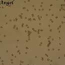
The fungus produces hyaline dikaryotic mycelia that grow and thicken, then fragment forming teliospores (chlamydospores). Teliospores are round or semi-round, yellowish brown to olivaceous brown with one side lighter in color, covered with minute spikes, with diameter 5-9µm (http://www.flickr.com/photos/wamawakil/8046454453/). Spore masses are olive brown. Ustilago tritici can be cultured on potato dextrose agar (PDA) and malt agar, but its growth rate is relatively slow and the colony appearance has a doubtful value as a feature of the fungal diagnosis (Curtis et al. 2002, Mathur and Cunfer 1993).
Occurrence and Impact:
Ustilago tritici (Pers.) Rostr. is the pathogen causing loose smut of wheat (Triticum spp.) that occurs wherever wheatis cultivated all over the world (http://www.agroatlas.ru/en/content/diseases/Secalis/Secalis_Ustilago_tritici/map/). Cool and moist environments are most suitable for the fungus growth (Curtis et al. 2002, Mathur and Cunfer 1993, Neergaard 1979).
This fungus has the potentiality of causing devastating losses that can reach 100% of the wheat yield (Mathur and Cunfer 1993).
Phylogeny:
Ustillago nuda causing loose smut of barely and U. maydis causing corn smut are relatives of U. tritici.
Ustilago nuda is considered the nearest relative of U. tritici. The cluster analysis of the teliospores of both species does not show significant differences. In addition, the ultrastructural studies did not present substantial differences. Moreover,the taxonomic criteria used in the routine identification are fragile to be followed when seeking differences between the two species.
DNA profile is used to analyze the phylogeny of the different species of Ustilago (Menziers et al. 2003). Inter simple sequence repeats technique and amplified fragment length polymorphisms are used to compare the DNA profiles. These techniques are used to separate the U. nuda and U. tritici as they have high morphological similarities (Sharifnabi et al. 2003a, b)
However, the traditional taxonomic criteria including spore size, shape, germination type, host range, and other features are still used in the diagnostic laboratories around the world.
Identification:
The assessment of U. tritici in the plant tissues by fluorescence microscopy and real-time PCR indicates that prediction of the fungus presence could be made at the second leaf stage of wheat seedlings. That is depending upon the fluorescence emitted during the reaction of each PCR cycle and the amplification of the product (Wunderler et al. 2012).
Symptoms and Signs:
There are no visible symptoms of infection during the life span of plants till maturity. At early maturity stage, signs of the pathogen start to appear in wheat fields on a few number of wheat heads (smutted heads) that carry millions of teliospores. These spores are capable to infect a whole wheat field and even the neighboring ones. This late symptom can be seen clearly at the field inspection on a higher level than the healthy un-matured heads.
The smutted heads consist of a powdery mass of the fungus teliospores covered by a delicate grayish membrane that will burst releasing the spores to be blown by wind and fall on the inflorescences of healthy plants and germinate to invade the ovary of the flower (http://wheatdoctor.cimmyt.org/index.php?option=com_content&task=view&id=103&Itemid=43&lang=en). Infection of healthy heads is rarely partial (Agrios 2005, Mathur and Cunfer 1993).
Transmission and Life Cycle:
Germination of teliospores on the flowers of wheat heads produces dikaryotic mycelium that penetrates the ovary invading all parts of the seed. The mycelium overwinters until seed germination, then it regains its activity growing intercellularly in the tissues of embryo then the seedling till it invades the whole plant with no visible sign of its growth. When plants reaches the head emergence stage, the mycelium starts to invade the spikelets and young kernels, then the mycelium thickens and fragment forming millions of teliospores turning the kernels into powdery black masses covered by grayish delicate membranes. Eventually these membranes will rupture releasing the spores (Agrios 2005, Curtis et al. 2002, Mathur and Cunfer 1993).
The smutted heads will develop sooner than healthy ones allowing spores to be blown by the wind to fall on healthy flowers and germinate forming promycelia. The promycelia infect ovaries of newly emerging heads of healthy plants and establish in the seeds' tissues as dormant mycelium till the new growing season and seeds germination starting again its life cycle (Agrios 2005, Curtis et al. 2002, Mathur and Cunfer 1993).
Management of Loose Smut:
1.Growing resistant varieties.
2.Using certified seeds.
3.Seed production fields should be isolated from commercial fields.
4.Seed treatment with hot water or solar heat (not useful for commercial use).
ØHot water treatment: seeds are soaked in 20-30ᵒC water for 4-6 hrs, then dipped in 49ᵒC water for 2 min, and left to air dry on polyethylene sheets before sowing.
ØSolar heat treatment: seeds are soaked in water for 4 hrs, then spread on a polyethylene sheets to be exposed to sunlight (about 40ᵒC) for another 4 hrs, seed should be left to air dry.
5.Field inspection and roguing smutted heads in the field at the early heading stage.
6.Clearing the field after harvesting from plant remains that may help spread the disease.
7.Seed certification schemes can help reduce the disease spread significantly. Tolerance levels of infested seeds ranges between 0.1-0.5% depending on the different classes of seeds.
8.Seed health testing through embryo count method (Mathur 1993) or crown test to avoid infested seed lots.
9.Seed treatment with systemic fungicides as carboxin, carbathiin, benomyl, triadimenol, triadimefon, pyracarbolid, terbutrazole, carbendazim, difenoconazole, etaconazole, ethyltrianol, flutriafol, furmecyclox, myclobutanil, or nuarimol.
10.Foliar application of broad spectrum systematic fungicides including conazole fungicides in a 3 time spray program has markedly reduced the disease incidence (Jones 1999).
11.Recent researches have been conducted to use PCR and ELISA techniques in the assessment of loose smut incidence in seed lots (Wunderle et al. 2012).
Ustilago tritici is a plant pathogen[1] infecting barley; rye and wheat.
Ustilago tritici is a plant pathogen infecting barley; rye and wheat.
Ustilago tritici est une espèce de champignons basidiomycètes de la famille des Ustilaginaceae.
C'est un champignon phytopathogène responsable de la maladie du charbon nu du blé. Ustilago nuda a une biologie similaire sur orge.
Selon Catalogue of Life (5 juillet 2015)[1] :
Le charbon se caractérise par la formation d'amas noir de chlamydospores dans les épis. Le charbon est dit nu car les enveloppes des fleurs sont détruites, seul persiste le rachillet principal.
Les spores tombées sur les papilles stigmatiques germent et forment un mycélium qui envahit l'ovule en environ trois semaines. Le mycélium reste ensuite à l'état latent jusqu'à ce que la semence germe. Il envahit alors la plantule. Les symptômes du charbon apparaissent entre la floraison et la maturité.
Ustilago tritici est une espèce de champignons basidiomycètes de la famille des Ustilaginaceae.
C'est un champignon phytopathogène responsable de la maladie du charbon nu du blé. Ustilago nuda a une biologie similaire sur orge.
Stuifbrand (Ustilago tritici, synoniemen: Ustilago nuda f. tritici, Ustilago segetum var. tritici) is een brandschimmel die behoort tot de basidiomyceten en tarwe kan infecteren. De aren verkleuren zwart door de vorming van chlamydosporen (teleutosporen). Door het ontsmetten van zaaizaad is de ziekte te voorkomen, waardoor de ziekte in de gangbare landbouw bijna niet voorkomt, maar nog wel in de biologische landbouw.
De aangetaste planten zijn langer en komen eerder in bloei dan de gezonde planten. De op de aangetaste aren gevormde teleutosporen worden na het breken van het dunne dekvlies door de wind verspreid en infecteren tijdens de bloei de stempels van de gezonde, bloeiende planten. De teleutospore vormt een basidium. Infectie treedt alleen op als twee verenigbare (compatibele) sporidia (basidiosporen) zich op de stempel onder de juiste omstandigheden ontmoeten en versmelten. Versmelting vindt plaats via een conjugatiebuis tot een tweekernige (dikaryotische) cel. Vervolgens ontstaat een appressorium dat de stempel binnendringt en wordt een promycelium gevormd, dat het vruchtbeginsel infecteert. De schimmel blijft in rust in het vruchtbeginsel. Tijdens de kieming en verdere groei van geïnfecteerde graankorrels verspreidt de schimmel zich door de plant en in de aar via de intercellulaire ruimten. In de aar worden de geelbruine tot olijfbruine, 5-9 µm grote teleutosporen gevormd worden. Alleen de aarspil wordt niet geïnfecteerd. Op de sporen zitten kleine stekeltjes.
Stuifbrand (Ustilago tritici, synoniemen: Ustilago nuda f. tritici, Ustilago segetum var. tritici) is een brandschimmel die behoort tot de basidiomyceten en tarwe kan infecteren. De aren verkleuren zwart door de vorming van chlamydosporen (teleutosporen). Door het ontsmetten van zaaizaad is de ziekte te voorkomen, waardoor de ziekte in de gangbare landbouw bijna niet voorkomt, maar nog wel in de biologische landbouw.
Głownia pszenicy (Ustilago tritici (Pers.) Rostr.) – gatunek grzybów z rodziny głowniowatych (Ustilaginaceae)[1]. Wywołuje chorobę pszenicy o nazwie głownia pyląca pszenicy[2].
Pozycja w klasyfikacji według Index Fungorum: Ustilago, Ustilaginaceae, Ustilaginales, Ustilaginomycetidae, Ustilaginomycetes, Ustilaginomycotina, Basidiomycota, Fungi[1].
Po raz pierwszy takson ten zdiagnozowany został w 1801 r. przez Ch.H. Persoona jako Uredo segetum ß tritici. Obecną, uznaną przez Index Fungorum nazwę nadał mu w 1842 r. E. Rostrup, przenosząc go do rodzaju Ustilago[1].
Wytwarzane przez głownię pszenicy teliospory zakażają zdrowe kłosy pszenicy. Z kiełkujących teliospor wyrastają czterokomórkowe podstawki, nie powstają z nich jednak sporydia. Pomiędzy dwoma haploidalnymi komórkami tej samej podstawki lub sąsiednich podstawek dochodzi do plazmogamii i powstaje dwujądrowa komórka (dikarion). Wyrasta z niej strzępka, która wrasta do zalążni zdrowego kwiatu. Kwiat rozwija się pozornie normalnie, następuje jego zapylenie, zapłodnienie i powstaje ziarniak, jednak w jego zarodku i w okolicach tarczki zarodkowej znajdują się strzępki patogena. Ziarno takie wygląda normalnie i optycznie nie można go odróżnić od ziarna zdrowego. Po jego wysianiu, podczas kiełkowania ziarniaków, strzępki patogena rosną wraz z pędem rośliny i poprzez łodygę dostają się na jego wierzchołek. W zawiązkach kłosków silnie rozrasta się grzybnia patogena niszcząc je. Zgrubiałe strzępki grzybni rozpadają się tworząc kuliste teliospory. Mają średnicę 5-9 μm i drobnobrodawkowatą powierzchnię. Nieco przed kiełkowaniem teliospor zachodzi w nich kariogamia, w wyniku czego z dikarionu powstaje diploidalna zygota. W czasie kiełkowania teliospor zachodzi w nich mejoza i znów powstają haploidalne komórki. Charakterystyczne jest oddalenie w czasie procesów plazmogamii i kariogamii[2].
Głownia pszenicy (Ustilago tritici (Pers.) Rostr.) – gatunek grzybów z rodziny głowniowatych (Ustilaginaceae). Wywołuje chorobę pszenicy o nazwie głownia pyląca pszenicy.
Ustilago tritici je grzib[3], co go nojprzōd ôpisoł Christiaan Hendrik Persoon, a terŏźnõ nazwã doł mu E. Rostr. 1890. Ustilago tritici nŏleży do zorty Ustilago i familije Ustilaginaceae.[4][5] Żŏdne podgatōnki niy sōm wymianowane we Catalogue of Life.[4]
Ustilago tritici je grzib, co go nojprzōd ôpisoł Christiaan Hendrik Persoon, a terŏźnõ nazwã doł mu E. Rostr. 1890. Ustilago tritici nŏleży do zorty Ustilago i familije Ustilaginaceae. Żŏdne podgatōnki niy sōm wymianowane we Catalogue of Life.
小麦散黑粉菌(学名:Ustilago tritici)是属于黑粉菌目黑粉菌科黑粉菌属的一种真菌,寄生在小麦等禾本科植物上,可引起小麦散黑穗病。该种分布于全世界各地。[1]


