El virus de la immunodeficiència felina (FIV o VIH felí) pertany al gènere Lentivirus, dins dels retrovirus. Difereix taxonòmicament del virus de la leucèmia felina (FeLV), però és extraordinàriament similar al virus de la immunodeficiència humana (VIH), causant de la SIDA:[1] tenen una estructura, cicle de vida i patogènesi similar. Tot i això, s'ha de tenir en compte que les persones no són susceptibles a la infecció per FIV.[2]
Va ser diagnosticat per primera vegada el 1986 dins d'una colònia de gats a Califòrnia. Des de llavors, molts casos de FIV han estat diagnosticats per tot el món.[3]
S’ha de tenir en compte que no només ataca els gats, sinó també els altres felins tals com lleons, tigres i pumes.[3]
Actua destruint les defenses del gat afectat (els limfòcits T), generant així una immunodeficiència que desemboca a la vegada en múltiples infeccions i complicacions cròniques, acabant en la mort de l’animal.[4]
L'estructura viral del virus de la immunodeficiència felina consisteix en un embolcall, core (amb l'ARN viral) i nucleocàpside. El core conté dues còpies del seu genoma ARN positiu monocatenari (en cada cas d'uns 9400 nucleòtids)[5] i diversos enzims.[6] El virió té un diàmetre de 80 a 100 nanòmetres i és pleomòrfic. El seu embolcall viral també té unes projeccions d'uns 8 nanòmetres que cobreixen uniformement la superfície.[5] Pel fet de tenir embolcall extern, s’inactiven ràpidament pels desinfectants habituals, es deshidraten en poc temps i són poc resistents a les condicions ambientals.[2]
Una de les característiques que diferencia als Lentivirus d'altres Retrovirus és la complexitat dels seus genomes virals.[6] Posseeix l'enzim transcriptasa inversa, el qual és capaç de crear una còpia d'ADN a partir d'ARN víric. Aquesta còpia d'ADN s'integra en forma de provirus en el genoma de la cèl·lula infectada i pot replicar-se juntament amb ella[7]
El gènere Lentivirus comprèn complexos retrovirus que contenen els tres gens essencials:
Els gens gag i pol no solen canviar entre les diferents soques. En canvi, en la regió SU (de la superfície o embolcall) del genoma del FIV, s’han identificat unes regions amb gran variabilitat (V1, V2, V3, V4 i V5). Són les diferències existents en les regions V3, V4 i V5 les que determinen els 5 subtipus filogenètics del virus A, B, C, D i E (A i B els més freqüents).[2] En un mateix gat infectat pot haver-hi diferents subtipus, la qual cosa indica una falta de protecció creuada entre aquests.[7]
Els tres gens (env, pol i gag) es troben flanquejats per repeticions terminals llargues (LTR) que posseeixen la informació necessària per a l’inici i la terminació de l'expressió gènica, i també per altres petits marcs oberts de lectura que codifiquen diverses proteïnes reguladores que modulen la seva infectivitat, tal com:
L’agrupació geogràfica dels subtipus es distribueix de la següent manera:
El VIF es troba present a la saliva o sang dels gats, de manera que les mossegades entre aquests o les ferides que es generen en les baralles, constitueixen una via de transmissió fonamental. És per això, que es tracta d'una malaltia predominant en el sexe masculí (per les lluites establertes per la territorialitat).[11]
S'havia considerat la no presència de transmissió transplacentària, pèro amb estudis experimentals s'ha refutat aquesta idea. Aquests estudis demostren que hi ha una transmissió de la infecció, des de la mare infectada (tan de forma aguda com crònica) als fetus dins de l'úter. A més a més, també han permès observar la possible infecció dels nounats durant el període lactant. Això no obstant, dins d'una mateixa camada pot donar-se que no tots els fetus s'infectin.[11][2]
Només una part de la cadellada resultarà infectada permanentment i la proporció de gatets que resultin infectats dependrà de la càrrega viral de la mare durant el període de gestació i el part. Si la mare presenta una infecció aguda, fins al 70% dels gatets poden resultar infectats, mentre que si la mare és asimptomàtica i està infectada de forma crònica, gairabé cap gatet de la cadellada resultarà infectat.[2]
Es coneix de forma experimental altres formes de transmissió més ocasionals, tot i que no han estat documentades com a tals. Aquestes consisteixen en una infecció per la boca, nas, vagina i recte. Malgrat tot, no hi ha indicis que indiquin aquestes vies com a fonts importants de transmissió del virus.[2]
El FIV és un virus que es replica en moltes cèl·lules del SI tals com els limfòctis TCD4 i TCD8 positius, els Limf B, monòcits, macròfags, astròcits i cèl·lules de la microglia.[2]
Això comporta una deficiència de leucòcits, febre o anèmia (entre altres coses) a llarg termini. L'evidència més important d'aquest tropisme és que l'animal perd la capacitat d'autoprotegir-se enfront de les infeccions, sent qualsevol bacteri, virus, fong o protozou el causant de malalties. Són aquestes infeccions secundàries les responsables de la majoria de signes clínics associats a la malaltia de la immunodeficiència felina i la principal causa de mort en gats VIF positius. [12][13]
La molècula vírica que permet la penetració del virus a l'interior cel·lular és la glicoproteina gp-120, la qual s'uneix a un receptor de membrana present a les cèl·lules animals, el CD134. Aquesta primera unió implica un canvi conformacional de la proteïna vírica, fet que permet a la vegada una segona interacció d'aquesta amb el coreceptor CXCR4. Posteriorment a aquestes interaccions, es fusiona la envoltura vírica amb la membrana cel·lular produint-se la penetració vírica.[2]
Cal tenir en compte que no ha estat demostrat en tots els tipus cel·lulars susceptibles de ser infectats per FIV, la presència del coreceptor CXRCR4. Per això cal suposar l'existència d'altres tipus de molècules receptores o mètodes d'entrada cel·lular.[2]
El FIV és un virus que presenta la capacitat de sintetitzar l'enzima transcriptasa inversa (RT). De manera que un cop es troba a l'interior cel·lular, pot transformar el seu RNA víric a DNA, amb el fi d'intercalar aquest en el DNA de la cèl·lula infectada. Aleshores estarà en fase de provirus.[2]
En un principi, la RT actua trascrivint l'RNA víric en DNA, formant un híbrid RNA-DNA. Posteriorment, es polimeritzarà una altra cadena de DNA a partir de la cadena de DNA ja preformada prèviament, obtenint una doble cadena de DNA que pot ser intercalada en el DNA cel·lular.[2]
Un fet molt important a tenir present és que la RT no té capacitat correctora. Per aquest motiu, el FIV és un virus que pot mutar molt fàcilment i ràpidament generant múltiples soques. Per altra banda, aquesta variabilitat genètica ajuda a evitar la detecció viral per part del SI de l'hoste.[2]
Podem distingir tres fases:.[2]
Una vegada s’ha inoculat el virus FIV, per qualsevol de les vies de transmissió, seguirà els següents passos:
A causa d’aquesta virèmia inicial el virus es dissemina per la resta de l'organisme infectat afectant limfòcits, monòcits i macròfags de la medul·la òssia, tracte intestina, pulmó, ronyó i cervell. A nivell del cervell infecta a les cèl·lules endotelials, de la micròglia, astròcits i neurones.[2]
Aquesta fase pot durar entre 3 i 6 mesos, després aquesta virèmia va decreixent. La caiguda de la virèmia coincideix amb el desenvolupament de la immunitat de tipus cel·lular i humoral, tot i que aquesta immunitat no és capaç d'eliminar la infecció.[13]
Una vegada ha disminuït la càrrega viral en el plasma comença la fase asimptomàtica o de latència clínica. Aquesta fase pot durar mesos, anys o fins i tot tota la vida.[2]
És una fase asimptomàtica però encara que els animals aparentin estar sans, es produeix una debilitació gradual del sistema immunitari. Disminueix el quocient CD4+/CD8+ perquè hi ha una disminució dels limfòcits T CD4+ col·laboradors que són essencials per a la immunitat cel·lular. Aquests limfòcits T CD4+ no només disminueixen en quantitat sinó que també en funcionalitat, en especial en la seva capacitat de reaccionar contra antígens i en la síntesi de interleuquina 2 (IL-2).Per tant, la infecció per FIV no té un període de latència real, ja que la infecció viral progressa encara que el gat sigui asimptomàtic.[2]
Aquesta fase acaba en la fase amb símptomes de caràcter lleu, que seria l'inici de la fase d'immunodeficiència.[2]
La fase d'immunodeficiència de la malaltia és provocada per la mort dels limfòcits T CD4+ que provoca una baixada del quocient de limfòcits TCD4+ /TCD8+. S’anomena fase SIDA (Síndrome d'Immunodeficiència Adquirida) per la similitud amb el VHI (humana).[2]
En aquesta fase trobarem infeccions oportunistes, ja que, l’animal serà incapaç de desenvolupar una resposta immunitària adequada. Aquesta fase sol acabar en la mort per síndrome crònica de consumpció ( pèrdua de pes i deteriorament físic), malaltia neurològica, neoplàsies o infeccions oportunistes sistèmiques.[2] Davant d’aquest tipus d’infecció el temps estimat de vida són de cinc anys.[13]
Els animals infectats són infectants d'ençà que s’inicia la fase de virèmia i en totes les fases de la malaltia, inclosa la fase de latència clínica. Són capaços d'eliminar virus, encara que sigui en dosis molt petites.[2]
La infecció dura tota la vida de l'animal perquè és un retrovirus. Una de les principals característiques dels retrovirus és que una vegada infecten la infecció es manté al llarg de tota la vida de l'animal. Això és degut al fenomen de latència, per mitjà del qual s’integra el provirus al genoma de les cèl·lules de l'hoste, on roman sense expressar-se i ocult al sistema immunitari durant llargs períodes de temps.[2]
Però aquesta fase de latència pot revertir, generalment per una immunosupressió deguda a l'administració de fàrmacs com els corticoesteroides, donant lloc a una expressió vírica i a la progressió de la infecció fins a acabar amb la mort de l'animal.[2]
Perquè sigui un estat de latència verdader és necessari que el virus integrat no s’expressi, és a dir, que no es pugui detectar ni el virus, ni el seu RNA ni les seves proteïnes víriques en les cèl·lules sanguínies o en els teixits de l'animal. Per tant, en el cas del virus de la immunodeficiència felina no s’estableix un estat de latència verdadera, ja que encara que els gats infectats pel virus no presentin simptomatologia clínica el RNA o les proteïnes virals poden detectar-se en els teixits de l'animal, en el plasma, en el líquid cefaloraquidi, en els teixits limfoides i semen. Això és així perquè hi ha una contínua replicació vírica, però a nivells molt baixos.[2]
La resposta humoral és aquella que inclou la participació dels anticossos per atacar els antígens. En el cas de la immunodeficiència felina, en general aquests anticossos apareixen 2 o 4 setmanes després de la infecció, moment en què té lloc el pic en la virèmia inicial. En el cas que l’animal (felí) s’hagi vist exposat a una càrrega viral molt reduïda, els anticossos apareixeran passats mesos o anys.
Els nivells d’aquests anticossos es mantenen més o menys constants durant llargs períodes de temps, tant de temps que poden romandre fins i tot anys. Tot i així, hi ha un descens d’aquests anticossos que s’observa en les etapes finals de la infecció.[2]
En aquestes etapes es produeix el següent:
Els anticossos més rellevants són aquells que impedeixen l’entrada del virus, impedint així la patogènia. En el cas del FIV ens trobem amb anticossos neutralitzants que impedeixen la unió de les glucoproteïnes víriques als seus receptors cel·lulars. Aquests anticossos, actuen doncs, sobre les glucoproteïnes de la càpside del virus.[2]
Aquests anticossos tan efectius es transmet la mare a les seves cries a través del calostre, per tant, els gatets adquireixen una immunitat passiva contra el FIV durant el temps que romanguin aquests anticossos, en aquest cas, 1, 2 o 3 mesos tot i que s'ha observat algun cas de fins a 4 mesos.[2]
En la resposta cel·lular intervenen els limfòcits T, els quals s’encarreguen de moderar la destrucció d’agents patògens.
Aquesta resposta és essencial per dos motius:
La resposta cel·lular dels limfòcits T CD8+ citotòxics es produeix 1 o 2 setmanes després de la infecció per FIV i abans que tingui lloc l’aparició dels anticossos neutralitzants propis de la resposta humoral. Els limfòcits T CD8+ citotòxics s’encarreguen d’induir apoptosis per diferents mecanismes en les cèl·lules infectades pel virus.[2]
Tot i que aquesta la resposta cel·lular es manté durant tot el període asimptomàtic (quan encara no han tingut lloc els símptomes clínics), no és capaç d’eliminar la infecció i els gats queden infectats tota la seva vida.[8]
Hi ha dos tipus: els Th1 i els Th2. Els Th1 s’encarreguen de destruir cèl·lules infectades mentre que els Th2 estimulen els limfòcits B per tal que alliberin anticossos.
A causa de la seva actuació, es considera que els limfòcits TCD4 són els grans mediadors de la resposta immunitària. Per tal de realitzar correctament les seves funcions, necessita poder “comunicar-se” amb les altres cèl·lules i ho fa a través de les CITOQUINES. El FIV modifica la síntesi d’aquestes citoquines, alterant així la resposta immunitària fent prevaldre la resposta humoral (Th2) davant de la cel·lular (Th1). Aquest fet condueix al fet que els animals (gats) infectats estiguin més predisposats a patir infeccions secundàries, per exemple, de Toxoplasma gondii. Aquestes infeccions secundàries són les responsables de moltes de les malalties associades a la immunodeficiència felina. [1][2][8]
Trobem nivells incrementats de:
Té lloc una disminució de:
A part, es produeix un marcat increment dels nivells de IL-10.[2]
Es produeix un increment d’aquest coeficient, ja que en fases avançades de la infecció per FIV els nivells de IL-10 són alts i els nivells de IL-12 són baixos. Aquest increment pot ser responsable de dos fets: per una banda de la disminució de la resposta davant patògens secundaris; i per una altra banda, de la patogènia produïda pel FIV, ja que provoquen que hi hagi un canvi de la resposta de tipus cel·lular al tipus humoral.
Tot això, comporta a un declivi en la funcionalitat immunitària.[2]
La marca característica de la patogènia d’aquesta malaltia és la desorganització progressiva de la funció immunològica. La immunosupressió es produeix per la destrucció de limfòcits T CD4+, l’activació de certa subpoblació de limfòcits T CD8+ citotòxics (inversió del quocient CD4+/CD8+ que provoca limfopènia) i alteracions en la funcionalitat de monòcits, macròfags, neutròfils, cèl·lules Natural Killer (NK) i limfòcits CD4+ i CD8+. El FIV es replica en limfòcits CD4+ i CD8+, en macròfags, en astròcits i en cèl·lules de la micròglia.[2]
Algunes soques del FIV es repliquen preferentment en limfòcits, i només mínimament en macròfags, mentre que altres soques són capaces de replicar-se igualment de bé en els dos tipus cel·lulars. Se sap que el reservori més gran de cèl·lules infectades en la sang perifèrica durant les primeres fases de la infecció són els limfòcits CD4+, però el canvi en el tropisme víric i la utilització de receptors en la infecció crònica encara és tema de debat. Es creu que durant les infeccions, els principals afectats són els limfòcits i que a mesura que persisteix l’atac del virus, aquest afecta cada cop més a macròfags. Pel que fa als receptors, el FIV no s’uneix a través de CD4 sinó a través de l’homòleg de CD9, així com de receptors de quimiocina alfa CXCR4.[14]
El FIV pot aïllar-se de limfòcits ja entre els 10 i 14 dies després de la infecció; la virèmia creix ràpidament fins al dia 21. Durant les primeres setmanes d’infecció disminueixen tant les cèl·lules CD4+ com les CD8+, mentre que els limfòcits B no es veuen afectats. La quantitat de virus arriba al màxim entre les setmanes 7 i 8 i és llavors quan les cèl·lules CD4+ disminueixen com a resultat de destrucció de les que són infectades, disminució de la seva producció i apoptosis prematura. Després la virèmia disminueix gradualment fins que la càrrega vírica torna a créixer en les etapes terminals.[9] En aquest moment hi ha una profunda depressió de la resposta a antígens timus dependents, però la resposta a antígens timus independents és normal.[14]
La limfopènia inicial va seguida d’una forta resposta immunitària caracteritzada per la producció d’anticossos anti-FIV i un repunt de les cèl·lules CD8+ per damunt dels nivells previs de la infecció, ja que aquestes es recuperen però les CD4+ no. Això ocasiona una inversió persistent en relació CD4/CD8. Amb el pas del temps, les dues poblacions cel·lulars disminueixen gradualment. Els limfòcits↵experimenten un increment en l'apoptosi espontània i les cèl·lules CD4 que↵sobreviuen a vegades presenten menor↵reactivitat als mitògens.[15]
A part de la disminució quantitativa de les cèl·lules CD4+, els gats infectats per FIV mostren una disfunció de les cèl·lules immunitàries (per exemple pèrdua de la capacitat de proliferar dels limfòcits en resposta a un estímul). A més, pot detectar-se una significativa pertorbació de la producció de citoquines que contribueix a la immunodeficiència. Es va demostrar que la immunitat mediada per cèl·lules resulta molt més afectada que la immunitat humoral. Les situacions d’inflamació crònica, neoplàsies i infeccions per organismes intracel·lulars són més habituals que les infeccions oportunistes controlades per anticossos. Sembla que els gats infectats per FIV responen de forma adequada a les vacunes i que desenvolupen amb freqüència una hipergammaglobulinèmia policlonal característica d’una estimulació inespecífica de la immunitat humoral.[9]
Altres alteracions que es donen en la FIV són la disminució en la producció de IL-2 i l'alta transcripció de IL-10, la qual pot ser que contribueixi a la immunodeficiència. Hi ha altes concentracions d’immunocomplexes circulants en el sèrum i dipositats en els glomèruls renals. Els gats afectats per FIV semblen tenir un nombre normal de limfòcits B, així com concentracions normals de IgM i IgA.[14]
El mecanisme pel qual apareixen tumors possiblement és de tipus indirecte, per una menor vigilància de la resposta cel·lular citotòxica o per la hiperplàsia crònica dels limfòcits B. No obstant això, no s’ha descartat totalment un possible paper directe del virus en l’oncogènesi dels limfomes, ja que se sap que un gat amb immunodeficiència té augmentada per cinc la possibilitat de tenir limfoma.[2]
Per tant, més que estar directament relacionat amb l’aparició de tumors, el virus inhibeix els factors supressors tumorals i activa els oncògens. El tumor més freqüent és el limfoma de cèl·lules B i les varietats més habituals trobades són els limfomes en cap i coll, el limfoma renal i l’alimentari, amb els símptomes corresponents a cada localització.[16]
Altres tumors descrits en associació al FIV són els carcinomes mamaris, de cèl·lules escamoses, trastorns mieloproliferatius i fibrosarcomes.[16]
El FIV és capaç de provocar lesions inflamatòries de teixit nerviós en aproximadament el 30% dels gats infectats. Aquestes lesions afecten el nucli caudat, al mesencèfal i a la porció rostral del tronc cerebral. El FIV també es relaciona amb desmielinització en les columnes dorsals de la medul·la espinal, vacuolització de les beines de mielina en les arrels de nervis espinals i infiltració perivascular i perineuronal de cèl·lules mononuclears, així com amb nòduls glials. Les síndromes neurològiques es manifesten amb demència, convulsions, espasmes facials, trastorns del somni, canvis de comportament (agressivitat o por) i conducta compulsiva o atàxia. En menor grau les lesions deriven d’inefccions secundàries amb Toxoplasma i Cryptococcus.[16][2]
És freqüent que durant la fase de virèmia, els virus travessin la barrera hematoencefàlica, sols o vehiculats per limfòcits i monòcits, infectant cèl·lules endotelials, cèl·lules de la micròglia, astròcits i neurones, donant lloc a alteracions neurològiques i del comportament. El virus de la FIV pot infectar les cèl·lules de la micròglia de forma molt ràpida després de la fase de virèmia i replicar-se en elles a pesar de la resposta inflamatòria que es produeix. Posteriorment, el virus ja no podrà ser eliminat de les cèl·lules de la micròglia i, per tant, el cervell quedarà com un reservori del virus, podent reactivar-se la replicació vírica en qualsevol moment.[2]
En la patogènia nerviosa dels retrovirus felins podrien estar implicades les noves variants víriques neurotròpiques que es produeixen, cada cop més adaptades al cervell: la replicació vírica es dóna en les cèl·lules glials i en macròfags cerebrals majoritàriament, causant d’aquesta manera dany al sistema nerviós central i lesions al còrtex cerebral.[10] També podria estar implicada la glicoproteïna d’embolcall gp120, amb un marcat caràcter neurotòxic en augmentar les concentracions de calci intracel·lular en les neurones que infecten. Els limfòcits T CD8+ també estarien relacionats, ja que activen l’alliberació de neurotoxines per als macròfags, com l’òxid nítric, que mata a les cèl·lules nervioses. Això és un altre exemple de com la immunitat dels animals infectats pot estar col·laborant indirectament amb la mateixa patogènia dels retrovirus.[2]
Els símptomes, les alteracions laboratorials i els llargs períodes asimptomàtics que acompanyen la infecció per FIV són insuficients per poder diagnosticar de ciència certa la malaltia, per tant necessitem recórrer a les proves diagnòstiques. Les diverses proves diagnòstiques es poden dividir en dos subgrups:
Tenint en compte aquest conjunt de proves diagnòstiques, per poder diagnosticar la infecció per FIV se segueixen una sèrie de regles generals que són les següents:
Per tant les tres proves diagnòstiques més importants per poder interpretar i confirmar una infecció per FIV són la ELISA, i en el cas que doni positiu aplicarem o la PCR o el Western blot. Les característiques principals d’aquestes proves en relació al virus de la immunodeficiència felina són:
Segons els resultats que s'obtinguin de l'aplicació d'aquestes tres proves les conclusions a les que arribarem són les que podem observar a les imatges de la dreta:
Podem distingir diferents signes clínics segons la fase de la infecció en què es trobi l’animal:
El virus de la immunodeficiència felina (FIV o VIH felí) pertany al gènere Lentivirus, dins dels retrovirus. Difereix taxonòmicament del virus de la leucèmia felina (FeLV), però és extraordinàriament similar al virus de la immunodeficiència humana (VIH), causant de la SIDA: tenen una estructura, cicle de vida i patogènesi similar. Tot i això, s'ha de tenir en compte que les persones no són susceptibles a la infecció per FIV.
Va ser diagnosticat per primera vegada el 1986 dins d'una colònia de gats a Califòrnia. Des de llavors, molts casos de FIV han estat diagnosticats per tot el món.
S’ha de tenir en compte que no només ataca els gats, sinó també els altres felins tals com lleons, tigres i pumes.
Actua destruint les defenses del gat afectat (els limfòcits T), generant així una immunodeficiència que desemboca a la vegada en múltiples infeccions i complicacions cròniques, acabant en la mort de l’animal.
Felinní imunodeficientní virus (FIV) je retrovirus, způsobující získanou ztrátu imunity u koček.
Původcem onemocnění je RNA virus z čeledi Retroviridae (Lentivirus). Poprvé byl izolován v roce 1986 v Kalifornii. Virion je sférického až elipsovitého tvaru o velikosti 105-125 nm. Je příbuzný k viru HIV, viru maedi-visna, viru infekční anémie koní či viru artritidy a encefalopatie koz. Virus je specifický pro kočky, některé lentiviry jsou přenosné i z kočkovitých šelem. Virus FIV má vysokou afinitu k lymfocytům, peritoneálním makrofágům, astrocytům a makrofágům CNS.
FIV (felint immundefektvirus) er en lentivirus, der påvirker tamme huskatte over hele verden og er den virus, der forårsager katteaids. Ca. 11%[1] af katte på verdensplan, og omkring 2,5% af alle katte i USA[2] er inficeret med FIV. FIV adskiller sig taxonomisk fra to andre katte-retrovira, felin leukæmi-virus (FeLV) og katteskummende virus (FFV), og er mere tæt forbundet med human immundefektvirus (hiv). FIV har fem undertyper (der er identificeret), som baseret på nukleotidsekvens-forskelle, der koder for den virale envelop (ENV) eller polymerase (POL). FIV er den eneste ikke-primatære lentivirus, der forårsager en aids-lignende syndrom, men FIV giver ikke altid en dødsdom til katte; de kan leve forholdsvis sundt som bærere og sendere af sygdommen i mange år. Der findes en vaccine, selv om dens effekt er fortsat usikker, og katte vil blive testet positiv for FIV-antistoffer efter vaccination.
FIV blev først opdaget i 1986 i en koloni af katte, der havde en høj forekomst af opportunistiske infektioner og degenerative betingelser, og er siden blevet identificeret som en endemisk sygdom i huskatte, gældende for hele verden[2].
De primære former for FIV-smitte forekommer ved dybe bide-sår og skrammer, hvor kattens infecerede spyt bliver ført ind i en anden kats blodårer. FIV kan også overføres fra gravide hunkatte til deres fostre i livmoderen[3]. Dette adskiller sig fra FeLV, der kan spredes via mere afslappet ikke-aggressiv kontakt, da virus er også til stede ved slimhindeinfektioner som dem i munden, endetarmen og skeden. Tilfældig kontakt kan derfor ikke udelukkes som en potentiel transmission.
Når dyrlæger vil kontrollere en kats historie og stamtræ for at se om sygdommen blev overført arveligt, og eventuelt administrere en blodprøve for FIV-antistoffer. FIV rammer 2-3% af alle katte i USA og derfor er testning let tilgængelig. Det skal bemærkes, at denne test identificerer de katte som er påført FIV-antistof, og detekterer ikke selve virussen. Derfor finder en positiv test nødvendigvis ikke at katten er bærer af FIV.
Falske positiver opstår når katten bærer FIV-antistof (som er uskadeligt), men ikke selve virus. Den hyppigste forekomst af dette er når killingerne er testet efter indtagelse af antistoffer fra modermælken, og test af katte der tidligere er blevet vaccineret for FIV. Af denne grund er hverken killinger under 8 uger eller katte, der tidligere er blevet vaccineret, testes.
Killinger og unge katte der testes positive for FIV-antistof, kan med en test, hvor forekomst af et negativt resultat på et senere tidspunkt på grund af seroreversion, forudsat at de aldrig er blevet smittet med FIV og aldrig er blevet vaccineret med FIV-vaccinen.
Katte der er blevet vaccineret vil testes positiv for FIV-antistof for resten af deres liv på grund af serokonversionen, selv om de ikke er smittet. Derfor er tests af adopterede katte er noget usikre, da det er umuligt at vide hvorvidt de er blevet vaccineret i fortiden. Af disse grunde bør en positiv FIV-antistoftest i sig selv aldrig bruges som kriterier for aktiv dødshjælp.[kilde mangler]
Test kan gennemføres i en dyrlæges rum med resultater på få minutter, der giver mulighed for hurtig konsultation. Tidlig opdagelse hjælper med at bevare kattens sundhed og forhindrer spredning af smitte til andre katte. Med ordentlig pleje kan smittede katte lever langt og sundt liv.
FIV (felint immundefektvirus) er en lentivirus, der påvirker tamme huskatte over hele verden og er den virus, der forårsager katteaids. Ca. 11% af katte på verdensplan, og omkring 2,5% af alle katte i USA er inficeret med FIV. FIV adskiller sig taxonomisk fra to andre katte-retrovira, felin leukæmi-virus (FeLV) og katteskummende virus (FFV), og er mere tæt forbundet med human immundefektvirus (hiv). FIV har fem undertyper (der er identificeret), som baseret på nukleotidsekvens-forskelle, der koder for den virale envelop (ENV) eller polymerase (POL). FIV er den eneste ikke-primatære lentivirus, der forårsager en aids-lignende syndrom, men FIV giver ikke altid en dødsdom til katte; de kan leve forholdsvis sundt som bærere og sendere af sygdommen i mange år. Der findes en vaccine, selv om dens effekt er fortsat usikker, og katte vil blive testet positiv for FIV-antistoffer efter vaccination.
FIV blev først opdaget i 1986 i en koloni af katte, der havde en høj forekomst af opportunistiske infektioner og degenerative betingelser, og er siden blevet identificeret som en endemisk sygdom i huskatte, gældende for hele verden.
Das Feline Immundefizienz-Virus (FIV) ist ein Virus aus der Familie der Retroviren (Retroviridae). Das Virus löst bei Katzen eine Immunschwächekrankheit aus, die als Felines Immundefizienzsyndrom oder umgangssprachlich als Katzen-AIDS bezeichnet wird, da sie der Erkrankung AIDS beim Menschen stark ähnelt. Menschen können sich jedoch mit FIV nicht infizieren. FIV gehört innerhalb der Retroviren zur Gattung der Lentiviren und wurde 1986, also vier Jahre nach der Entdeckung des Humanen Immundefizienz-Virus (HIV), zum ersten Mal beschrieben. Die Erkrankung ist bisher nicht wirkungsvoll behandelbar, verläuft aber oft über lange Zeit symptomlos. Langfristig wird jedoch das Immunsystem zerstört und Sekundärinfektionen führen zum Tod. Bisher wurden neun verschiedene Stämme des Virus aus elf verschiedenen Katzenarten isoliert, darunter spezifische Stämme aus Löwen und Pumas. Auch in der Tüpfelhyäne, die nicht zur Familie der Katzen gehört, wurde FIV gefunden. Neben dem Felinen Coronavirus, dem Erreger der felinen infektiösen Peritonitis (FIP) und dem Felinen Leukämievirus (FeLV), dem Erreger der Katzenleukämie, gehört das Virus zu den Auslösern der klinisch bedeutsamsten viralen Infektionskrankheiten bei Hauskatzen.
Die ersten FIV-Virusstämme wurden 1986 aus Hauskatzen isoliert. In einem Haushalt in Petaluma, Kalifornien, in dem sehr viele Katzen lebten, kam es zu einem Ausbruch einer Immunschwächekrankheit. Diese Tiere wurden auf das Feline Leukämievirus (FeLV) getestet, waren aber alle negativ. Man entnahm ihnen Blutproben und injizierte diese in zwei gesunde Tiere, die nach vier bis sechs Wochen Fieber, eine Abnahme der Zahl der weißen Blutkörperchen (Leukopenie) und eine Lymphknotenschwellung entwickelten. Aus den mononukleären peripheren Blutzellen (PBMCs) dieser Tiere wurde anschließend das erste FIV isoliert.
Bald darauf wurde festgestellt, dass auch Serumproben von wild lebenden Katzen wie afrikanischen Löwen und Geparden, asiatischen Löwen und Tigern, südamerikanischen Jaguaren und nordamerikanischen Pumas mit den Antigenen von FIV und EIAV, einem Pferdelentivirus, kreuzreagierten. Diese Reaktionen deuteten auf eine Infektion mit FIV hin. Serologische Untersuchungen dieser Art (zum Beispiel mittels ELISA) sind bis heute die wichtigste Methode, um eine Infektion mit FIV nachzuweisen. Zunächst wurden die Antigene der Hauskatzen auch für Tests der Seren von wild lebenden Katzenarten verwendet. Mit zunehmender Charakterisierung der speziesspezifischen FIV-Stämme wurden deren Antigene verwendet, was die Sensitivität der Tests stark erhöhte. Gleichzeitig wurde erkannt, dass FI-Viren eine große und evolutionär gesehen alte Gruppe von Retroviren sind.
Nach der ersten Beschreibung aus Nordamerika wurde nach und nach in Hauskatzen aus aller Welt ebenfalls FIV nachgewiesen. Die weltweite Prävalenz von FIV-infizierten Hauskatzen in den Regionen und Ländern liegt zwischen zwei und 30 Prozent. Da die Hauskatze bereits vor Hunderten von Jahren von Europa aus mit den Händlern und Entdeckern in alle Welt verbreitet wurde, kann davon ausgegangen werden, dass auch FIV Hauskatzen bereits seit langer Zeit infiziert waren. Auch eingefrorene Katzenseren – die ältesten getesteten Seren stammen von 1968 aus Japan und den USA – lieferten positive Serotests. Die Zahlen über die Verbreitung schwanken in Abhängigkeit von der Vorselektion des Probenmaterials und der Populationsdichte.[2][3][4] Auch in verschiedenen geografischen Regionen treten große Schwankungen auf. Die Löwen in der Serengeti sind praktisch zu 100 Prozent seropositiv, während die Löwen in Namibia und freilebende Asiatische Löwen durchweg seronegativ sind. Pumas in Wyoming sind fast zu 100 Prozent positiv, während Pumas in Montana nur zu 20 Prozent positiv sind. Weil die evolutionäre Entwicklung von Lentiviren schneller verläuft als die von Katzenarten, lassen die Verbreitung und Ähnlichkeit der FIV-Stämme Rückschlüsse auf Ausbreitung, Territorien, Wanderungsverhalten und Populationsdynamiken der verschiedenen Katzenarten zu.[5] Die Erfassung und Analyse dieser Daten steht jedoch noch am Anfang.
Die Bezeichnung des Virus mit FIV bezieht sich in den meisten Fällen auf das Isolat aus Hauskatzen. Die Standardnomenklatur für die Bezeichnung von Stämmen aus verschiedenen Spezies ist ein hintenangestelltes Kürzel, das sich aus dem ersten Buchstaben des Gattungsnamens und aus den ersten beiden Buchstaben des Artnamens zusammensetzt. Das FIV der Hauskatze (Felis catus) wird daher auch als FIVfca bezeichnet, das des Afrikanischen Löwen (Panthera leo) heißt FIVple und das von Puma concolor FIVpco. Das Puma-FIV wird manchmal auch als PLV und Löwen-FIV mit LLV bezeichnet, aber diese beiden Virusstämme sind im Bezug auf einen eigenen Namen die einzigen Ausnahmen von der Nomenklatur.
Die bisher bekannten FIV-Stämme sind sehr divergent, aber monophyletisch, also aus einer Stammform entstanden. Für drei der FIV-Stämme aus den verschiedenen Tierarten konnten Subtypen ermittelt werden. Das FIV der Hauskatze ist bisher am besten untersucht und weist fünf Subtypen auf, die weltweit in unterschiedlicher Häufigkeit vorkommen und die mit A bis E bezeichnet werden. Die Unterteilung in fünf Untergruppen erfolgte nach Vergleichen der DNA-Sequenz des env-Gens, das die Hüllproteine codiert. Die Untergruppen A bis C sind weltweit verbreitet, D kommt vor allem in Ostasien vor und E nur in Südamerika.
Auch von FIVple wurden drei Untergruppen bestimmt, bezeichnet mit A bis C. Diese Unterteilung erfolgte aufgrund von Sequenzunterschieden im pol-Gen, das die viralen Enzyme (Protease, Integrase und Reverse Transkriptase) codiert. Bei FIVpci wurden aufgrund der Unterschiede in pol zwei Untergruppen ermittelt und mit A und B bezeichnet. Die Unterschiede der DNA-Sequenz sind zwischen den einzelnen FIV-Stämmen zum Teil erheblich und liegen zum Beispiel für das pol-Gen von FIVple, FIVfca und FIVpco bei 30 Prozent.
Die bekannten FIV-Stämme bilden einen eigenen Cluster innerhalb der Lentiviren und können grob in Alt- und Neuweltspezies unterteilt werden. Die engste Verwandtschaft besteht zu den Lentiviren der Rinder und Pferde.
FIV ist aufgebaut wie andere Lentiviren, die Immunschwächesyndrome bei Säugetieren hervorrufen. Das vollständige Virion hat einen Durchmesser von 105 bis 125 Nanometer, ist sphärisch oder ellipsoid geformt und besitzt in der Virushülle kurze, wenig definierte Vorsprünge (Spikes), die aus den viralen Glykoproteinen gp95 und gp44 bestehen. Es hat wie andere Retroviren eine Dichte von 1,15 bis 1,17 Gramm pro Kubikzentimeter. Die Viruspartikel (Virionen) werden durch übliche alkohol- oder chlorhaltige Desinfektionsmittel zerstört und durch kurzzeitiges (wenige Minuten) Erhitzen auf 60 Grad Celsius inaktiviert.
Das Virusgenom ist diploid. Es besteht aus zwei identischen einzelsträngigen aus jeweils etwa 9400 Nukleotiden bestehenden RNA-Molekülen in Plus-Strang-Orientierung. Es besitzt die typische genomische Struktur der Retroviren, die aus den Genen gag-pol-env besteht, und hat wie andere Lentiviren zusätzliche (zusätzliche) Gene. Diese sind vif, vpr und rev. Es fehlen tat, vpu, vpx und nef, FIV ist damit weniger komplex als HIV. FIV besitzt eine Desoxyuridinpyrophosphatase (dUTPase), die in Lentiviren der Nicht-Primaten vorkommt und bisher außer für FIV noch für EIAV und das Visna-Maedi-Virus (VMV) beschrieben wurde. Die dUTPase wird in der pol-Region codiert, das fertige Enzym baut dUTP zu dUMP und Pyrophosphat ab, was dem Virus wahrscheinlich dabei hilft, einen falschen Einbau von dUTP in das Genom zu verhindern. Von dem proviralen Genom werden sechs verschiedene Spleißvarianten der mRNA erzeugt.
Die Pathogenität der FIV-Stämme ist für Katzen in freier Wildbahn nur schwer zu bestimmen. Epidemiologische Studien, in denen die Überlebensraten mit den Infektions- und Reproduktionsraten verglichen wurden, konnten keine statistisch signifikanten Nachteile für die infizierten Tiere feststellen. Viele der vorkommenden Stämme können somit als nicht pathogen angesehen werden. In Gefangenschaft dagegen, wenn sich die durchschnittliche Lebenserwartung der Tiere deutlich erhöht, kommt es zu der Entwicklung der Krankheitssymptome. Die geringe Pathogenität der FIV-Stämme bei wild lebenden Katzen deutet auf eine lange Pathogen-Wirt-Wechselwirkung hin, die wie bei SIV vermutlich seit etwa ein bis zwei Millionen Jahren besteht.[6] In welchen Vorläuferspezies sich FIV zunächst entwickelt hat, ist nicht bekannt. Übertragungen zwischen verschiedenen Katzenarten kommen in freier Natur nur selten vor, in Gefangenschaft dagegen häufiger.[7]
Das Feline Immundefizienz-Virus ist sehr wirtsspezifisch, Übertragungen von Katze zum Menschen sind nicht bekannt.[8] FIV infiziert wie HIV-1 hauptsächlich CD4-positive T-Lymphozyten. Im Vergleich zu HIV-1, einem der beiden bei Menschen vorkommenden Verwandten, hat FIV jedoch ein etwas breiteres Spektrum an Zellen, die es infizieren kann. Neben den CD4-positiven T-Zellen, Monozyten, Makrophagen und Gliazellen infiziert FIV auch CD8-positive T- und B-Zellen. Als primärer Rezeptor für das externe Glykoprotein (gp95) von FIV dient nicht wie bei HIV-1 CD4, sondern CD134.[9][10] Für die Interaktion zwischen FIV gp95 und CD134 wird CXCR4 als essentieller Korezeptor benötigt.[11] Das Hüllprotein gp95 des Virus bindet mit seinen Vorsprüngen (engl. spikes) an CD134, was eine Konformationsänderung in gp95 zur Folge hat, die eine Interaktion mit CXCR4 ermöglicht. Diese Interaktion mit dem Corezeptor regt die Fusion der Virushülle mit der Zellmembran an und ermöglicht den Eintritt in die Zelle. Da auch Virusstämme beschrieben wurden, die CD134 nicht benötigen, ist die Charakterisierung der Rezeptoren noch nicht vollständig abgeschlossen.
Es ist bisher in keinem einzigen Fall gelungen, das FI-Virus in menschlichen Zellen oder Zelllinien zur Vermehrung zu bringen. Der „Block“, also die Barriere, die das Virus daran hindert, einen vollständigen Replikationszyklus zu durchlaufen, liegt weder beim Zelleintritt noch bei der Überwindung der Kernmembran, sondern besteht darin, dass das in der DNA integrierte und nachweisbare Provirus den kritischen Schritt der Transkription nicht mehr durchläuft. Daher können nach der Infektion der Zelle keine weiteren Viruspartikel gebildet werden. Der Block ähnelt damit dem von EIAV in menschlichen Zellen und HIV in Mäusezellen.
Nach einer Erstinfektion produziert die Katze sofort virusspezifische Antikörper und cytotoxische T-Zellen, ist jedoch trotz der heftigen Immunreaktion nicht in der Lage, die Infektion vollständig zu überwinden. Das Virus verbleibt daher in allen bisher untersuchten Fällen dauerhaft im Körper.
Relativ viel Aufmerksamkeit hat die Entwicklung eines FIV-Impfstoffs erhalten, der 2002 in den USA zugelassen wurde. Man erhofft sich aus den Erfahrungen Erkenntnisse für die Entwicklung eines Impfstoffs gegen HIV.[12] Die Entwicklung eines solchen Impfstoffs wurde seit Entdeckung des FI-Virus vorangetrieben,[13] und es wurden verschiedene Impfstofftypen getestet, darunter inaktivierte Viren, mit Viren infizierte Zellen, DNA-Impfstoffe und virale Vektoren. Es ist unklar, ob diese unter Laborbedingungen erbrachten Ergebnisse unter Feldbedingungen mit einer Vielzahl verschiedener FIV-Stämme reproduzierbar sind.
Wie bei HIV ist die Entwicklung einer wirksamen Impfung gegen FIV aufgrund der hohen Anzahl und Variationen der Virus-Stämme schwierig. Für sogenannte „single strain“-Vakzine, also Impfstoffe, die nur gegen eine vorkommende Virusvariante schützen, konnte bereits eine gute Wirksamkeit gegen homologe FIV-Stämme nachgewiesen werden. Mit der Entwicklung einer „dual-subtype“ FIV-Vakzine (Name: Fel-O-Vax FIV) wurde es möglich, Katzen auch gegen weitere FIV-Stämme zu immunisieren. Der Impfstoff besteht aus inaktivierten (abgetöteten) FIV-Partikeln der beiden Stämme Petaluma subtype A und Shizuoka subtype D.[14] Unter Laborbedingungen trat bei 82 Prozent der Katzenprobanden eine Immunität gegenüber FIV durch die Verabreichung des Impfstoffs auf.[15] Eine generelle Immunisierung gegen Primärisolate aus der freien Wildbahn scheint jedoch nach wie vor nur unzureichend möglich zu sein. Bisher konnten außerdem nur wenige Erkenntnisse aus der Impfstoffentwicklung für die Entwicklung eines HIV-Impfstoffs genutzt werden.[12] Wichtigster Kritikpunkt an dem zur Verfügung stehenden Impfstoff ist die Tatsache, dass geimpfte Tiere serologisch nicht von infizierten Tieren zu unterscheiden sind.[16] An der Entwicklung eines Tests zur Unterscheidung wird gearbeitet.
Durch das vergleichbare Krankheitsbild und die Möglichkeit einer Immunisierung spielt die Hauskatze dennoch eine zunehmend wichtige Rolle bei der Erforschung von HIV und Aids.[17]
Auf der Basis von FIV werden virale Vektoren für die Gentherapie beim Menschen entwickelt.[18] Als ein Vorteil von FIV wird dabei das fehlende Krankheitsbild beim Menschen angesehen. Die FIV-Vektoren werden auch in der Grundlagenforschung eingesetzt.
Hauptsächlich wird FIV über Bisswunden übertragen.[19] Dagegen ist die vertikale Übertragung (von Mutter zu Kind in utero oder über Milch) oder zwischen Katzen in einem stabilen Haushalt selten.[20] Experimentell ist FIV auch parenteral übertragbar, also intravenös, intraperitoneal oder subkutan.[21] Die Übertragung über Schleimhäute (Nase, Mund, Vagina, Rectum) ist experimentell ebenso möglich aber weitaus weniger effektiv als beim menschlichen HIV.[22] Viele Studien, die sich mit der Infektion von FIV-negativen (FIV-)Katzen in Haushalten mit FIV-positiven (FIV+) Katzen beschäftigten ergaben jedoch widersprüchliche Resultate. Hierbei wurde allerdings nicht auf die Aggression zwischen den Katzen geachtet, auch waren die Beobachtungszeiträume sehr kurz.[23]
Einige Studien befassten sich mit vertikaler Übertragung des Virus entweder nach experimentellen oder natürlichen Infektionen.[24][25][26]
In einer Studie[27] konnte keine vertikale oder horizontale Übertragung des Virus unter 25 Katzen (sechs FIV+, 19 FIV-) und 48 Katzenjunge (30 von FIV+ Katzen geboren) beobachtet werden (geschlossene Kolonie, Beobachtung von neun Monaten). Eine Studie zeigte die Übertragung eines natürlich infizierten Muttertiers an das Kind. Ebenfalls konnte bei chronisch experimentell infizierten Muttertieren gezeigt werden, dass etwa die Hälfte der Kinder ebenfalls FIV+ waren.[28] Infektion über die Milch fand laut Sellon et al. bei über 60 % der Katzenjungen von akut infizierten Katzen statt.[29] Bei vielen der oben genannten Studien handelt es sich allerdings um experimentell herbeigeführte Infektionen, die offenbar im Wesentlichen leichter infektiös sind als natürliche.
In einer Studie mit natürlich infizierten FIV+ Katzen konnte keine Übertragung von fünf FIV+ Katzen auf ihre 19 Katzenjungen festgestellt werden. Ebenfalls konnte keine Übertragung auf FIV- Katzen bei einer natürlichen Exposition mit FIV+ Katzen über 38 Monate festgestellt werden. Deshalb halten es die Autoren für unwahrscheinlich, dass bei natürlich infizierten Katzen es zu einer vertikalen oder horizontalen Übertragung kommen kann (ausgenommen über Bisswunden). Bei der Integration von neuen Katzen sollte demnach darauf geachtet werden, dass diese keine Aggressionen zeigen.[30]
Werden Katzenjunge mit FIV infiziert, kommt es zu einem transienten Fieber (einige Tage bis hin zu zwei Wochen) und Neutropenie (Abnahme der weißen Blutkörperchen), die vier bis acht Wochen nach der Infektion beginnt. Dies wird begleitet von einer Lymphadenopathie (Schwellung der Lymphknoten), die bis zu neun Monate anhalten kann. Die meisten Katzen erholen sich von dieser initialen Phase und bleiben lebenslang Träger des Virus. Bei einer Anzahl von Katzen kommt es nach einer langen, asymptomatischen Phase zu Symptomen, die dem menschlichen AIDS ähnlich sind. Hierbei kann es zu Infektionen der Atemwege, des Gastrointestinaltraktes, der Harnröhre oder der Haut kommen. Ebenso sind Anämien, Neoplasien (Tumoren) oder neurologische Schäden möglich. Die letzteren können zu Demenz und Verhaltensstörungen führen.[31]
Die Prognose ist stark abhängig vom Alter der Katze während der Infektion. Neugeborene Katzen entwickeln Thymusatrophie, also einen Gewebsschwund des Thymus, der zu schweren Immundefekten und frühem Tod führt. Ältere Katzen zeigen oft rückläufige Infektionen oder mildere Verlaufsformen. Ebenso ist der FIV-Subtyp und die Exposition anderer Pathogene für die Länge der asymptomatischen Phase wichtig.[32][33]
Zusammenfassend kann man sagen, dass die meisten natürlicherweise infizierten FIV-Katzen keine schweren klinischen Symptome zeigen und noch Jahre nach der Infektion ohne gesundheitliche Probleme leben können.[33]
Es gibt bereits zahlreiche antivirale Medikamente, die die Replikation des FIV behindern. Der Wirkstoff AMD3100 zeigte gute Verträglichkeit bei Katzen und konnte die FIV-Replikation senken. Die Komponenten Zidovudin, Stavudin, PMEA, Dideoxycytidin, Fozivudin, WHI-07, Stampidin und Lamivudin konnten Virusreplikation in vivo und vitro verhindern und senkten den Virustiter bei chronisch infizierten Katzen. Atazanavir, Tipranavir, Lopinavir und TL-3 konnten ebenfalls die Virusreplikation hemmen, reduzierten neurodegenerative Effekte und wurden von den Katzen vertragen.[34]
Das Feline Immundefizienz-Virus (FIV) ist ein Virus aus der Familie der Retroviren (Retroviridae). Das Virus löst bei Katzen eine Immunschwächekrankheit aus, die als Felines Immundefizienzsyndrom oder umgangssprachlich als Katzen-AIDS bezeichnet wird, da sie der Erkrankung AIDS beim Menschen stark ähnelt. Menschen können sich jedoch mit FIV nicht infizieren. FIV gehört innerhalb der Retroviren zur Gattung der Lentiviren und wurde 1986, also vier Jahre nach der Entdeckung des Humanen Immundefizienz-Virus (HIV), zum ersten Mal beschrieben. Die Erkrankung ist bisher nicht wirkungsvoll behandelbar, verläuft aber oft über lange Zeit symptomlos. Langfristig wird jedoch das Immunsystem zerstört und Sekundärinfektionen führen zum Tod. Bisher wurden neun verschiedene Stämme des Virus aus elf verschiedenen Katzenarten isoliert, darunter spezifische Stämme aus Löwen und Pumas. Auch in der Tüpfelhyäne, die nicht zur Familie der Katzen gehört, wurde FIV gefunden. Neben dem Felinen Coronavirus, dem Erreger der felinen infektiösen Peritonitis (FIP) und dem Felinen Leukämievirus (FeLV), dem Erreger der Katzenleukämie, gehört das Virus zu den Auslösern der klinisch bedeutsamsten viralen Infektionskrankheiten bei Hauskatzen.
Feline immunodeficiency virus (FIV) is a lentivirus that affects cats warldwide. Frae 2.5% up tae 4.4%[1][2] o cats warldwide are infectit wi FIV.
Feline immunodeficiency virus (FIV) is a lentivirus that affects cats warldwide. Frae 2.5% up tae 4.4% o cats warldwide are infectit wi FIV.
Feline immunodeficiency virus (FIV) is a Lentivirus that affects cats worldwide, with 2.5% to 4.4%[1][2] of felines being infected.
FIV was first isolated in 1986, by Niels C Pedersen and Janet K. Yamamoto at the UC Davis School of Veterinary Medicine in a colony of cats that had a high prevalence of opportunistic infections and degenerative conditions and was originally called Feline T-lymphotropic virus.[3] It has since been identified in domestic cats.[4] It has been suggested FIV originated in Africa and has since spread to feline species worldwide.
FIV compromises the immune system of cats by infecting many cell types, including CD4+ and CD8+ T lymphocytes, B lymphocytes, and macrophages. FIV can be tolerated well by cats, but can eventually lead to debilitation of the immune system in its feline hosts by the infection and exhaustion of T-helper (CD4+) cells.
FIV and HIV are both lentiviruses. However, humans cannot be infected by FIV, nor can cats be infected by HIV. FIV is transmitted primarily through deep bite wounds, where the virus present in the infected cat's saliva enters the body tissues of another cat. FIV-positive cats can share water bowls, food bowls (for both wet and dry cat food), and use the same litter box with low danger of transmitting the disease. A vigilant pet owner who treats secondary infections can allow an infected cat to live a reasonably long life. The chance that an FIV-infected cat will pass the virus to other cats within a household is low, unless there is fighting between cats, or wounds present that could allow entry of the virus from infected to non-infected cat.
Newborn kittens may test positive for up to six months and most thereafter will gradually test negative. It is thought that this is due to antibodies transferred to the kittens via the mother's milk. However these antibodies are transient so subsequent testing will be negative. Once they have received vaccinations against FIV, they will, in the future, always test positive, as the various blood tests detect and show the antibodies that have developed in response to the vaccination.
FIV is known in other feline species, and in fact is endemic in some large wild cats, such as African lions. Three main clades of FIV are recognized as of 2006, FIV-Ple (lion), FIV-Fca (domestic cat), and FIV-Pco (puma).[5] The host boundaries are usually well-kept due to the limited types of APOBEC3 enzymes viral infectivity factor can neutralize.[6]
Consensus in the United States on whether there is a need to euthanize FIV-infected cats has not been established. The American Association of Feline Practitioners (an organization in the United States), as well as many feral cat organizations, recommends against euthanizing FIV-positive cats, or even spending funds to test for the virus. [7]
The virus gains entry to host cells through the interaction of its own envelope glycoproteins with the target cells' surface receptors. First, the SU glycoprotein binds to CD134, a receptor on the host cell. This initial binding changes the shape of the SU protein to one that facilitates interaction between SU and the chemokine receptor CXCR4.[8] This interaction causes the viral and cellular membranes to fuse, allowing the transfer of the viral RNA into the cytoplasm, where it is reverse transcribed and integrated into the cellular genome through nonhomologous recombination. Once integrated into the host cell's genome, the virus can lay dormant in the asymptomatic stage for extended periods of time without being detected by the immune system or can cause lysis of the cell.[9][10]
CD134 is predominantly found on activated T cells and binds to OX40 ligand, causing T-cell stimulation, proliferation, activation, and apoptosis (3). This leads to a significant drop in cells that have critical roles in the immune system. Low levels of CD4+ and other affected immune system cells cause the cat to be susceptible to opportunistic diseases once the disease progresses to feline acquired immune deficiency syndrome (FAIDS).[11]
The primary mode of transmission is via deep bite wounds, in which the infected cat's saliva enters the other cat's tissues. FIV may also be transmitted from pregnant females to their offspring in utero; however, this vertical transmission is considered to be relatively rare, based on the small number of FIV-infected kittens and adolescents.[12][11] This differs from FeLV, which may be spread by more casual, non-aggressive contact, such as mutual grooming and sharing of food bowls.
Risk factors for infection include male sex, adulthood, and outdoor access. One case study conducted in São Paulo found that 75% of FIV-infected cats were males. Higher rates of infection in males than females occurs due to biting being more frequently engaged in by males defending their territory.[10]
FIV progresses through similar stages to HIV. The initial stage, or acute phase, is accompanied by mild symptoms such as lethargy, anorexia, fever, and lymphadenopathy (swelling of the lymph nodes).[11] This initial stage is fairly short and is followed by the asymptomatic stage. Here the cat demonstrates no noticeable symptoms for a variable length of time. Some cats stay in this latent stage for only a few months, but for some it can last for years. Factors that influence the length of the asymptomatic stage include the pathogenicity of the infecting virus and FIV subtype (A–E), the age of the cat, and exposure to other pathogens. Finally, the cat progresses into the final stage (known as the feline acquired immune deficiency syndrome (FAIDS) stage), wherein the cat is extremely susceptible to secondary diseases that inevitably are the cause of death.[10]
Veterinarians will check a cat's history, look for clinical signs, and possibly administer a blood test for FIV antibodies. FIV affects 2–3% of cats in the US and testing is readily available. This testing identifies those cats that carry the FIV antibody but does not detect the actual virus.
"False positives" occur when the cat carries the antibody (which is harmless) but does not carry the virus. The most frequent occurrence of this is when kittens are tested after ingesting the antibodies from mother's milk (passive immunity), and when testing cats that have been previously vaccinated for FIV (active immunity). For this reason, neither kittens under eight weeks nor cats that have been previously vaccinated are tested. Kittens and young cats that test positive for the FIV antibody via passive immunity test negative later in life due to seroreversion, provided they have never been infected with FIV and have never been immunized with the FIV vaccine.
Cats that have been vaccinated will test positive for the FIV antibody for the rest of their lives owing to seroconversion, even though they are not infected. Therefore, testing of strays or adopted cats is inconclusive, since it is impossible to know whether or not they have been vaccinated in the past. For these reasons, a positive FIV antibody test by itself should never be used as a criterion for euthanasia.[13]
Tests can be performed in a vet's office with results in minutes, allowing for quick consultation. Early detection helps maintain the cat's health and prevents spreading infection to other cats. With proper care, infected cats can live long and healthy lives.
In 2006, the United States Department of Agriculture issued a conditional license for a new treatment aid termed Lymphocyte T-Cell Immunomodulator (LTCI).[14] Lymphocyte T-Cell Immunomodulator is manufactured and distributed exclusively by T-Cyte Therapeutics, Inc.[15]
Lymphocyte T-Cell Immunomodulator is intended as an aid in the treatment of cats infected with feline leukemia virus (FeLV) and/or feline immunodeficiency virus (FIV), and the associated symptoms of anemia (reduced oxygen-carrying ability in the blood), opportunistic infection, lymphocytopenia, granulocytopenia, or thrombocytopenia (low levels of lymphocytes, granulocytes, and platelets respectively, the first two are types of white blood cell). The absence of any observed adverse events in several animal species suggests that the product has a very low toxicity profile.
Lymphocyte T-Cell Immunomodulator is a potent regulator of CD-4 lymphocyte production and function.[16] It has been shown to increase lymphocyte numbers and Interleukin 2 production in animals.[17] It is a single-chain polypeptide and a strongly cationic glycoprotein, and is purified with cation exchange resin. Purification of protein from bovine-derived stromal cell supernatants produces a substantially homogeneous factor, free of extraneous materials. The bovine protein is homologous with other mammalian species and is a homogeneous 50 kDa glycoprotein with an isoelectric point of 6.5. The protein is prepared in a lyophilized (freeze-dried) 1 microgram dose. Reconstitution in sterile diluent produces a solution for subcutaneous injection.
As with HIV, the development of an effective vaccine against FIV is difficult because of the high number of, and differences between, variations of the virus strains. "Single-strain" vaccines, i.e., vaccines that only protect against a single virus variant, have already demonstrated a good efficacy against homologous FIV strains. A dual-subtype vaccine for FIV released in 2002 called Fel-O-Vax made it possible to immunize cats against more FIV strains. It was developed using inactivated isolates of two of the five FIV subtypes (or clades): A Petaluma and D Shizuoka.[18] The vaccine was shown to be moderately protective (82% of cats were protected) against subtype A FIV,[19] but a later study showed it to offer no protection against subtype A.[20] It has shown 100% effectiveness against two different subtype B FIV strains.[21][22] Vaccination will cause cats to have positive results on FIV tests, making diagnosis more difficult. For these reasons the vaccine is considered "non-core", and the decision to vaccinate should be made after discussion with a veterinarian and consideration of the risks vs. the effectiveness.[23]
FIV displays a similar structure to the primate and ungulate lentiviruses. The virion has a diameter from 80 to 100 nanometers and is pleomorphic. The viral envelope also has surface projections that are small, 8 nm, and evenly cover the surface.[9]
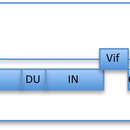
The FIV virus genome is diploid. It consists of two identical single-strands of RNA in each case about 9400 nucleotides existing in plus-strand orientation. It has the typical genomic structure of retroviruses and includes LTR, vif, pol, gag, orfA, env, and rev genes.[24][25][26] The Gag polyprotein is cleaved into matrix (MA), capsid (CA) and nucleocapsid (NC) proteins. Cleavage between CA and NC releases a nine amino acid peptide, while cleavage at the C-terminus of NC releases a 2kDa fragment (p2). The Pol polyprotein is translated by ribosomal frame-shifting, a feature shared with HIV. Cleavage of Pol by the viral protease releases the protease itself (PR), reverse transcriptase (RT), deoxyuridine triphosphatase (dUTPase or DU) and integrase (IN). The Env polyprotein consists of a leader peptide (L), surface (SU) and transmembrane (TM) glycoproteins. In common with other lentiviruses, the FIV genome encodes additional short open reading frames (ORFs) encoding the Vif and Rev proteins. An additional short ORF termed orfA (also known as orf2) precedes the env gene. The function of OrfA in viral replication is unclear, however the orfA-encoded product may display many of the attributes of HIV-1 accessory gene products such as Vpr, Vpu or Nef.
Among these subtypes, genetic sequences are mostly conserved; however, wide-ranging genetic differences exist between species specific FIV subtypes. Of FIV's genome, Pol is the most conserved across FIV strains along with gag. On the contrary, env, vif, orfa, and rev are the least conserved and exhibit the most genetic diversity among FIV strains.[27]
The capsid protein derived from the polyprotein Gag is assembled into a viral core (the protein shell of a virus) and the matrix protein also derived from Gag forms a shell immediately inside of the lipid bilayer. The Env polyprotein encodes the surface glycoprotein (SU) and transmembrane glycoprotein (TM). Both SU and TM glycoproteins are heavily glycosylated, a characteristic that scientists believe may mask the B-cell epitopes of the Env glycoprotein giving the virus resistance to the virus neutralizing antibodies.[9]
Like HIV-1, FIV has been engineered into a viral vector for gene therapy.[28] Like other lentiviral vectors, FIV vectors integrate into the chromosome of the host cell, where it can generate long-term stable transgene expression. Furthermore, the vectors can be used on dividing and non-dividing cells.[28][29] FIV vectors could potentially be used to treat neurological disorders like Parkinson's disease, and have already been used for transfer RNAi, which may find use as gene therapy for cancer.[30]
The exact origins and emergence of FIV in felids is unknown; however, studies of viral phylogenetics, felidae speciation, and FIV occurrence alludes to origins in Africa. Analysis of viral phylogenetics shows phylogenetic trees with a starburst phylogenetic pattern which is usually demonstrated by viruses that are a recent emergence with rapid evolution.[31] However, differences in topology, branch lengths, high genetic divergence suggest a more ancient origin in felidae species. Fossil records indicate extant felids arose from a common ancestor in Asia approximately 10.8 million years ago, and since then thirty eight species from eight distinct evolutionary lineages have spread and successfully inhabited every continent but Antarctica.[24] Despite felidae origins in Asia, FIV is absent from felidae species in Asia except for the Mongolian Pallas cat; however, FIV is highly endemic in Africa with four out of five felids having seropositive PCR results.[32] Due to the widespread occurrence and interspecies divergence of FIV strains in Africa, it's suggested that FIV arose in Africa before disseminating worldwide. The high genetic diversity and divergence between FIV strains in African felidae species and the presence of hyena FIV-Ccr, is consistent with a long residence time giving rise to increased opportunities for inter-species transmission among species. Additionally, lentiviruses are also highly endemic in Africa infecting not only felids, but also primates, and ungulate species. This suggests to the origins of all lentiviruses and supports FIV origins in Africa; however, further research is needed.[33][34]
The spread of FIV from Africa might have occurred during two points of felidae migration. The earliest migration across the Bering Strait into North America occurred approximately 4.5 million years ago during a period of low sea levels.[35] Early felids in North America descended into seven species of the ocelot lineage, two species of the puma lineage, and four of the modern species of lynx.[36] The most recent migration of Asian lions and jaguars across Eurasia into North and South America occurred during the Pliocene/early Pleistocene.[35] These migrations events increased opportunities for FIV transmission among felids and established infections globally for felidae species.
Comparisons of FIV subtypes illustrate rapid evolution and highlights divergence in FIV strains. FIV-Pco, which is specific to American pumas, has two highly divergent subtypes.[37] Several studies have demonstrated subtypes A and B to have long branch lengths and low geographic similarities which indicates the possibility of two separate FIV introductions into populations coupled with a long residence time.[37] In the late Pleistocene, pumas fell victim to the ice age, went extinct in North America except for a small inbred population in Florida, and did not re-emerge until 10-12,000 years ago.[35][38] Phylogenetic analysis of FIV-Pco strains in Central, South, and North America show Central and South American strains are more closely related to North American strains than to each other.[37][39] This suggests FIV-Pco was already present in South American pumas which repopulated North America.[39] In African lions, FIV-Ple has diverged in to six subtypes A-F which exhibit distinct geographical endemicity to some degree.[40] Approximately 2 million years ago, African lions arose and dispersed throughout Africa, Asia, and North, Central, and South America. Modern lions currently reside only on the African continent except for a small population in India.[35] There is no documented disease association of FIV, but seroprevalence in free- ranging lion populations are estimated to be roughly 90%.[41] Phylogenetic analysis of FIV-Ple subtypes A, B, and C show high intra and interindividual genetic diversity and sequence divergence comparable to genetic differences to strains from other Felidae species.[25] These findings indicate these strains evolved in geographically distant lion populations; however, recent occurrences of these strains within populations in Serengeti National Park suggests recent convergence in the same population.
In domestic cats, FIV-Fca is pathogenic and can lead to feline AIDS symptoms and subsequent death. Phylogenetic analysis shows FIV to be a monophyletic branch that diverges into three subtypes A, B, and C.[27] Domestic cats arose more recently than other felidae species approximately around 10,000 years ago from a subspecies of wildcat Felis silvestris which inhabited East Asia. Genetic analysis indicates lower genetic diversity of FIV in the domestic cat compared to wild Felidae species, higher evolutionary rates, and higher mortality rates when compared to FIV-Ple and FIV-Pco.[42] This suggests the emergence of FIV in domestic cats was recent since newly emerged viruses tend to have higher evolutionary rates with little to no co-adaption between virus and new host species occurring.[27] Additionally, seroprevalence studies show companion cats to have a 4–12% occurrence while feral cats have an 8–19% prevalence which is much lower compared to wild felidae species which supports the hypothesis of FIV's recent emergence in this species.[43][44]
FIV and feline leukemia virus (FeLV) are sometimes mistaken for one another though the viruses differ in many ways. Although they are both in the same retroviral subfamily (orthoretrovirinae), they are classified in different genera (FeLV is a gamma-retrovirus and FIV is a lentivirus like HIV-1). Their shapes are quite different: FeLV is more circular while FIV is elongated. The two viruses are also quite different genetically, and their protein coats differ in size and composition. Although many of the diseases caused by FeLV and FIV are similar, the specific ways in which they are caused actually differ. Also, while the feline leukemia virus may cause symptomatic illness in an infected cat, an FIV infected cat can remain completely asymptomatic its entire lifetime.
Feline immunodeficiency virus (FIV) is a Lentivirus that affects cats worldwide, with 2.5% to 4.4% of felines being infected.
FIV was first isolated in 1986, by Niels C Pedersen and Janet K. Yamamoto at the UC Davis School of Veterinary Medicine in a colony of cats that had a high prevalence of opportunistic infections and degenerative conditions and was originally called Feline T-lymphotropic virus. It has since been identified in domestic cats. It has been suggested FIV originated in Africa and has since spread to feline species worldwide.
Virusa imunodeficito de katoj (ankaŭ Sindromo de akirita perdo de la imuneco, popole Kata aidoso, enkutimiĝinta siglo FIV, angle Feline Acquired Immune Deficiency Syndrome - FAIDS) estas infekta malsano trafanta katojn.
Viruso FIP estas retroviruso kaŭzanta disigon de la imuneco de katoj. Ĝi apartenas en grupon de lentovirusoj simile kiel viruso de HIV, kiu kaŭzas aidoson ĉe homo. Tial oni kutimas nomigi ĝin ankaŭ kiel katan aidoson.
La vituso FIV troviĝas en la sango, salivoj kaj lakto. En ekstera medio ĝi estas nestabila. Tial transporto de la infekto okazas per rekta kontakto inter katoj. Seksa transporto de la viruso ĝis nun ne estis pruvita. Katidoj jam povas kontaĝi jam en la utero, dum nasko, per salivoj aŭ per lakto. Relative pli ofte la kontaĝo okazas ĉe virkatoj dum bataloj je teritorio.
La viruso kaŭzas malaltigitan defendkapablon de la kato, sekve de kio povas ataki la organismon ordinaraj bakterioj aŭ tiniaj infektoj. Tio montriĝas ĉe katoj per pligrandiĝintaj limfaj ganglioj, inflamaj malsaniĝoj de gingivoj, konjunktivito, inflamoj de haŭto, nazkataro, kronikaj diareoj, febroj, malviglado, dum pli longa paso aperas glaŭkomoj kaj diversaj specoj de tumoroj. Ĉe kelkaj unuopuloj aperas diversaj neŭrologiaj abnormalaĵoj kondukantaj al devigaj movoj, timo aŭ nepriregebla agresiemo.
Dum ne ekzistas fidinda kuracmetodo kondukanta al resaniĝo de la kata aidoso. Estas necese izoli de la ceteraj la trafitajn bestojn pro risko de transporto de la malsano. Ne estas bezone mortigi la katon tuj diagnozinte la malsanon, ĉar la kato kapablas kun bona zorgemo supervivi eĉ kun la viruso ankoraŭ kelke da plenvaloraj jaroj. Tuj kiam komenciĝos montriĝi longdaŭraj sekundaraj infektoj kaj la besto suferas, poste estas konsiderinda eŭtanazio. La terapio konsistas precipe en:
En Eŭropo ne ekzistas vakcinaĵo kontraŭ FIV, sed en Usono estis en la jaro 2002 aprobita vakcina preparaĵo.
Virusa imunodeficito de katoj (ankaŭ Sindromo de akirita perdo de la imuneco, popole Kata aidoso, enkutimiĝinta siglo FIV, angle Feline Acquired Immune Deficiency Syndrome - FAIDS) estas infekta malsano trafanta katojn.
El virus de inmunodeficiencia felina, VIF o FIV (por su sigla en inglés) es un lentivirus (de la familia Retroviridae) que afecta a los gatos domésticos mundialmente, y es el agente causante del sida felino. Aproximadamente un 11 %[1] de los gatos del mundo están infectados con el virus. Este virus difere taxonómicamente de otros dos retrovirus felinos, el virus de leucemia felina (FeLV en inglés) y el espumavirus felino (FFV en inglés) y está más emparentado con el virus de inmunodeficiencia humana (VIH). Dentro de los VIF, se identificaron 5 subtipos basándose en las diferencias de la secuencia de codificación de la cobertura viral. El VIF es el único lentivirus no primate que causa un síndrome similar al del sida, pero no siempre la muerte del gato, pueden vivir relativamente familiares como portadores y transmisores de la enfermedad por muchos años. Hay disponible una vacuna aunque su eficacia permanece incierta, y los gatos siguen dando positivo a las pruebas de anticuerpos del VIF después de ser vacunados.[2]
El VIF fue descubierto en 1986 en una colonia de gatos que tenía una alta prevalencia de infecciones oportunistas y condiciones degenerativas, y fue identificada como enfermedad endémica en la población mundial de gatos domésticos.[3]
El virus de inmunodeficiencia felina, VIF o FIV (por su sigla en inglés) es un lentivirus (de la familia Retroviridae) que afecta a los gatos domésticos mundialmente, y es el agente causante del sida felino. Aproximadamente un 11 % de los gatos del mundo están infectados con el virus. Este virus difere taxonómicamente de otros dos retrovirus felinos, el virus de leucemia felina (FeLV en inglés) y el espumavirus felino (FFV en inglés) y está más emparentado con el virus de inmunodeficiencia humana (VIH). Dentro de los VIF, se identificaron 5 subtipos basándose en las diferencias de la secuencia de codificación de la cobertura viral. El VIF es el único lentivirus no primate que causa un síndrome similar al del sida, pero no siempre la muerte del gato, pueden vivir relativamente familiares como portadores y transmisores de la enfermedad por muchos años. Hay disponible una vacuna aunque su eficacia permanece incierta, y los gatos siguen dando positivo a las pruebas de anticuerpos del VIF después de ser vacunados.
El VIF fue descubierto en 1986 en una colonia de gatos que tenía una alta prevalencia de infecciones oportunistas y condiciones degenerativas, y fue identificada como enfermedad endémica en la población mundial de gatos domésticos.
Kasside immuunpuudulikkuse viirus (FIV) on retroviirus. FIV-ga on maailmas haigestunud kuni 4,4% kassidest.[1][2]
FIV erineb taksonoomiliselt kahest kaslaste retroviirusest: leukeemiaviirusest (FeLV) ja FFV-st. Mõlemad on seotud inimese immuunpuudulikkuse viirusega (HIV). FIV-is on identifitseeritud viis alamtüüpi nukleotiidijärjestuste erinevusi, mis kodeerivad viiruse ümbrist või polümeraasi. FIV on ainuke mitteprimaatset päritolu AIDSi-laadset haigust põhjustav retroviirus, aga FIV ei ole kassidele letaalne ning nad võivad elada aastaid tervislikult, kuigi on haiguse kandjad. Vaktsiin on saadaval, aga selle mõju on veel teadmata. Pärast vaktsineerimist on kassid FIV-positiivsed.[3]
Haiguse isoleeris esimesena Niels Pedersen 1986. aastal UC Davise veterinaariakoolis, kus kassikoloonias olid suur infektsioonide esinemissagedus ja esialgu kutsuti haigust kasside T-lümfotsüütseks viiruseks (FTLV).[4] Seda on seni avastatud kodukasside populatsioonides üle maailma.[5]
FIV võib kahjustada kasside immuunsüsteemi. FIV nakatab erinevaid peremeesrakke, kaasa arvatud CD4+ ja CD8+ T-lümfotsüüte, B-lümfotsüüte ja makrofaage. Kassid võivad FIVd hästi taluda, kuid see võib lõpuks viia immuunsüsteemi nõrgenemiseni T-helperi (CD4+) nakatumise ja ammendumise tõttu. Võrreldes inimese HIVga, mille nakatumise protsent on umbes 50%, on kasside nakatumise protsent väga väike, vähem kui 5%.
FIV ja HIV on retroviirused. Seevastu inimesed ei ole võimelised nakatuma FIV-sse ja kassid pole võimelised nakatuma HIV-sse. FIV-d kantakse edasi sügavate hammustushaavade kaudu: nakatunud kassi süljes sisalduv viirus siseneb teise kassi keharakkudesse. FIV-positiivsed kassid saavad jagada omavahel vett, sööki ja käia samas liivakastis, ilma et haigus edasi kanduks. Valvas loomaomanik, kes ravib sekundaarseid infektsioone, võimaldab kassil elada majapidamises pikka aega. Oht, et FIV-ga nakatunud kass nakataks teist kassi, on väga väike, kui ei esine kasside vahelist võitlemist või avatud haavu, mis võimaldavad kassidel üksteisele nakkust edasi kanda.
Vastsündinud kassipoegadel võib haigus osutuda positiivseks kuni kuus kuud, aga pärast testimist negatiivseks, sest antikehad kanduvad kassipoegadele ema piima kaudu. Kuue kuu möödudes on uued testid negatiivsed, kuna antikehad ei ole püsivad. Kassipoegi vaktsineerides on testitulemused positiivsed, kuna vereproov näitab antikehade teket.
FIV on tuntud ka teiste kaslaste hulgas ja see on endeemiline suuremate kaslaste hulgas, näiteks Aafrika lõvi.
Viirus saab siseneda peremeesrakku koostoimes viituse glükoproteiinide ümbrise ja märklaudrakkude pinna retseptorite kaudu. Kõigepealt seondub SU glükoproteiin CD134ga, peremeesraku retseptoriga. Esialgne seondumine muudab SU-valgu kuju selliseks, mis hõlbustab SU ja kemokiini retseptori CXCR4 interaktsiooni[6]. See interaktsioon põhjustab viiruse ja rakkude membraanide sulandumise ja võimaldab viiruse RNA kandumist tsütoplasmasse, kus see pöördtranskribeeritakse ja integreeritakse raku genoomi läbi mittehomoloogilise rekombinatsiooni. Peremeesraku genoomis võib viirus püsida pikka aega asümptomaatilises staadiumis, ilma et immuunsüsteem seda avastaks või põhjustaks rakkude lüüsimist.[7][8]
CD134 on leitav aktiveeritud T-rakkudes ja seostunult OX40 ligandiga, põhjustades T-raku stimulatsiooni, proloferatsiooni, aktivatsiooni ja apoptoosi. T-raku aktivatsioon toob kaasa märkimisväärse kahanemise rakkudes, millel on immuunsüsteemis kriitiline roll. Immuunsüsteemi rakud ja madal CD4+ tase muudavad kassid vastuvõtlikuks haiguste suhtes ning haigus võib progresseeruda omandatud immuunpuudulikkuse sündroomiks (FAIDS)[9].
Peamiselt levib viirus sügavate hammustushaavade kaudu, kus haigustkandva kassi sülg siseneb teise kassi kudedesse. FIV võib ka tiinetelt kassidelt edasi kanduda oma järglastele emaka kaudu, kuigi seda peetakse väga haruldaseks.[3][9] FIV erineb FeLvst kuna see võib levida sama kammi kasutamisest ja toidukausside jagamisest.
Infektsiooni riskiteguriteks on täiskasvanud isased kassid, kes saavad õues käia. Sao Paulos läbi viidud uuringus leiti, et 75% FIV-sse nakatunud kassidest olid isased. Suurem infektsiooniprotsent esineb isastel seetõttu, et nad hammustavad üksteist tihemini ja kaitsevad oma territooriumi.[8].
FIV progresseerub sarnaselt nagu HIV inimestel. Esimese või akuutse faasiga kaasnevad sümptomid, nagu letargia, anoreksia, palavik ja lümfadenopaatia[9]. Esimene faas on lühike ja sellele järgneb asümptomaatiline staadium, kus kassil ei esine pikemat aega ühtegi märgatavat sümptomit. Mõned kassid püsivad selles staadiumis kuid, teised aga mitu aastat. Tegurid, mis mõjutavad asümptomaatilise staadiumi pikkust, hõlmavad viiruse patogeensust ja FIV alamtüüpi (A-E), kassi vanust ja kokkupuudet teiste patogeenidega. Pärast seda liigub kass viimasesse staadiumisse (FAIDS staadium), kus kass on äärmiselt vastuvõtlik sekundaarsetele haigustele ja paratamatult lõpeb isendi surmaga[8].
Veterinaarid kontrollivad kassi ajalugu ja testivad verd FIV antikehade leidmiseks.
Valed positiivsed tulemused ilmuvad, kui kass kannab antikehi (mis on kahjutud), aga ei kanna viirust. Kõige sagedamini juhtub seda, kui kassipojad on tarbinud ema piima ja kui kasse on vaktsineeritud FIV vastu. Sellel põhjusel alla kaheksa nädala vanuseid kassipoegi ja hiljuti vaktsineerituid kasse ei testita.
Kassipojad ja noored kassid, kes on testitud positiivseks FIV antikehade suhtes, võivad osutuda negatiivseks tingimusel, et nad ei ole kunagi FIVga nakatunud ja neil pole kunagi kasutatud FIV-vaktsiini.
Kassid, keda on varem vaktsineeritud, osutuvad FIV-positiivseteks kuni elu lõpuni, kuigi nakatumist ei ole toimunud. Seetõttu ei anna kodutute ja adopteeritud kasside testimine tavaliselt õigeid, kuna on võimatu teada, kas neid on varem vaktsineeritud või mitte. Seega ei tohiks kunagi kasutada eutanaasia põhjusena positiivset FIV antikehade testi[10].
Teste saab teha loomaarsti kabinetis mõne minutiga. Varajane tuvastamine aitab säilitada kassi tervist ning takistab haiguse edasikandumist teistele kassidele. Õige hoolitsusega on kassid võimelised elama pikka ja täisväärtuslikku elu.
Lümfotsüütide T-rakkude immunomodulaator on ette nähtud kasside leukeemia viiruse (FeLv) või kasside immuunpuudulikkuse viiruse (FIV) raviks ning sellega seotud oportunistliku infektsiooni, aneemia raviks. Kui kõrvalnähtuseid ei täheldata, on tootel väga madal toksilisuse profiil.
Lümfotsüütide T-rakkude immunomodulaator on CD-4 lümfotsüütide tootmise ja funktsiooni regulaator[13]. See on näidanud suurendavat lümfotsüütide arvu ja Interleukin2 tootmist loomades[14]. See on üheahelaline polüpepriid ja on tugevalt katioonne glükoproteiin ning seda puhastatakse katioonvahetusvaiguga. Veiselt saadud stroomarakkude supernatandidest pärineva valgu puhastamine annab homogeense teguri, mis ei sisalda võõrkehasid. Veiste valk on homoloogne teiste imetajateliikidega ja on homogeenne 50kDa glükoproteiin, mille isoelektriline punkt on 6,5. Valk valmistatakse lüofiliseeritud 1 mikrogrammi annuses. Lahustumine steriilses lahustis annab naha alla süstitava lahuse.
Nagu HIV puhul, on ka FIV vastu efektiivse vaktsiini väljatöötamine keeruline viirustüvede suure arvukuse ja variatsioonide tõttu. "Ühe tüve" vaktsiinid ehk vaktsiinid, mis kaitsevad ainult ühe viirusevariandi vastu, on näidanud juba head mõju homoloogsete FIV-tüvede vastu. 2002. aastal avaldatud kaksik-alamtüüpi vaktsiin Fel-O-Vax FIV vastu tegi võimalikuks kassi immuniseerida teiste FIV-alamtüüpide vastu. See arendati välja, kasutades inaktiveeritud isolaate 2–5 FIV alamtüübist: Petaluma ja D Shizuoka[15]. Vaktsiin osutus A FIV-alamtüübi suhtes mõõdukalt tõhusaks (82% kassidest said kaitstud)[16], aga hiljem tehtud uuring näitas, et see ei paku kaitset alamtüübi A vastu[17]. Samas on vaktsiin näidanud 100% efektiivsust kahe erineva B FIV-alamtüübi vastu[18][19]. Vaktsiinid näitavad positiivseid FIV-testi tulemusi kassidel ning seetõttu on diagnoosimine keeruline. Nendel põhjustel on vaktsiin "mitte keskne" ja vaktsineerimise otsus oleks vaja teha pärast loomaarstiga konsulteerimist[20].
FIV-l on sarnane struktuur primaatide ja kabiloomade retroviirustega. Virioni diameeter varieerub 80 nanomeetrist 100 nanomeetrini ja on pleomorfne. Viiruse ümbrisel on ka väljaulatuvad osad, mis on väikesed, 8 nm, ja katavad pinna ühtlaselt[7].
Viiruse genoom on diploidne. See koosneb kahest identsest üheahelalisest RNAst, mis on ligikaudu 9400 nukleotiidi pikk ja plussahelaga orientatsioonis. Sellel on tüüpiline retroviiruse genoomistruktuur, kaasa arvatud gag-, pol- ja env-geenid. Gag polüproteiin on kleepunud maatriksi, kapsiidi ja nukelokapsiidi valkude külge. Kapsiidi ja nukleokapsiidi vaheline lõikamine vabastab üheksa aminohappe peptiidi, samal ajal kui lõikamine nukleokapsiidi C-otsas vabastab 2kDa fragmendi. Pol polüproteiin transleeritakse ribosomaalse raaminihke kaudu, sama omadus on ka HIV-l. Pol lõikamine viiruse proteaasiga vabastab proteaas pöördtranskriptaasi, desoksüuridiintrifosfataasi ja integraasi. Env polüproteiin koosneb liiderpeptiidist, pinna- ja transmembraandstest glükoproteiinidest. Sarnaselt teiste retroviirustega kodeerib FIV genoomi täiendavaid lühikesi lugemisraame.
Polüproteiini Gagist pärinev kapsiidivalk liigub viiruse südamikku ja maatriksivalk formeerub kestaks lipiidide kaksikkihi sees. Env polüproteiin kodeerib pealmist glükoproteiini ja transmembraanset glükoproteiini. Mõlemad glükoproteiinid on palju glükoseeritud ning teadlased usuvad, et see võib anda viirusele resistentsuse viirust neutraliseerivate antikehade suhtes[7].
Nagu HIV-1 on ka FIV kasutusel viirusvektorina geeniteraapias[21]. Nagu teised retroviiruselised FIV-vektorid integreeruvad kromosoomi läbi peremeesraku, kus see võimaldab genereerida pikaajalise stabiilse transgeense ekspressiooni. Samuti saab kasutada vektoreid rakutsüklis ja väljaspool rakutsüklit[21][22]. FIV-vektoreid võib tõenäoliselt kasutada tulevikus, et ravida neuroloogilisi haigusi nagu Parkinsoni tõbi. Vektoreid on juba kasutatud RNAi ülekandmiseks, mis võivad kasutust leida vähiravis[23].
6. "Mapping of Receptor Binding Interactions with the Fiv surace Glycoprotein (SU); Implications Regarding Immune survelliance and cellular Targets of Infection"doi10.4137/RRT.S9429PMC3523734PMID23255871. Retrieved 2017-08-18
7. "Vaccination against the feline immunodeficiency virus: The road not taken"doi10.1016/j.cimid.2007.07.007, retrieved 15 November 2011
8. "Clinical aspects of feline immunodeficiency and feline leukemia virus infection"doi10.1016/j.vetimm.2011.06.003, retrieved 16 November 2011
9. Current HIV Researchdoi10.2174/157016210790416361PMC3721975
10. doi10.1016/j.jfms.2009.05.006PMID19481037
11. LTCI Product Information, retrieved 28 July 2012
12. T-Cyte Therapeutics, Inc., retrieved 28 July 2012
13. Beardsley, et al. "Induction of T-Cell Maturation by a Cloned Line of Thymic Epithelium (TEPI) Immunology 80: pp. 6005-6009, (Oct. 1983).
14. US patent 7196060, Beardsley, Terry R., "Method to enhance hematopoiesis", published 2005-05-19, issued 2007-03-27
15. doi10.1016/j.jfms.2008.03.002PMID18455463
16. doi10.1079/AHR200487PMID15984343
17. doi10.1136/vr.158.16.561PMID16632531
18. doi10.1016/j.vetmic.2005.02.014PMID15899558
19. doi10.1016/j.jfms.2004.08.005PMID15686976
20. doi10.1016/j.jfms.2008.03.002PMID18455463
21. doi10.1038/nm0398-354PMID9500613
22. doi10.1128/JVI.00958-06PMC1617215PMID16973543
23. doi10.2174/156652308786848030PMID19075624
Kasside immuunpuudulikkuse viirus (FIV) on retroviirus. FIV-ga on maailmas haigestunud kuni 4,4% kassidest.
FIV erineb taksonoomiliselt kahest kaslaste retroviirusest: leukeemiaviirusest (FeLV) ja FFV-st. Mõlemad on seotud inimese immuunpuudulikkuse viirusega (HIV). FIV-is on identifitseeritud viis alamtüüpi nukleotiidijärjestuste erinevusi, mis kodeerivad viiruse ümbrist või polümeraasi. FIV on ainuke mitteprimaatset päritolu AIDSi-laadset haigust põhjustav retroviirus, aga FIV ei ole kassidele letaalne ning nad võivad elada aastaid tervislikult, kuigi on haiguse kandjad. Vaktsiin on saadaval, aga selle mõju on veel teadmata. Pärast vaktsineerimist on kassid FIV-positiivsed.
Haiguse isoleeris esimesena Niels Pedersen 1986. aastal UC Davise veterinaariakoolis, kus kassikoloonias olid suur infektsioonide esinemissagedus ja esialgu kutsuti haigust kasside T-lümfotsüütseks viiruseks (FTLV). Seda on seni avastatud kodukasside populatsioonides üle maailma.
Kissan immuunikatovirus (lyh. FIV) on kissoilla esiintyvä retroviruksiin kuuluva lentivirus, joka aiheuttaa kissoille elinikäisen immuunikatotaudin infektoimalla auttaja-T-soluja ja makrofageja. Kissa saa virustartunnan useimmiten puremasta, sillä virus erittyy sylkeen. Kissanpennut voivat saada tartunnan myös virusta kantavalta emolta istukan tai emonmaidon kautta. Taudin oireet esiintyvät tavallisimmin useita vuosia tartunnan saamisesta.
Immuunikatovirusta tavataan sekä kesyillä että villeillä kissaelämillä. Villeistä lajeista leijona on laajimmin virusta kantava laji; lähes kaikilla Afrikan leijonilla on FIV.[1]
Kissan immuunikatovirus voidaan diagnosoida vasta-aineiden määrityksellä verestä.
Kissan immuunikatovirus (lyh. FIV) on kissoilla esiintyvä retroviruksiin kuuluva lentivirus, joka aiheuttaa kissoille elinikäisen immuunikatotaudin infektoimalla auttaja-T-soluja ja makrofageja. Kissa saa virustartunnan useimmiten puremasta, sillä virus erittyy sylkeen. Kissanpennut voivat saada tartunnan myös virusta kantavalta emolta istukan tai emonmaidon kautta. Taudin oireet esiintyvät tavallisimmin useita vuosia tartunnan saamisesta.
Immuunikatovirusta tavataan sekä kesyillä että villeillä kissaelämillä. Villeistä lajeista leijona on laajimmin virusta kantava laji; lähes kaikilla Afrikan leijonilla on FIV.
Le virus de l'immunodéficience féline (FIV ou FIV, de l'anglais feline immunodeficiency virus) est un virus qui cause le syndrome d'immunodéficience acquise, une maladie virale grave qui atteint les félins et notamment le chat. Il s'agit d'un lentivirus (sous-groupe des rétrovirus, proche du VIH).
Cette maladie rend le chat vulnérable aux infections est équivalente au sida humain, bien qu'y a aucun risque de transmission du chat à l'humain. La présence du FIV est également observée chez le lynx et le puma.
Il n'existe pas de traitement spécifique, mais l'animal peut cependant vivre plusieurs années avec le virus. La prévention s'opère à travers l'évitement de la contamination et l'évitement des contacts entre chats sains et chats atteints.
L'identification du virus responsable du syndrome d'immunodéficience félin est rapportée dans un article scientifique publié en 1987[2].
Le FIV est présent sur tous les continents[2]. Il concerne environ 11 % de la population féline domestique à travers le monde, bien que ce pourcentage soit certainement sous-estimé puisqu'entre 10 et 15 % des chats FIV ne sont pas séronégatifs[3]. Les mâles sont davantage concernés que les femelles, car plus susceptibles d'être impliqué dans des bagarres avec morsures[2]. Les individus les plus à risque sont ceux qui vivent dans des zones à haute densité de chats et ceux qui ont la possibilité de se promener librement à l'extérieur[2].
Quatre sous-types (A, B, C et D) de virus ont été identifiés. 80 % des virus isolés appartiennent aux sous-types A et B. Des recombinaisons entre différents sous-types ont également été observés[3].
Le virus de l'immunodéficience féline s'attaque aux cellules immunitaires que sont les lymphocytes T, les macrophages et les cellules microgliales[3].
Le virus se transmet entre chats principalement par les morsures, mais également par les rapports sexuels. Il peut aussi être transmis par la mère à ses petits[3].
Le virus ne survit que quelques heures dans l'environnement[4].
À noter que des chatons infectés par la mère et donc testés positifs, peuvent se révéler être négatifs après quelques mois. Jusqu'à 5 % des chatons porteurs d’anticorps réagissant positivement au test du virus du FIV seront testés négatifs par la suite
La durée d'incubation est de 4 à 6 semaines.
Des symptômes peuvent parfois se manifester durant la première phase qu'est la phase aiguë, qui survient entre un et trois mois après l'infection : l'animal présente alors de la fièvre, de la fatigue, entérite, stomatite, dermatite, conjonctivite, affection respiratoire, et gonflement des ganglions lymphatiques[4],[5]. Cependant, les symptômes restant discrets, cette période n'est pas toujours remarquée par les propriétaires[4]. La seconde période consiste en une phase asymptomatique où le chat ne manifeste aucun signe clinique[5],[6]. Cette période peut durer de quelques mois à plusieurs années, en fonction de l'âge auquel le chat a été infecté, le sous-type de FIV, et l'exposition à d'autres pathogènes[5]. La phase asymptomatique peut également durer jusqu'à la mort de l'animal,Enfin, la troisième et dernière phase consiste en une période symptomatique pendant laquelle le chat est touché par des infections opportunistes, une néoplasie, une dépression médullaire et des troubles neurologiques[5].
Les chats atteints par le FIV sont globalement plus sensibles aux infections dont les effets peuvent durer plus longtemps et être plus sévères[6]. Les chats positifs au FIV ont cinq fois plus de risques de développer un lymphome ou une leucémie ; ils sont d'ailleurs surreprésentés parmi les chats atteints de lymphomes[5]. La moitié des chats déclarent une immunodéficience.
Certaines pathologies sont courantes chez les chats atteints par le virus de l'immunodéficience féline comme par exemple :
L'espérance de vie de l'animal ne semble pas significativement modifiée[4].
Les anticorps spécifiques du FIV ne sont détectables qu'à partir d'entre deux et dix mois après la contamination[4].
Le FIV peut être diagnostiqué grâce à des tests rapides, des tests Elisa ou par technique PCR. En pratique, le vétérinaire utilise généralement un test rapide qui se base sur une prise de sang, et la détection d'anticorps anti-GP40 à partir de sang total, de plasma ou de sérum. Le résultat est obtenu en 10 à 15 minutes. Un test salivaire est également possible[7].
Le dépistage du FIV peut être proposé à n'importe quel âge en cas de suspicion, comme dès les premières consultations vaccinales d'un chaton, pour écarter toute transmission par la mère[7]. Il est conseillé de réaliser un test lors de l'acquisition d'un chat, et régulièrement auprès des chats à risque[4]. Tester tous les chats du foyer en cas de test positif est également à privilégier. En cas de test négatif, il est recommandé de réaliser un second test afin de confirmer le premier[4].
Il n'existe pas de traitement contre le FIV. La principale prise en charge consiste à diminuer les risque de contracter des infections autres qui profiteraient de l'immunodéficience pour se développer[4]. Un suivi régulier du poids et des paramètres sanguin est recommandé, ainsi qu'un traitement précoce des affections pouvant toucher l'individu[4].
Une fois la maladie déclenchée, les soins sont ponctuels et visent les maladies opportunistes au fur et à mesure qu'elles atteignent le chat. Les traitements symptomatiques à la cortisone comportent de nombreux effets secondaires. D'autre part, la cortisone associée à l'interféron réduirait sérieusement l'effet bénéfique de l'interféron, puisque schématiquement, la cortisone est un immunosuppresseur, à l'inverse de l'interféron qui est un "immunobooster". Il existe aussi un traitement thérapeutique à l’interféron oméga félin injectable (Virbagen félin). Ce traitement, en cure de plusieurs injections, est très coûteux (environ 300-400 euros par cure), mais il améliore grandement les conditions de vie de l’animal et allonge sa durée de vie.
La prévention passe par la réduction des risques de morsures[4], à travers la limitation des déplacements extérieurs des chats, et l'évitement des contacts entre chats sains et ceux positifs aux FIV[8].
Un vaccin contre le FIV est commercialisé en Australie[9] et un autre en Amérique du Nord, qui a une efficacité partielle et une utilisation controversée. En Europe, il n'y a pas de projet de commercialisation de ce vaccin qui a été développé avec des souches virales présentes essentiellement en Amérique du Nord[10].
Le FIV provoque le sida chez les chats de la même manière que le VIH chez les humains : en décimant les lymphocytes T qui luttent contre les infections. Voilà pourquoi, en 2011, une équipe dirigée par Eric Poeschla de la clinique Mayo (Minnesota) a inséré un gène de macaque rhésus - producteur d'une molécule antivirale - dans des ovules de chattes non encore fécondés. Pour suivre le transfert du gène sous un microscope et certaines lumières, les scientifiques ont ajouté une protéine luminescente issue d'une méduse. Résultat à la génération suivante : des chatons qui brillent dans le noir et produisent eux-mêmes la protéine antivirale. Bientôt, l'équipe procédera à des tests pour voir si les chats génétiquement modifiés sont vraiment immunisés contre le FIV. Paula Canon, spécialiste de thérapie génique à l'université de Californie du Sud, explique que ces travaux sont « une étape essentielle » dans les recherches génomiques sur le sida - pour la santé des chats, mais aussi des humains.
Le FIV observé chez les félins non domestiques est génétiquement différent du FIV observé chez les animaux domestiques et la transmission interspécifique est très rare[11]. Le virus a été détecté chez le lynx (Lynx rufus) et chez le puma (Puma concolor) en Californie et en Floride[12].
En Europe, le FIV est signalé pour la première fois en 2016 chez le lynx en Suisse, où un individu capturé dans le canton de Soleure a été reconnu porteur du virus ; au début de 2017, deux individus porteurs également sont capturés dans le canton du Jura, à Saint-Ursanne et à Longeau[13].
Le virus de l'immunodéficience féline (FIV ou FIV, de l'anglais feline immunodeficiency virus) est un virus qui cause le syndrome d'immunodéficience acquise, une maladie virale grave qui atteint les félins et notamment le chat. Il s'agit d'un lentivirus (sous-groupe des rétrovirus, proche du VIH).
Cette maladie rend le chat vulnérable aux infections est équivalente au sida humain, bien qu'y a aucun risque de transmission du chat à l'humain. La présence du FIV est également observée chez le lynx et le puma.
Il n'existe pas de traitement spécifique, mais l'animal peut cependant vivre plusieurs années avec le virus. La prévention s'opère à travers l'évitement de la contamination et l'évitement des contacts entre chats sains et chats atteints.
Galar víreasach i gcait, atá gaolmhar le víreas easpa imdhíonachta daonna (HIV) ach scartha uaidh. Ní chuireann an víreas ná an galar isteach ar dhaoine. Tarlaíonn raon comharthaí cliniciúla sa chat de réir mar a sháraíonn orgánaigh eile a chóras imdhíonach. Tarlaíonn athlasadh ainsealach don bhéal de bharr fiabhrais gan stad, agus leanann cailliúint thapa meáchain é sin. Ní féidir diagnóis chruinn a dhéanamh ar na comharthaí seo gan an fhuil a thástáil.
Feline immunodeficiency virus (disingkat FIV, juga dikenal sebagai feline AIDS) adalah spesies virus dalam genus Lentivirus yang menyebabkan penurunan sistem imun pada kucing. Sebanyak 11% kucing di dunia terinfeksi dengan FIV. Menurut penelitian lainnya, 2,5% kucing di Amerika Serikat terinfeksi FIV. Virus ini berada pada famili retrovirus, sama seperti Feline leukemia virus (FeLV) dan Feline foamy virus (FFV). Terdapat vaksin untuk virus ini walaupun kemanjurannya tetap tidak menentu.[1]
Virus FIV pertama kali diisolasi tahun 1986 pada koloni kucing yang mengalami infeksi oportunistik dan kondisi yang merosot, dan telah diidentifikasikan sebagai penyakit endemik pada kucing domestik di dunia. Virus FIV ditransmisikan melalui luka gigitan yang dalam, di mana virus dalam air liur kucing terinfeksi masuk ke dalam jaringan kucing lain.
Virus ini menyerang sistem kekebalan kucing, seperti Human immunodeficiency virus (HIV) yang menyerang sistem kekebalan manusia. FIV menginfeksi banyak tipe sel, termasuk limfosit CD4+ dan CD8+, limfosit T, limfosit B, dan makrofag.
Feline immunodeficiency virus (disingkat FIV, juga dikenal sebagai feline AIDS) adalah spesies virus dalam genus Lentivirus yang menyebabkan penurunan sistem imun pada kucing. Sebanyak 11% kucing di dunia terinfeksi dengan FIV. Menurut penelitian lainnya, 2,5% kucing di Amerika Serikat terinfeksi FIV. Virus ini berada pada famili retrovirus, sama seperti Feline leukemia virus (FeLV) dan Feline foamy virus (FFV). Terdapat vaksin untuk virus ini walaupun kemanjurannya tetap tidak menentu.
Virus FIV pertama kali diisolasi tahun 1986 pada koloni kucing yang mengalami infeksi oportunistik dan kondisi yang merosot, dan telah diidentifikasikan sebagai penyakit endemik pada kucing domestik di dunia. Virus FIV ditransmisikan melalui luka gigitan yang dalam, di mana virus dalam air liur kucing terinfeksi masuk ke dalam jaringan kucing lain.
Virus ini menyerang sistem kekebalan kucing, seperti Human immunodeficiency virus (HIV) yang menyerang sistem kekebalan manusia. FIV menginfeksi banyak tipe sel, termasuk limfosit CD4+ dan CD8+, limfosit T, limfosit B, dan makrofag.
Il virus dell'immunodeficienza felina (spesso abbreviato in FIV, Feline Immunodeficiency Virus) è un Virus della famiglia dei retrovirus, appartenente alla sottofamiglia dei Lentivirinae, genere lentivirus. Nel gatto, questo agente patogeno costituisce la causa di un'immunodeficienza, denominata sindrome da immunodeficienza acquisita felina e volgarmente chiamata AIDS del gatto, data la forte somiglianza alla sindrome da immunodeficienza acquisita che colpisce l'uomo. Questa peculiarità non implica affatto la possibilità della trasmissione del morbo da felini all'uomo: l'essere umano non è contagiabile dal virus dell'immunodeficienza felina. La prima descrizione della patologia risale al 1986, quattro anni dopo la scoperta ufficiale dell'HIV. La malattia finora è classificata come incurabile, ma spesso sul breve periodo si rivela asintomatica. A più lungo termine, però, si avvia il processo di deterioramento del sistema immunitario dell'organismo ospite, il quale, in seguito a infezioni secondarie, muore. Finora sono stati identificati nove diversi ceppi del virus da undici diverse razze di gatto in isolamento, compresi i ceppi specifici da leoni e puma. Anche esemplari di iena maculata sono risultati positivi ai test sul virus, nonostante essi non appartengano alla stessa famiglia del gatto.
Il Feline Immunodeficiency Virus è un lentivirus che colpisce il sistema immunitario del gatto compromettendone l'efficiente funzionamento e rendendo quindi il gatto affetto da FIV più suscettibile a infezioni secondarie.
Il virus si trova nei gatti domestici di tutto il mondo ed infetta anche felini selvaggi come i leopardi delle nevi, i leoni, le tigri, i giaguari, le pantere e le linci rosse della Florida. Benché sia stato isolato solamente nel 1987, è noto che FIV esiste da molto tempo. L'infezione è molto comune nei gatti che vivono fuori casa o che sono liberi di girovagare. I gatti maschi sono 2 volte più colpiti delle femmine. Infatti, i gatti liberi di girovagare hanno più probabilità di altri di contrarre l'infezione, essendo maggiormente esposti al rischio di ferite da morso. Diversamente da FeLV, nei rifugi per gatti l'infezione da FIV è rara in proporzione alle ferite da morso che ci si aspetterebbe in un luogo simile.
L'infezione da FIV nei gatti, come quella da HIV negli esseri umani, è caratterizzata da tre stadi. Quello iniziale o acuto dell'infezione da FIV è spesso caratterizzato da febbre, linfonodi gonfi e predisposizione ad infezioni della pelle o dell'intestino. Questo stadio generalmente si sviluppa alla quarta - sesta settimana dopo l'esposizione al virus.
Il secondo stadio è latente, non si notano i sintomi della malattia, e può durare per molti anni, durante i quali il sistema immunitario può essere lentamente cancellato.
Quando l'immunodeficienza diventa grave, si sviluppa il terzo stadio dell'infezione. Il terzo stadio è quello terminale e si riscontra più comunemente nei gatti tra i 5 e i 12 anni (FeLV colpisce gatti tra 1-5 anni). Durante questo stadio clinico terminale, il sistema immunitario del gatto non funziona correttamente dal momento che il virus ne ha ucciso le cellule essenziali. Pertanto, gli animali nel terzo stadio della malattia sono particolarmente predisposti alle infezioni, di solito croniche, che possono essere batteriche, da miceti o da parassiti. Spesso sono causate da microrganismi che normalmente non causano gravi malattie nei gatti, ma il sistema immunitario ormai limitato non riesce a contrastarle, per cui si determina la proliferazione dell'infezione che si moltiplica rapidamente causando la malattia. Queste si definiscono infezioni “opportunistiche”.
Il contagio avviene tramite scambi di sangue o altri fluidi organici tra un gatto malato ed uno sano: più frequentemente nei gatti il contagio avviene attraverso l'accoppiamento o tramite ferite da morso: per questo i gatti maschi interi, con libero accesso all'esterno, sono i soggetti più a rischio. A differenza di FeLV, FIV non si trasmette con un prolungato e stretto contatto. Poiché si trova nella saliva, il virus viene inoculato attraverso le ferite da morso. Il contagio tramite la cassettina della lettiera, le scodelle del cibo e dell'acqua, oppure perché i gatti dormono vicini o giocano insieme è estremamente improbabile.
Si può verificare se la gatta viene infettata durante la gestazione o l'allattamento. La trasmissione dell'infezione nell'utero o attraverso il latte è molto rara. Le gatte già infette dal FIV prima della gravidanza di solito partoriscono gattini non infetti.
Il virus determina la sindrome da immunodeficienza acquisita felina, simile all'AIDS umano. La malattia determina nel lungo periodo una maggiore suscettibilità del gatto ad infezioni secondarie, perlopiù opportunistiche, e all'insorgenza di neoplasie.
Il virus dell'immunodeficienza felina (spesso abbreviato in FIV, Feline Immunodeficiency Virus) è un Virus della famiglia dei retrovirus, appartenente alla sottofamiglia dei Lentivirinae, genere lentivirus. Nel gatto, questo agente patogeno costituisce la causa di un'immunodeficienza, denominata sindrome da immunodeficienza acquisita felina e volgarmente chiamata AIDS del gatto, data la forte somiglianza alla sindrome da immunodeficienza acquisita che colpisce l'uomo. Questa peculiarità non implica affatto la possibilità della trasmissione del morbo da felini all'uomo: l'essere umano non è contagiabile dal virus dell'immunodeficienza felina. La prima descrizione della patologia risale al 1986, quattro anni dopo la scoperta ufficiale dell'HIV. La malattia finora è classificata come incurabile, ma spesso sul breve periodo si rivela asintomatica. A più lungo termine, però, si avvia il processo di deterioramento del sistema immunitario dell'organismo ospite, il quale, in seguito a infezioni secondarie, muore. Finora sono stati identificati nove diversi ceppi del virus da undici diverse razze di gatto in isolamento, compresi i ceppi specifici da leoni e puma. Anche esemplari di iena maculata sono risultati positivi ai test sul virus, nonostante essi non appartengano alla stessa famiglia del gatto.
Kattenaids, ook wel felien immunodeficiëntievirus (FIV) (Engels: feline immunodeficiency virus) genoemd, is een ziekte bij katten, andere katachtigen en hyena's. Het is een retrovirus dat in 1986 voor het eerst is beschreven in de Verenigde Staten. Wereldwijd is inmiddels 11 % van de katten besmet.
Besmetting vindt vaak plaats door (bijt)wonden toegebracht bij gevechten tussen katten. Er zijn echter ook aanwijzingen dat normaal contact tussen katten (via oraal, rectaal en vaginale afscheiding) de ziekte kan overbrengen. De ziekte vertoont sterke gelijkenissen met aids bij mensen, hoewel katten geen hiv kunnen oplopen en mensen geen fiv. Katten worden dan ook vaak gebruikt voor onderzoek naar aids.
FIV infecteert verschillende typen immuuncellen, waaronder monocyten, macrofagen, B- en T-lymfocyten. Chronische infectie wordt gekenmerkt door een sterke vermindering van CD4+ T-helpercellen en verlaging van de ratio van CD4+:CD8+ cellen, tijdens de asymptomatische fase. Daalt de hoeveelheid T-helpercellen onder een bepaalde grens, dan treedt immuundysfunctie op. Deze symptomatische fase ('kattenaids') kenmerkt zich door secundaire en opportunistische infecties.[1]
Hoewel oorspronkelijk werd gedacht dat de afname van CD4+ T-lymfocyten te wijten is aan het doden van deze cellen door FIV, wordt dit veroorzaakt door chronische activatie van het immuunsysteem.[1]
Kattenaids, ook wel felien immunodeficiëntievirus (FIV) (Engels: feline immunodeficiency virus) genoemd, is een ziekte bij katten, andere katachtigen en hyena's. Het is een retrovirus dat in 1986 voor het eerst is beschreven in de Verenigde Staten. Wereldwijd is inmiddels 11 % van de katten besmet.
Besmetting vindt vaak plaats door (bijt)wonden toegebracht bij gevechten tussen katten. Er zijn echter ook aanwijzingen dat normaal contact tussen katten (via oraal, rectaal en vaginale afscheiding) de ziekte kan overbrengen. De ziekte vertoont sterke gelijkenissen met aids bij mensen, hoewel katten geen hiv kunnen oplopen en mensen geen fiv. Katten worden dan ook vaak gebruikt voor onderzoek naar aids.
FIV infecteert verschillende typen immuuncellen, waaronder monocyten, macrofagen, B- en T-lymfocyten. Chronische infectie wordt gekenmerkt door een sterke vermindering van CD4+ T-helpercellen en verlaging van de ratio van CD4+:CD8+ cellen, tijdens de asymptomatische fase. Daalt de hoeveelheid T-helpercellen onder een bepaalde grens, dan treedt immuundysfunctie op. Deze symptomatische fase ('kattenaids') kenmerkt zich door secundaire en opportunistische infecties.
Hoewel oorspronkelijk werd gedacht dat de afname van CD4+ T-lymfocyten te wijten is aan het doden van deze cellen door FIV, wordt dit veroorzaakt door chronische activatie van het immuunsysteem.
Do tej pory znane szczepy FIV są bardzo rozbieżne, lecz monofiletyczne, co oznacza, że powstały z jednej formy podstawowej. Dla trzech szczepów tego wirusa udało się wyróżnić ich podtypy. Jak do tej pory FIV kotów domowych jest najlepiej zbadane i zawiera pięć podtypów, które występują na całym świecie z różną częstotliwością, a oznaczane są literami od A do E. Podział na pięć podgrup dokonywany jest na podstawie porównania sekwencji DNA genu env. Podgrupy A do C są rozpowszechnione na całym świecie, D występuje przede wszystkim we wschodniej Azji, a E tylko w Ameryce Południowej.
Również dla FIVple wyszczególniono podgrupy określane od A do C. Podział na te podgrupy nastąpił na podstawie różnic sekwencyjnych w genie pol, który koduje wirusowe enzymy (proteazy, integrazy i odwrotne transkryptazy). Dla FIVpci wyszczególniono na podstawie różnic w pol dwie podgrupy oznaczane jako A i B. Różnice sekwencji DNA są po części znaczące pomiędzy poszczególnymi szczepami FIV i kształtują się np. dla genu pol w FIVple, FIVca FIVca FIVpco na poziomie 30 procent.
Znane szczepy FIV tworzą swoją własną grupę w obrębie lentiwirusów i można je podzielić na stare i nowe gatunki. Najbliżej spokrewnione są z lentiwirusami bydła i koni.
FIV ma budowę podobną do innych lentiwirusów, które wywołują objawy niedoboru odporności u ssaków. Kompletny wirion ma średnicę 105 do 125 nanometrów, ma kulisty lub owalny kształt oraz posiada w osłonce wirusa krótkie, nieokreślone elementy (ang. spikes), które składają się z wirusowych glikoprotein gp95 i gp44. Ma on tak jak inne retrowirusy gęstość 1,15–1,17 g/cm³. Cząstki wirusowe rozpadają się w wyniku działania zwykłych środków odkażających zawierających alkohol i chlor, i dezaktywują się poprzez krótkie ogrzewania do 60 °C.
Genom wirusa jest podwójny. Składa się z dwóch identycznych pojedynczych molekuł RNA składających się każda po około 9400 nukleoidów. Genom ten posiada typową genomową strukturę retrowirusów, która składa się z genów gag-pol-env i tak jak inne lentiwirusy posiada dodatkowe geny. Są nimi vif, vpr, ref. Nie zawierają one tat, vpu, vpx i nef, dlatego FIV jest mniej złożony niż wirus HIV. FIV posiada deoksyurydynopirofosfatazę (dUTPaza), która nie występuje u lentiwirusów naczelnych. dUTPaza kodowana jest w pol-regionie; enzym redukuje dUTP do dUMP oraz pirofosforanu, co prawdopodobnie uniemożliwia błędne włączenie dUTP do genomu.
Chorobotwórczość szczepów FIV u kotów żyjących na wolności jest trudna do określenia. Badania epidemiologiczne, w których porównano częstość przeżycia z częstotliwością występowania choroby i wskaźnikiem rozrodu, nie wykazały statystycznie istotnych negatywnych skutków dla zakażonych zwierząt. Wiele występujących szczepów nie może być zatem określanych jako patogenne. Niska patogenność szczepów tego wirusa u dziko żyjących kotów wskazuje na długie wzajemne oddziaływanie patogenu z gospodarzem, zachodzące już od około miliona bądź dwóch milionów lat[6]. Nie wiadomo jeszcze, u których gatunków rozwinął się pierwotnie FIV. Przenoszenie wirusa między różnymi gatunkami kotów na wolności występuje rzadko, w niewoli natomiast częściej[7].
Zespół niedoboru immunologicznego kotów jest specyficzny dla kotowatych i ryzyko zarażenia u ludzi określane jest jako minimalne[8]. FIV infekuje, podobnie jak HIV-1, głównie limfocyty T CD4+. W porównaniu do HIV-1, jednego z najbardziej zbliżonych do FIV spośród występujących u ludzi wirusów, może on zarażać szersze spektrum komórek. Obok limfocytów T CD4+, monocytów, makrofagów i komórek glejowych, infekuje on także limfocyty T i B CD8+. Za pierwotny receptor glikoproteiny (gp95) uważane są nie cząsteczki CD4, jak przy HIV-1, lecz CD134[9][10]. Do interakcji pomiędzy glikoproteiną 95 i CD134 potrzebny jest CXCR4 jako główny koreceptor. Glikoproteina 95 wirusa wiąże się za pośrednictwem wypustek (ang. spikes) z cząsteczką CD134, co prowadzi do zmiany konformacji w gp95 umożliwiającej interakcję z CXCR4[11]. Ta interakcja z koreceptorem pobudza syntezę osłonek wirusowych z błoną komórkową i umożliwia wniknięcie do komórek. Ponieważ opisane zostały także szczepy wirusów, które nie wymagają CD132, rola receptorów nie jest całkowicie wyjaśniona.
Dotychczas w żadnym przypadku nie powiodło się wszczepienie wirusa zespołu niedoboru immunologicznego do ludzkich komórek czy też linii komórkowych w celu rozmnożenia. Przyczyną, dla której wirus nie mógł przejść całego cyklu replikacji był fakt, że wykrywalny prowirus zintegrowany w DNA nie pokonuje krytycznego etapu transkrypcji. Dlatego po zainfekowaniu komórek nie może dojść do syntezy pełnych cząstek wirusa. Blokada ta przypomina tym samym tę spotykającą EIAV w komórkach ludzkich i HIV w komórkach mysich.
W organizmie kota wkrótce po infekcji produkowane są przeciwciała i powstają cytotoksyczne komórki T, jednak pomimo gwałtownej reakcji immunologicznej nie dochodzi do całkowitego zwalczenia infekcji. Wirus pozostaje przez to na stałe w organizmie – we wszystkich dotychczas badanych przypadkach.
Stosunkowo dużo uwagi poświęca się opracowaniu szczepionki przeciwko temu wirusowi, która została dopuszczona w 2002 roku w Stanach Zjednoczonych. Po doświadczeniach z tą szczepionką narodziły się nadzieje na opracowanie szczepionki przeciwko HIV[12]. Prace nad tą szczepionką przyspieszyły po odkryciu wirusa[8], badano wiele różnych typów szczepionek, w tym inaktywowane wirusy, komórki zainfekowane wirusami, szczepionki DNA i wektory wirusowe. Nie jest jasne, czy otrzymane w warunkach laboratoryjnych rezultaty potwierdzą się w praktyce, zwłaszcza biorąc pod uwagę dużą liczbę różnych szczepów wirusa.
Rozwój skutecznej szczepionki przeciwko wirusowi FIV jest tak jak w przypadku wirusa HIV utrudniony przez dużą liczbę wirusów i wariacje ich szczepów. Dla tzw. szczepionki "pojedynczego szczepu" ("single strain"), a więc chroniącej tylko przed jednym z wariantów wirusa, udowodniono duża skuteczność także wobec homologicznych szczepów FIV. Wraz z rozwojem szczepionki przeciwko FIV "dual-subtype" (Fel-O-Vax FIV) stało się możliwe uodpornienie kotów na dalsze szczepy tego wirusa. Szczepionka składa się z inaktywowanych cząstek FIV szczepów Petaluma subtype A i Shizuoka subtype D[13]. W warunkach laboratoryjnych zaobserwowano uodpornienie 82% badanych kotów po podaniu szczepionki[14]. W przypadku dwóch różnych podtypów szczepu B wirusa wykazano skuteczność 100%[15][16]. Całkowite uodpornienie zwierząt żyjących na wolności przeciwko pierwotnym szczepom wydaje się być jednak wciąż nieosiągalne. Dotychczas można było tylko opierać się na niektórych doświadczeniach z badań nad szczepionką przeciwko HIV[12]. Najbardziej krytykowaną stroną dostępnej szczepionki jest fakt, że zaszczepione zwierzęta nie różnią się serologicznie od zainfekowanych[17]. Trwają prace nad testem pozwalającym na ich rozróżnienie.
Zbliżony obraz kliniczny niedoboru odporności u kotów i AIDS oraz możliwość uodpornienia przeciwko FIV sprawiają, że wirus ten stał się użytecznym modelem w badaniach nad HIV/AIDS[18].
W oparciu o FIV opracowywane są wektory wirusowe na potrzeby ludzkiej terapii genowej[19]. Jest to możliwe dzięki braku patogenności FIV wobec ludzi. Wektory wirusa znajdują także zastosowanie w podstawowych badaniach naukowych.
A AIDS Felina é uma infecção causada pelo vírus da imunodeficiência felina (FIV, na sigla em inglês para feline immunodeficiency vírus). Trata-se de um retrovírus complexo que, quando manifestada no gato, é similar aquela observada em humanos com Aids, sobretudo no que diz respeito ao aumento da susceptibilidade a infecções oportunistas[1]. A AIDS Felina não tem cura. Ela só é transmitida entre gatos, ou seja, é uma enfermidade que não acomete outras espécies, incluindo o homem[2].
Assim como ocorre com a AIDS em humanos, há gatos que são portadores assintomáticos do vírus. Eles passam anos sem apresentar sintomas de modo que a doença só poderia ser detectada por exames de sangue, como o teste ELISA ou o PCR (do inglês “reação em cadeia da polimerase”), ambos realizados no Brasil[2].
A transmissão da AIDS felina entre gatos é dada principalmente por mordidas ou arranhões de gatos infectados por FIV. Também pode ser transmitida por via transplacentária, pela amamentação. Geralmente, os gatos FIV positivo são machos e de rua, onde a concentração é alta e a taxa de estresse é contínua, o que pode ocasionar o aparecimento do vírus[1].
Na ocorrência de anormalidades em relação à saúde do felino, a detecção de um possível gato seropositivo, é feita a partir do surgimento de sintomas como diarreia persistente, problemas respiratórios, febre, emagrecimento, anemia, infecções e mais raramente a morte[1]. Os principais sintomas são a úlceração da mucosa oral e a perda de peso decorrente da dor na ingestão de alimentos.[3]
A prevenção poderá ser feita através da administração da vacina da FIV. No entanto, esta vacina pode não evitar a infeção e protege apenas para algumas estirpes do vírus. No entanto, a vacinação reduz a expressão da doença e poderá até ser vantajosa em animais FIV positivo.[4] Através de cuidados com a higiene do animal, alimentação saudável, castração[5] e visitas ao médico veterinário, é possível reduzir as chances de contaminação em gatos domésticos.
Algumas pessoas confundem a AIDS Felina (FIV) com a leucemia felina (FELV). Apesar de ambas as doenças estarem associadas a retrovírus da mesma família e causarem condições secundárias similares, trata-se de enfermidades distintas. O FIV é menos patogénico enquanto que o FELV é mais patogénico e, em casos raros, poderá ser eliminado pelo organismo no início da infeção.[6]
Os gatos tendem a ser discriminados quando se descobre que têm a doença. Porém, a luta contra o vírus deve ser válida. Gatos seropositivos têm chances de ter uma qualidade de vida normal. Além disso, estudo publicado pela Preventive Veterinary Medicine indicou que a AIDS felina não afeta a longevidade dos gatos: que gatos com AIDS tendem a viver tanto quanto gatos negativos para AIDS[7].
A AIDS Felina é uma infecção causada pelo vírus da imunodeficiência felina (FIV, na sigla em inglês para feline immunodeficiency vírus). Trata-se de um retrovírus complexo que, quando manifestada no gato, é similar aquela observada em humanos com Aids, sobretudo no que diz respeito ao aumento da susceptibilidade a infecções oportunistas. A AIDS Felina não tem cura. Ela só é transmitida entre gatos, ou seja, é uma enfermidade que não acomete outras espécies, incluindo o homem.
Assim como ocorre com a AIDS em humanos, há gatos que são portadores assintomáticos do vírus. Eles passam anos sem apresentar sintomas de modo que a doença só poderia ser detectada por exames de sangue, como o teste ELISA ou o PCR (do inglês “reação em cadeia da polimerase”), ambos realizados no Brasil.
Vírusová imunodeficiencia mačiek (aj Syndróm získanej straty imunity, hovorovo Mačací AIDS, zaužívaná skratka FIV, po anglicky Feline Acquired Immune Deficiency Syndrome – FAIDS) je infekčná choroba postihujúca mačky.
Vírus FIV je retrovírus vyvolávajúci rozvrátenie obranyschopnosti mačiek. Patrí do skupiny lentovírov podobne ako vírus HIV, ktorý vyvoláva AIDS u človeka. Preto sa zvykne označovať aj ako mačací AIDS.
Vírus FIV sa nachádza v krvi, slinách, mledzive a mlieku. Vo vonkajšom prostredí nie je stabilný. Prenos infekcie sa uskutočňuje preto priamym stykom medzi mačkami. Pohlavný prenos vírusu nebol dosiaľ preukázaný. Mačiatka sa môžu infikovať už v maternici, počas pôrodu, slinami, mledzivom alebo mliekom. Relatívne častejšie dochádza k nákaze u kocúrov pri bitkách o teritórium.
Vírus spôsobuje zníženú obranyschopnosť mačky, následkom čoho môžu organizmus napadnúť bežné bakteriálne alebo plesňové infekcie. U mačiek sa prejavujú zväčšené lymfatické uzliny, zápalové ochorenia ďasien, očných spojiviek a kože, nádcha, chronické hnačky, teploty, chátranie, pri dlhšom priebehu glaukóm a rôzne druhy nádorov. U niektorých jedincov sa prejavujú rôzne neurologické abnormality vedúce k nutkavým pohybom, prejavom strachu alebo nezvládnuteľnej agresivite.
Zatiaľ neexistuje spoľahlivá liečebná metóda vedúca k vyliečeniu mačacieho AIDS, choré mačky však pri dobrej starostlivosti môžu prežiť niekoľko plnohodnotných rokov. Postihnuté zvieratá treba izolovať od ostatných kvôli riziku prenosu choroby. Nie je potrebné mačku usmrtiť hneď po diagnostikovaní choroby, mačka dokáže pri vhodnej starostlivosti prežiť s vírusom aj niekoľko rokov. Hneď ako sa začnú prejavovať dlhotrvajúce sekundárne infekcie a zviera trpí, potom treba zvážiť eutanáziu. Terapia spočíva najmä v :
V Európe neexistuje očkovacia látka proti FIV. V Spojených štátoch bol však v roku 2002 schválený očkovací preparát.
Vírusová imunodeficiencia mačiek (aj Syndróm získanej straty imunity, hovorovo Mačací AIDS, zaužívaná skratka FIV, po anglicky Feline Acquired Immune Deficiency Syndrome – FAIDS) je infekčná choroba postihujúca mačky.
Feline immunodeficiency virus (FIV) är en typ av lentivirus som drabbar kattdjur.
Viruset finns hos tamkatter över hela världen. Även lejon och andra vildkatter har viruset men utvecklar sällan några symtom. Viruset attackerar immunsystemet hos värden, bland annat CD4 celler som har en viktig roll i organiseringen av immunsystemet.
Virus Feline immunodeficiency, còn gọi là Vi rút suy giảm miễn dịch mèo (viết tắt là FIV) là một lentivirus ảnh hưởng đến mèo trên toàn thế giới. Từ 2,5% đến 4,4%[1][2] tổng số mèo trên toàn thế giới bị nhiễm FIV. FIV khác về mặt phân loại với hai loại retrovirus mèo khác, virus bạch cầu mèo (FeLV) và vi rút bọt xốp (FFV), và có liên quan chặt chẽ hơn với virus suy giảm miễn dịch ở người (HIV). Trong vi rút FIV, năm loại phụ đã được xác định dựa trên sự khác biệt trình tự nucleotit mã hóa được bản chất virus (env) hoặc polymerase (pol). FIV là lentivirus không thuộc nhóm linh trưởng duy nhất gây ra một hội chứng giống AIDS, nhưng FIV không gây tử vong cho mèo, và các con mèo mang bệnh có thể sống tương đối khỏe mạnh, đóng vai trò như các vật chủ mang và truyền bệnh trong nhiều năm. Một loại thuốc chủng ngừa đã có sẵn mặc dù hiệu quả của nó vẫn chưa được chắc chắn. Mèo sẽ có kết quá xét nghiệm là dương tính với kháng thể FIV sau khi được tiêm phòng.[3]
FIV lần đầu tiên được chiết tách vào năm 1986 bởi Tiến sĩ Smith tại trường thú y UC Davis, với nguồn gốc là từ một nhóm mèo có tỷ lệ nhiễm trùng cơ hội và điều kiện thoái hóa cao và ban đầu virus này được gọi với cái tên là Feline T-lymphotropic Virus (FTLV).[4] Virus này đã được xác định có trong các quần thể mèo nhà trên toàn thế giới.[5]
Virus Feline immunodeficiency, còn gọi là Vi rút suy giảm miễn dịch mèo (viết tắt là FIV) là một lentivirus ảnh hưởng đến mèo trên toàn thế giới. Từ 2,5% đến 4,4% tổng số mèo trên toàn thế giới bị nhiễm FIV. FIV khác về mặt phân loại với hai loại retrovirus mèo khác, virus bạch cầu mèo (FeLV) và vi rút bọt xốp (FFV), và có liên quan chặt chẽ hơn với virus suy giảm miễn dịch ở người (HIV). Trong vi rút FIV, năm loại phụ đã được xác định dựa trên sự khác biệt trình tự nucleotit mã hóa được bản chất virus (env) hoặc polymerase (pol). FIV là lentivirus không thuộc nhóm linh trưởng duy nhất gây ra một hội chứng giống AIDS, nhưng FIV không gây tử vong cho mèo, và các con mèo mang bệnh có thể sống tương đối khỏe mạnh, đóng vai trò như các vật chủ mang và truyền bệnh trong nhiều năm. Một loại thuốc chủng ngừa đã có sẵn mặc dù hiệu quả của nó vẫn chưa được chắc chắn. Mèo sẽ có kết quá xét nghiệm là dương tính với kháng thể FIV sau khi được tiêm phòng.
FIV lần đầu tiên được chiết tách vào năm 1986 bởi Tiến sĩ Smith tại trường thú y UC Davis, với nguồn gốc là từ một nhóm mèo có tỷ lệ nhiễm trùng cơ hội và điều kiện thoái hóa cao và ban đầu virus này được gọi với cái tên là Feline T-lymphotropic Virus (FTLV). Virus này đã được xác định có trong các quần thể mèo nhà trên toàn thế giới.
貓免疫缺陷病毒(英文:Feline Immunodefiency Virus,簡稱:FIV)屬於反轉錄病毒科慢病毒屬,受到感染的貓會罹患貓後天免疫缺乏症候群,俗稱貓愛滋病。雖然稱為貓愛滋病,但與人類的愛滋病不同的是,貓愛滋病並非透過性行為傳染,主要是透過打架時、含病毒唾液經傷口傳染;另一個不同在於,感染FIV的貓之死亡率並沒有人類的愛滋病來得高,通常就算患病,不使用藥物的情況下仍然能夠活至正常壽命。目前已有對抗此病毒的疫苗。
對養貓者來說,必須注意貓是否有感染FIV,有感染的與沒有感染也沒有注射疫苗的貓若會打架、則必須隔離。有養貓的家庭在撿到流浪貓時必須把家貓與流浪貓先行隔離檢疫,在確定沒有FIV等傳染病以後才能接觸。
FIV病毒並不會感染人體,但「貓愛滋」的名稱很容易造成無知者棄貓,因此在媒體報導或宣導貓愛滋時強調貓愛滋對人類無害,媒體應該遵守此基本職業道德。
此病毒只會對貓科動物有作用,包括貓、老虎、美洲豹、豹、獅子等。然而,與家貓不同的是,其他的貓科動物受到感染後並不會出現症狀。
罹患貓愛滋的貓有食慾不振、體重減輕、皮毛狀況不佳等症狀,還會有下痢及口腔、牙齦、皮膚、肺部的慢性感染。有些會有貧血或癲癇樣的神經症狀。
貓免疫缺陷病毒(英文:Feline Immunodefiency Virus,簡稱:FIV)屬於反轉錄病毒科慢病毒屬,受到感染的貓會罹患貓後天免疫缺乏症候群,俗稱貓愛滋病。雖然稱為貓愛滋病,但與人類的愛滋病不同的是,貓愛滋病並非透過性行為傳染,主要是透過打架時、含病毒唾液經傷口傳染;另一個不同在於,感染FIV的貓之死亡率並沒有人類的愛滋病來得高,通常就算患病,不使用藥物的情況下仍然能夠活至正常壽命。目前已有對抗此病毒的疫苗。
對養貓者來說,必須注意貓是否有感染FIV,有感染的與沒有感染也沒有注射疫苗的貓若會打架、則必須隔離。有養貓的家庭在撿到流浪貓時必須把家貓與流浪貓先行隔離檢疫,在確定沒有FIV等傳染病以後才能接觸。
FIV病毒並不會感染人體,但「貓愛滋」的名稱很容易造成無知者棄貓,因此在媒體報導或宣導貓愛滋時強調貓愛滋對人類無害,媒體應該遵守此基本職業道德。
此病毒只會對貓科動物有作用,包括貓、老虎、美洲豹、豹、獅子等。然而,與家貓不同的是,其他的貓科動物受到感染後並不會出現症狀。
猫後天性免疫不全症候群(ねここうてんせいめんえきふぜんしょうこうぐん)とは、FIV(feline immunodeficiency virus = ネコ免疫不全ウイルス)により、ネコに引き起こされる諸症状のこと。俗にネコエイズとも言う。FIVに遺伝的に近縁なウイルス(FIV関連ウイルス(FIV-related virus))がライオン、ピューマなどでも分離されているが、これらの野生動物がFIV関連ウイルスで発症したという明確な報告はない。猫免疫不全ウイルス感染症の病態の一つである。
FIVウイルスの主な感染経路は、ケンカなどによる咬み傷からの体液の接触感染が考えられる。自然界では人間のように交尾によって感染したと断定できる報告はない[1]。出産時の母子感染も確認されている。HIVと同じレトロウイルス科レンチウイルス属に分類されるが、ネコおよびネコ属に特異的なウイルスであり、犬や人に感染することは無い。
疫学的には、国内のFIV抗体陽性率は約12%とされているが、野良猫におけるFIVの保有率はこれよりも高いと考えられる[2]。 2006年10月にアメリカのワシントンで開催された国際ネコレトロウイルス研究シンポジウムにおいて、フロリダ大学のグループは興味深い疫学調査結果を発表している。それは、アメリカの獣医病院に来院し検査を受けた67,963匹のネコを調べたところ、「FIV陽性ネコの生存率(survival rate)は、FIVに陽性と診断されてから1年でおよそ約20%が死亡する(発症していないネコの安楽殺を含む)ものの、それ以降はFIV陽性ネコ群と陰性ネコ群では生存率に大きな差は見られない。」というものである。 ドイツやオーストラリアでなされた疫学調査からも、FIV陽性ネコと陰性ネコの平均寿命に有意差は見られていない。
このことからも明らかなように、FIV陽性ネコのすべてが発症するわけではない。FIV感染により免疫が抑制されるが、HIVほどではない。またたとえ免疫が抑制されても、FIVの感染が直接病気を引き起こすわけではなく、FIV感染による免疫抑制に伴う他の病原体の感染により発症する。SPFネコにおける感染実験においてもFIV単独で発症した例はほとんどない。FIVの受容体(ウイルス感染に必要な分子)は、CD134という分子であり、これは抗原刺激に伴って発現されるCD4陽性T細胞の副刺激分子の一つである。従って様々な病原体、もしくは非病原性のウイルスや細菌などの抗原刺激によりFIVが増殖可能なCD4陽性T細胞が増加し、それに伴い体内のFIVの量も増えるものと考えられ、抗原刺激に乏しい(クリーンな)環境においてはネコの体内でのウイルス量は低いままであると考えられる。
ネコのエイズの進行には以下のステージがあり、一定の順序に従って発症していく[1]。各ステージはおおよそ括弧内の期間継続する。
家庭では、子猫のうちにウイルス検査を行い(潜伏期があるため、最後に他のネコと接触してから数か月おいて検査すると確実)、陰性であれば不妊・去勢手術を行い他のネコとの交尾・ケンカの機会を減らすことでほぼ確実に予防できる。
ウイルスの潜伏場は白血球のT細胞であり、採血できれば、簡易検査キットにより簡単に動物病院等で検査が可能である。この検査キットではウイルスに対する抗体を検出する。抗体が陽転するまで約1ヶ月かかるので、陽転するまでの間はたとえ感染していても陰性と判定される。感染の可能性が強く示唆される場合は、1ヶ月後以降(できれば2ヶ月後以降)に再検査することが必要である。
猫後天性免疫不全症候群(ねここうてんせいめんえきふぜんしょうこうぐん)とは、FIV(feline immunodeficiency virus = ネコ免疫不全ウイルス)により、ネコに引き起こされる諸症状のこと。俗にネコエイズとも言う。FIVに遺伝的に近縁なウイルス(FIV関連ウイルス(FIV-related virus))がライオン、ピューマなどでも分離されているが、これらの野生動物がFIV関連ウイルスで発症したという明確な報告はない。猫免疫不全ウイルス感染症の病態の一つである。
고양이 면역결핍바이러스(영어: Feline immunodeficiency virus, FIV)는 고양이의 면역 체계를 파괴시키는 레트로바이러스이다. 모든 고양이과 동물들에게 영향을 미친다. 전세계적으로 2.5%에서 4.4%의 고양이들이 고양이 면역결핍바이러스에 감염되었다.[1][2] 고양이 면역결핍바이러스는 분류학상으로 다른 두 고양이 RNA 종양 바이러스인 고양이 백혈병 바이러스 (FeLV)와 고양잇과 거품성 바이러스 (FFV)와 다르며 인간 면역 결핍 바이러스 (HIV)에 더 가깝다. 고양이 면역결핍바이러스 내에서 다섯 개의 종이 바이러스막과 중합효소 코딩의 뉴클레오타이드 순서의 차이로 구분된다. 고양이 면역결핍바이러스는 오직 비영장류 레트로바이러스로 AIDS 같은 증후군을 일으키지만 고양이 면역결핍바이러스는 일반적으로 고양이에게 치명적이지 않고 보균자로 남거나 전달자 질병의 증상으로 나타나 비교적 건강하게 살 수 있다.
FIV는 1986년 UC Davis 수의과 대학의 Niels C Pedersen과 Janet K. Yamamoto에 의해 기회 감염과 퇴행성 질환의 유병률이 높았고 원래 고양이 T-림프성 바이러스라고 불렸던 고양이 군집에서 처음 분리되었다.[3] 리포니아 대학교 수의과 대학 Smith 박사는 기회감염과 퇴행성 상태의 높은 감염성을 가진 고양이의 집단에서 떨어져 나왔다고 밝혔다. 이후 집고양이에서 확인되었다.[4]
FIV는 아프리카에서 기원한 것으로 알려져 있으며, 이후 전 세계적으로 고양이과 동물로 확산되었다.
고양이 면역결핍바이러스(영어: Feline immunodeficiency virus, FIV)는 고양이의 면역 체계를 파괴시키는 레트로바이러스이다. 모든 고양이과 동물들에게 영향을 미친다. 전세계적으로 2.5%에서 4.4%의 고양이들이 고양이 면역결핍바이러스에 감염되었다. 고양이 면역결핍바이러스는 분류학상으로 다른 두 고양이 RNA 종양 바이러스인 고양이 백혈병 바이러스 (FeLV)와 고양잇과 거품성 바이러스 (FFV)와 다르며 인간 면역 결핍 바이러스 (HIV)에 더 가깝다. 고양이 면역결핍바이러스 내에서 다섯 개의 종이 바이러스막과 중합효소 코딩의 뉴클레오타이드 순서의 차이로 구분된다. 고양이 면역결핍바이러스는 오직 비영장류 레트로바이러스로 AIDS 같은 증후군을 일으키지만 고양이 면역결핍바이러스는 일반적으로 고양이에게 치명적이지 않고 보균자로 남거나 전달자 질병의 증상으로 나타나 비교적 건강하게 살 수 있다.
FIV는 1986년 UC Davis 수의과 대학의 Niels C Pedersen과 Janet K. Yamamoto에 의해 기회 감염과 퇴행성 질환의 유병률이 높았고 원래 고양이 T-림프성 바이러스라고 불렸던 고양이 군집에서 처음 분리되었다. 리포니아 대학교 수의과 대학 Smith 박사는 기회감염과 퇴행성 상태의 높은 감염성을 가진 고양이의 집단에서 떨어져 나왔다고 밝혔다. 이후 집고양이에서 확인되었다.
FIV는 아프리카에서 기원한 것으로 알려져 있으며, 이후 전 세계적으로 고양이과 동물로 확산되었다.


