-
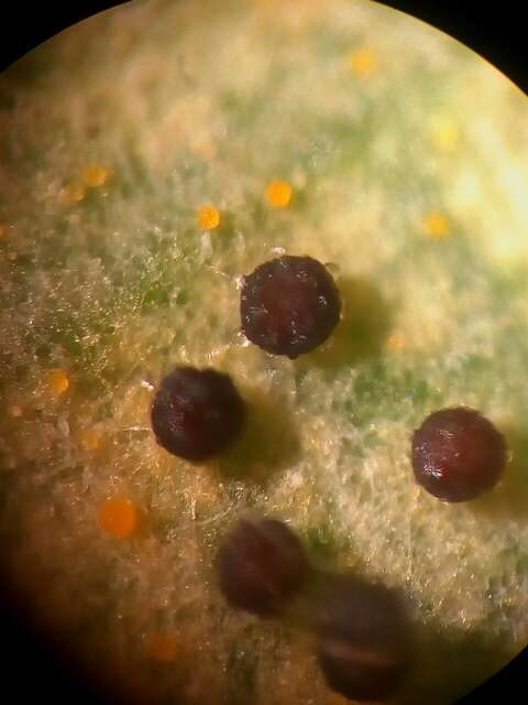
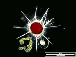
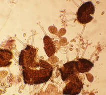
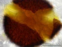
Cleisthotecium containing ascospores of the fungus that causes powdery meldew on leaves and stems of some trees. Scale bar indicates 500 µm.Sample from ponds situated in the vicinity of Lake Constance (Bodensee, Southern Germany). The image was built up using several photomicrographic frames with manual stacking technique. Images were taken using Zeiss Universal with Olympus C7070 CCD camera.
-
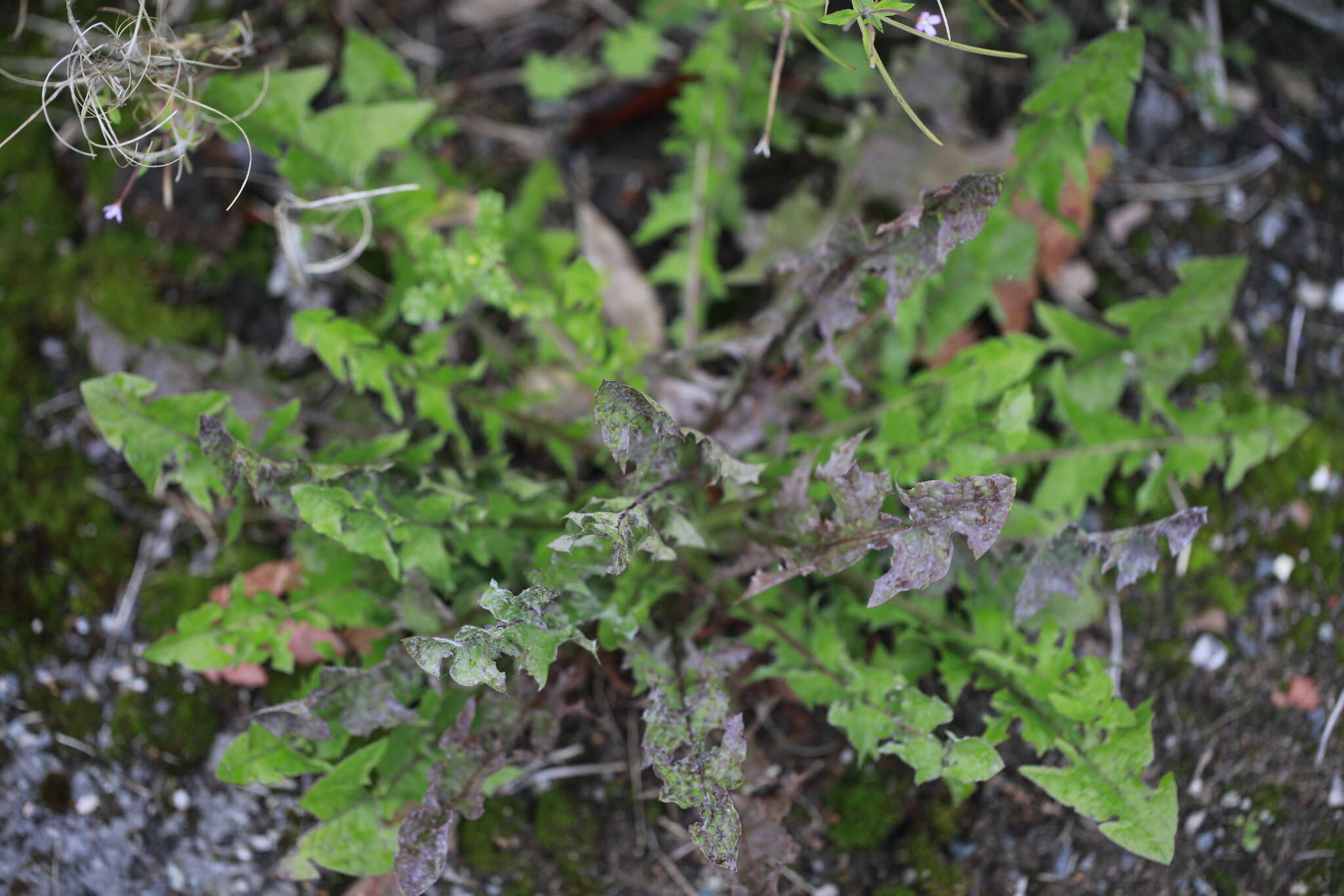
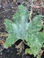
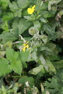
Midtsjælland, Danmark
-
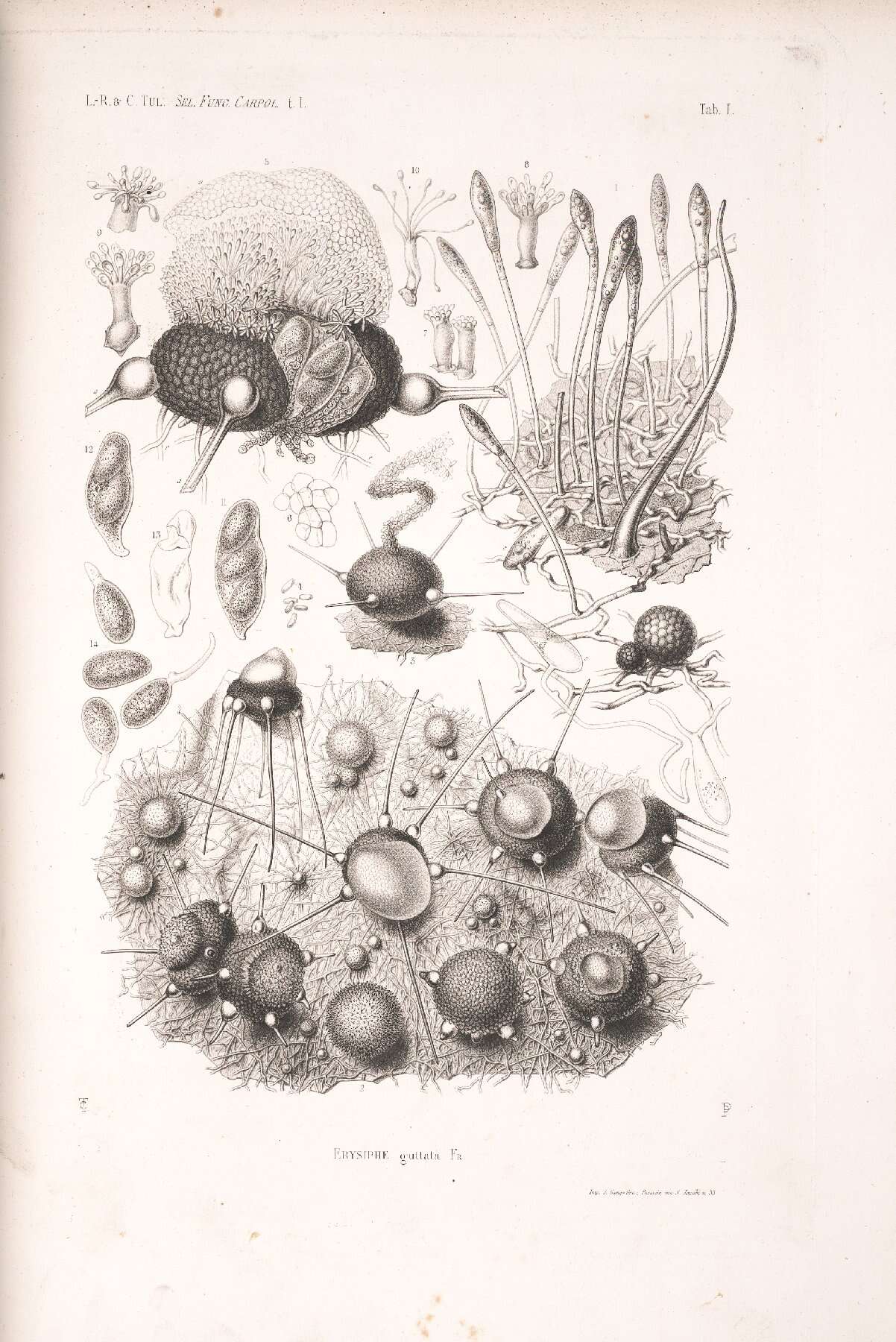
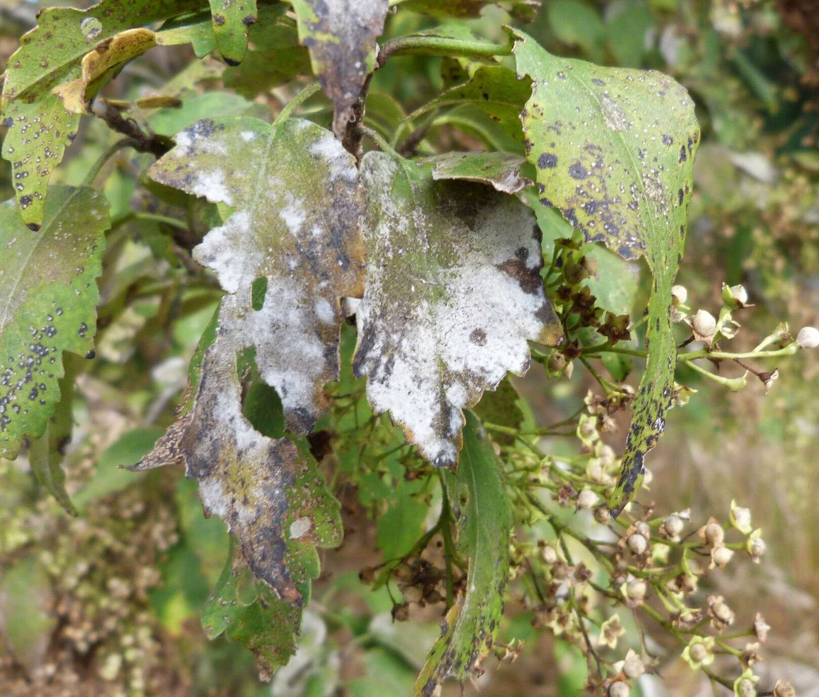
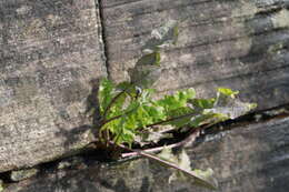
Mushroom Observer Image 949006: Phyllactinia guttata (Wallr.) Lev.
-
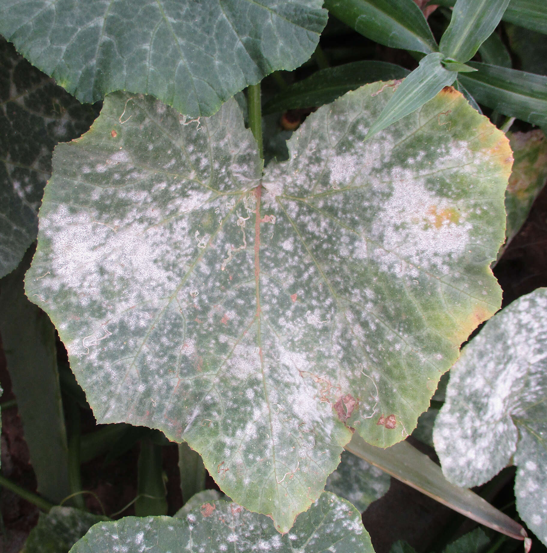
Horbury, England, United Kingdom
-
-
International Maize and Wheat Improvement Center
Flickr Group
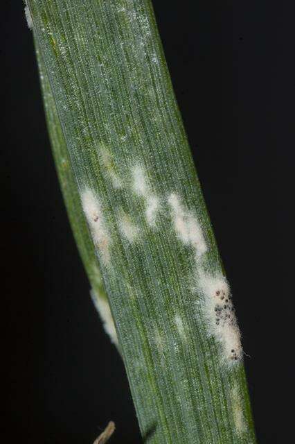
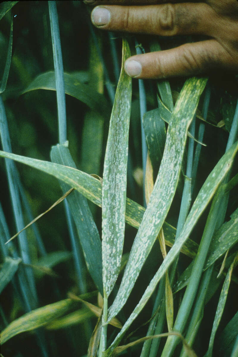
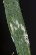
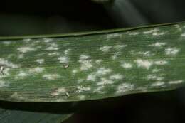
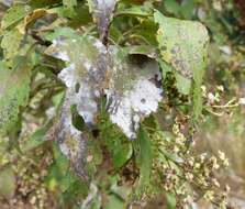
Wheat leaves showing fungal growths due to powdery mildew (
Blumeria graminis). These are the first visible symptoms of the disease and consist of white to pale gray, fuzzy or powdery colonies of mycelia and conidia, on the upper surfaces of leaves and leaf sheaths (especially on lower leaves), and sometimes on the spikes. This superficial fungal material can be rubbed off easily with the fingers. Host tissue beneath the fungal material becomes chlorotic or necrotic and, with severe infections, the leaves may die.For more information, see CIMMYT's Wheat Doctor:
wheatdoctor.cimmyt.org/index.php?option=com_content&t....Photo credit: CIMMYT.
-
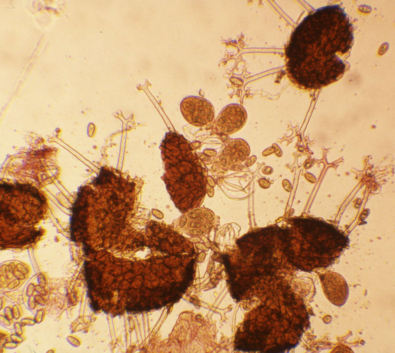
cleisthotecium open. Scale bar indicates 100 µm.Sample from ponds situated in the vicinity of Lake Constance (Bodensee, Southern Germany). The image was built up using several photomicrographic frames with manual stacking technique. Images were taken using Zeiss Universal with Olympus C7070 CCD camera.
-
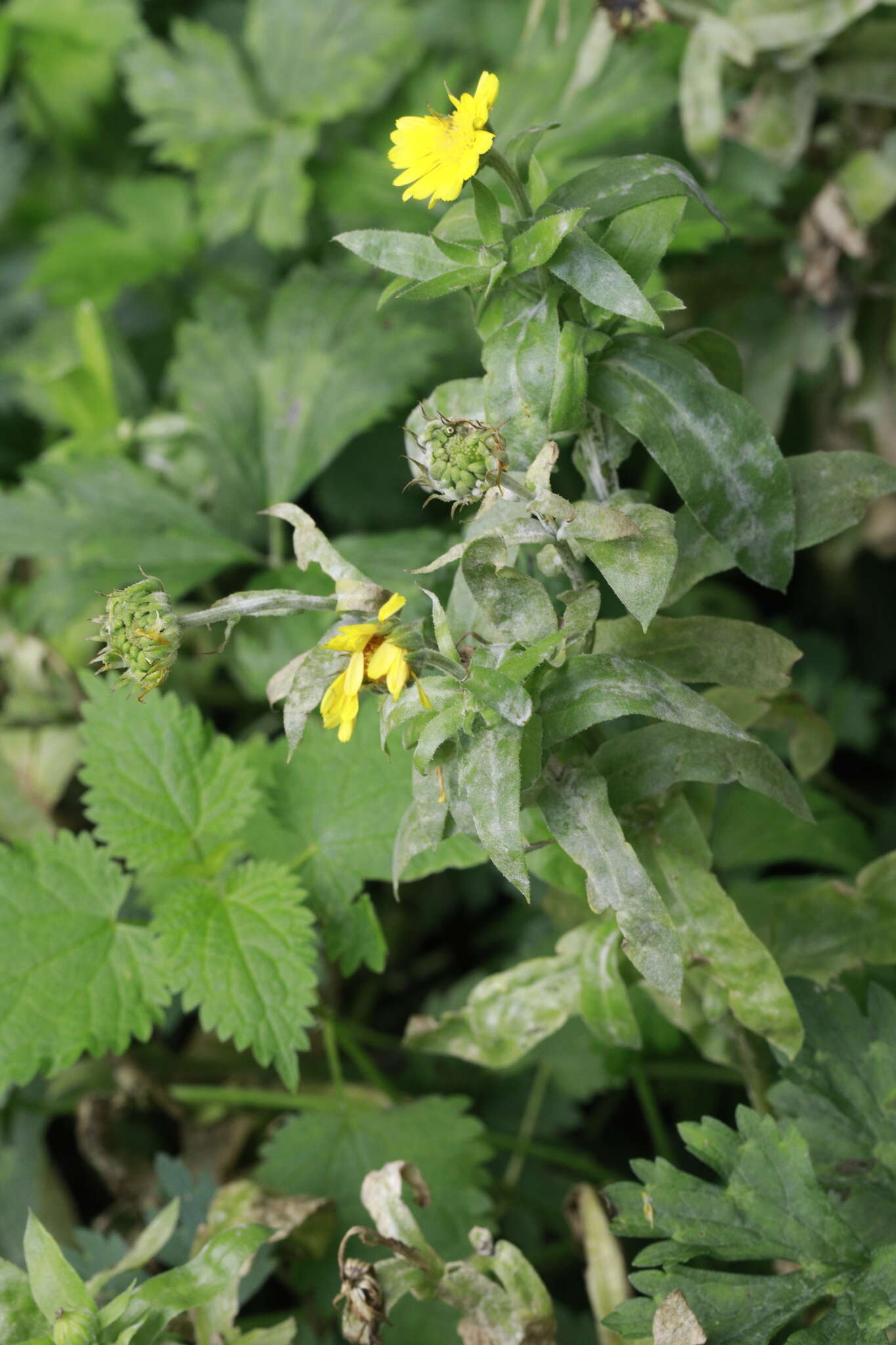
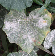
Midtsjælland, Danmark
-
International Maize and Wheat Improvement Center
Flickr Group
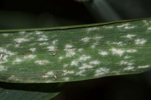
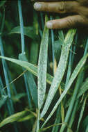
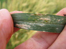
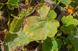
Wheat leaf showing powdery mildew caused by Blumeria graminis. These fungal growths are initially white to pale gray, while older fungal tissue turns yellowish gray. They consist of fuzzy or powdery colonies of mycelia and conidia, on the upper surfaces of leaves and leaf sheaths (especially on lower leaves), and sometimes on the spikes. This superficial fungal material can be rubbed off easily with the fingers. Host tissue beneath the fungal material becomes chlorotic or necrotic and, with severe infections, the leaves may die.For more information, see CIMMYT's Wheat Doctor: <http://wheatdoctor.cimmyt.org/index.php?option=com_content&task=view&id=115&Itemid=43&lang=en.Photo credit: Thomas Lumpkin/CIMMYT.
-
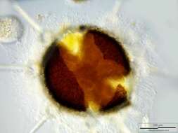
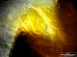
Closeup, showing one sporangium. Scale bar indicates 50 µm.Sample from ponds situated in the vicinity of Lake Constance (Bodensee, Southern Germany). The image was built up using several photomicrographic frames with manual stacking technique. Images were taken using Zeiss Universal with Olympus C7070 CCD camera.
-
International Maize and Wheat Improvement Center
Flickr Group
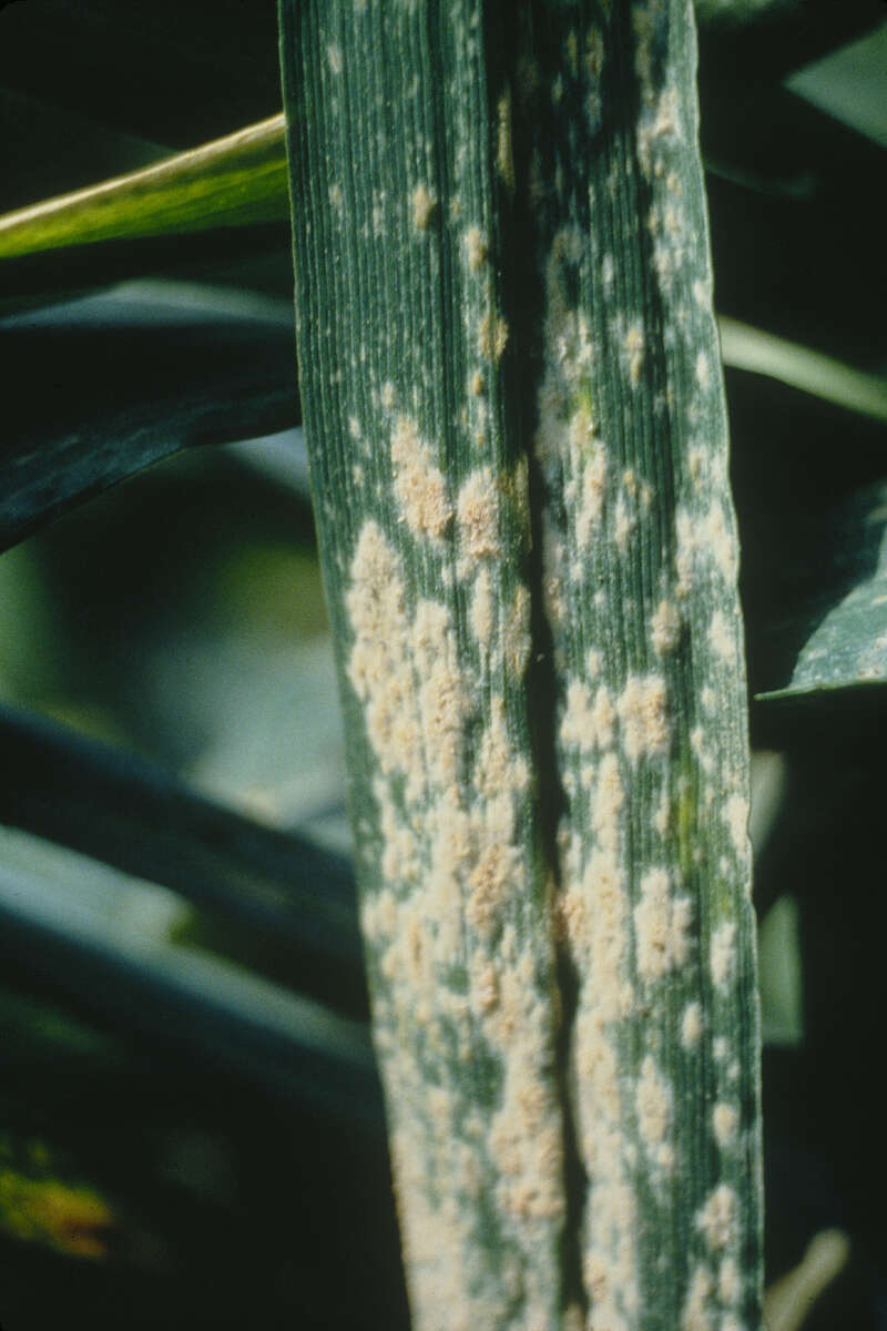
Wheat leaf showing fungal growths due to powdery mildew (
Blumeria graminis). These are initially white to pale gray, while older fungal tissue turns yellowish gray, as shown. They consist of fuzzy or powdery colonies of mycelia and conidia, on the upper surfaces of leaves and leaf sheaths (especially on lower leaves), and sometimes on the spikes. This superficial fungal material can be rubbed off easily with the fingers. Host tissue beneath the fungal material becomes chlorotic or necrotic and, with severe infections, the leaves may die.For more information, see CIMMYT's Wheat Doctor:
wheatdoctor.cimmyt.org/index.php?option=com_content&t....Photo credit: CIMMYT.
-
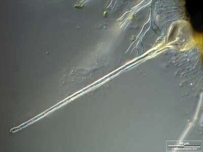
Hyaline appendage of the cleisthotecium. Scale bar indicates 50 µm.Sample from ponds situated in the vicinity of Lake Constance (Bodensee, Southern Germany). The image was built up using several photomicrographic frames with manual stacking technique. Images were taken using Zeiss Universal with Olympus C7070 CCD camera.
-
Open cleisthotecium showing sporangia. Scale bar indicates 100 µm.Sample from ponds situated in the vicinity of Lake Constance (Bodensee, Southern Germany). The image was built up using several photomicrographic frames with manual stacking technique. Images were taken using Zeiss Universal with Olympus C7070 CCD camera.
-
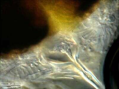
Closeup of the hyaline appendage of the cleisthotecium. Scale bar indicates 25 µm.Sample from ponds situated in the vicinity of Lake Constance (Bodensee, Southern Germany). The image was built up using several photomicrographic frames with manual stacking technique. Images were taken using Zeiss Universal with Olympus C7070 CCD camera.
-
-
-
-
-
-
-
-
-
-