nomes no trilho de navegação
Burkholderia mallei ist ein gramnegatives, stäbchenförmiges, aerobes Bakterium. Es ist eine pathogene Burkholderia-Art, die bei Mensch und Tier die Erkrankung Rotz auslösen kann, die beim Menschen zu Lungenentzündung, Sepsis sowie Haut- und Schleimhautinfektionen führt.[1] Der lateinische Name dieser meldepflichtigen Krankheit, malleus, gab dem früher Malleomyces mallei genannten Erreger den Namen. Burkholderia mallei unterscheidet sich von allen anderen Vertretern der Gattung Burkholderia durch seinen echten Parasitismus, seine Unbeweglichkeit und sein langsames Wachstum in Kultur. B. mallei steht ebenso wie B. pseudomallei auf der Liste für potenzielle Biowaffen-Agentien.
Seit seiner Entdeckung wurde der Erreger in zahlreiche systematische Gruppen eingeordnet: Bacillus, Corynebacterium, Mycobacterium, Peifferella, Loefflerella, Malleomyces, Actinobacillus, Pseudomonas. Der Gattung Burkholderia wird das Bakterium erst seit Anfang der 1990er Jahre zugeordnet.
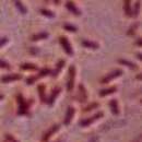
Aufgrund fehlender Begeißelung ist B. mallei als einziger Vertreter seiner Gattung nicht beweglich. Seine variable Form (Pleomorphismus) ist eine Mischung zwischen Stäbchen und Kugel. Das Bakterium misst 1,5–3 μm in der Länge und 0,5–1 μm in der Breite, die Enden sind stumpf oder abgerundet. Die Stäbchenform ist gerade oder leicht gekrümmt. In Probenmaterial und jungen Bakterienkulturen kommt der Erreger überwiegend in Stäbchenform vor, in älteren Kulturen dominiert ein pleomorphes Bild. B. mallei bildet keine Endosporen. Das Bakterium besitzt eine kapselähnliche Struktur (Exopolysaccharid-Kapsel), die nur im elektronenmikroskopischen Bild sichtbar wird.
Färbungen nach Gram sind möglich. Das Bakterium färbt sich oft nur schwach an oder zeigt gramlabiles Verhalten (gleichzeitiges Auftreten von grampositiven und -negativen Keimen im gleichen Präparat). Gespeichertes Poly-β-Hydroxybutyrat im Cytoplasma lässt das Bakterium granuliert und bipolar (Färbeunterschied zwischen Mitte und Enden des Bakteriums) erscheinen. Alternative Färbemethoden sind die Färbungen nach Wright, Giemsa oder mit Methylenblau. Die Kapsel ist lichtmikroskopisch nicht darstellbar.
Aus Gewebeschnitten ist der mikroskopische Nachweis von B. mallei schwierig, da der Erreger in kugeliger Form vorliegen kann. In Probenmaterial aus sekundär infizierten oder kontaminierten Bereichen kann der Rotzerreger mikroskopisch nicht von anderen Bakterien unterschieden werden.
Als obligat parasitär lebender Organismus kann sich der Erreger nur in infizierten Wirten vermehren. In klinischem Probenmaterial findet sich der Erreger oft außerhalb der Körperzellen (extrazellulär). Er kann gut aus frischen Hautläsionen isoliert werden, während er in älteren Läsionen (vor allem beim chronischen Rotz) nur spärlich vorkommt. Sputum, Speichel, Tränenflüssigkeit, Urin, Kot, Milch und Sperma können ebenfalls erregerhaltig sein. Aus Blut kann er nur während der Phase der disseminierten Infektion (Bakteriämie) nachgewiesen werden.
Ein optimales Wachstum findet im neutralen pH-Bereich, bei mesophilen Temperaturen (37 °C) und unter Anwesenheit von Sauerstoff (Aerobier) statt. Bei gleichzeitiger Anwesenheit von Nitrat kann auch ein Wachstum unter anaeroben Verhältnissen erfolgen (fakultativer Anerobier). Bei Temperaturen unter 5 °C und über 42 °C erfolgt keine Vermehrung des Erregers mehr.
Der Erreger kann auf üblichen, bluthaltigen Nährböden angezüchtet werden. Das Wachstum erfolgt nur langsam (48–72 h Inkubation) und es besteht die Gefahr der Überwucherung durch schneller wachsende Begleitkeime. Die Zugabe von Glycerin beschleunigt das Wachstum, sodass eine Voranreicherung mit Glycerin sinnvoll sein kann. Auf Glycerin-haltigen Nährböden wächst der Keim als weicher, feuchter, zartcremefarbener Schleier, bei dem die größenvariablen Einzelkolonien zum Zerfließen neigen. Mit zunehmendem Alter der Kultur wird die Bakterienschicht dicker, bernsteinfarben und bekommt eine zähe, klebrige Konsistenz. Zur Anzucht eignen sich auch Glycerin-Kartoffel-Medium oder Glycerin-Bouillon. Auf einfachem Nähragar oder in Gelatinekulturen erfolgt nur spärliches Wachstum. Traditionell wurde B. mallei auf Kartoffelscheiben angezüchtet. Charakteristisch für die Bakterienkulturen ist ein aromatischer Geruch. Zur Unterdrückung unerwünschter Begleitflora werden klinische Proben mit Penicillin vorbehandelt oder es erfolgt ein Zusatz bakterienhemmender Stoffe (Proflavin, Kristallviolett) zu den Nährmedien. Für die Anzucht des Erregers wurde ein selektives Medium auf der Basis von Glycerin, Serum (Pferd oder Esel) und Tryptose entwickelt, dem Polymyxin E, Bacitracin und Actidion zugesetzt werden.
B. mallei ist chemo-organotroph. Auf bluthaltigen Nährmedien wird keine Hämolyse beobachtet. Der Erreger besitzt die Enzyme Oxidase und Katalase. Indol-Test und Voges-Proskauer-Reaktion fallen negativ aus. Der Erreger kann Nitrat reduzieren. Photosynthetische Farbpigmente werden während des Wachstums nicht gebildet. Der Glukose-Abbau erfolgt überwiegend oxidativ. Obwohl die Gattung Burkholderia den Non-Fermentern (kein Abbau von Substrat durch Gärung) zugeordnet wird, gibt es Hinweise auf vereinzelte Fermentation von Glukose, Mannose, Arabinose und Fruktose. Die präzise Bestimmung der Spezies Burkholderia mallei auf biochemischer Basis ist bei der Verwendung handelsüblicher Testsysteme (Bunte Reihe) nicht möglich.
In der freien Umwelt ist B. mallei weder lebens- noch vermehrungsfähig, die Widerstandsfähigkeit (Tenazität) gegen äußere Einflüsse ist gering. Er ist empfindlich gegen Austrocknung, Hitze und Sonnenlicht. Direkte Sonneneinstrahlung tötet den Erreger innerhalb von 24 h.[2] In eingetrockneten Sekreten verliert er schon nach wenigen Tagen seine Infektiosität. An feuchten, dunklen Orten kann er jedoch bis zu 3 Monaten überleben, in Tränkewasser bleibt er 1 Monat lang kontagiös.
In Urin wird der Erreger innerhalb von 40 min, durch Magensaft innerhalb einer halben Stunde inaktiviert.[3] Fäulnis zerstört ihn erst nach 2–4 Wochen.[4]
Das Bakterium ist gegenüber zahlreichen Desinfektionsmitteln empfindlich, darunter Formaldehyd (2 %), Glutaraldehyd (2 %), Benzalkoniumchlorid, Iod, Bleichlorid, Kaliumpermanganat, Natriumhypochlorit (1 %) und Ethanol (70 %). Phenolhaltige Mittel zeigen dagegen wenig Wirksamkeit. Historisch bedeutsame Desinfektionsmittel waren Chlorkalk und Phenol.
Die Mikroorganismen werden durch Hitze inaktiviert. Ausreichend sind einmaliges Aufkochen, Temperaturen von 80 °C für 5 min oder 55 °C für 10 min.[4] UV-Bestrahlung tötet das Bakterium ebenfalls ab. Durch Kälte wird der Erreger nicht eliminiert.[2]
Die natürliche Wachstumsgrenze des Erregers liegt zwischen 20 °C und 45 °C.[5]
Antibiotika wie Ceftazidim, Imipenem, Aminoglykoside wie Streptomycin,[6] Amikacin, Tetracyclin, Doxycyclin und Sulfathiazol waren zumindest in vitro effektiv. Eine (achtwöchige) antibiotische Therapie des Rotz beim Menschen kann mit Imipenem und Doxycyclin erfolgen.
Burkholderia mallei ist ein gramnegatives, stäbchenförmiges, aerobes Bakterium. Es ist eine pathogene Burkholderia-Art, die bei Mensch und Tier die Erkrankung Rotz auslösen kann, die beim Menschen zu Lungenentzündung, Sepsis sowie Haut- und Schleimhautinfektionen führt. Der lateinische Name dieser meldepflichtigen Krankheit, malleus, gab dem früher Malleomyces mallei genannten Erreger den Namen. Burkholderia mallei unterscheidet sich von allen anderen Vertretern der Gattung Burkholderia durch seinen echten Parasitismus, seine Unbeweglichkeit und sein langsames Wachstum in Kultur. B. mallei steht ebenso wie B. pseudomallei auf der Liste für potenzielle Biowaffen-Agentien.
Burkholderia mallei is a Gram-negative, bipolar, aerobic bacterium, a human and animal pathogen of genus Burkholderia causing glanders; the Latin name of this disease (malleus) gave its name to the species causing it. It is closely related to B. pseudomallei, and by multilocus sequence typing it is a subspecies of B. pseudomallei.[1] B. mallei evolved from B. pseudomallei by selective reduction and deletions from the B. pseudomallei genome.[2] Unlike B. pseudomallei and other genus members, B. mallei is nonmotile; its shape is coccobacillary measuring some 1.5–3.0 μm in length and 0.5–1.0 μm in diameter with rounded ends.
Wilhelm Schütz and Friedrich Löffler first isolated B. mallei in 1882.[3] It was isolated from an infected liver and spleen of a horse.[4] This bacterium is also one of the first to be identified containing a type VI secretion system which is important for its pathogenicity.[5] In 1885, the German Botanist and Bacteriologist, Wilhelm Zopf (1846–1909) gave the pathogen its binomial name, after analyzing samples of the bacterium. He further refined his observations with the pathogen in 1886.[6]
Most organisms within the Burkholderiaceae live in soil; however, B. mallei does not. Because B. mallei is an obligate mammalian pathogen, it must infect a host mammal to live and to be transmitted from one host to another.[7]
B. mallei is very closely related to B. pseudomallei, being 99% identical in conserved genes when compared to B. pseudomallei. B. malllei has about 1.4 Mb less DNA than B. pseudomallei.[4] B. mallei may have actually evolved from a strain of B. pseudomallei after the latter had infected an animal. The bacterium would have lost the genes that were not necessary for living in an animal host. This suggestion has found support from studies that compare strains of B. mallei to B. pseudomallei and indicate that their two respective genomes are very similar. The genes that allowed the bacterium to survive in a soil environment, like genes that gave B. mallei the capacity to protect against bactericidals, antibiotics, and antifungals, were likely deleted. Thus, the reason that B. mallei is not found outside of a host is because it lacks the genes necessary for survival in the soil. Genome comparisons also seem to indicate that the B. mallei is still evolving and adapting to an intracellular lifestyle.[8]
The genome of B. mallei was sequenced in the United States by The Institute of Genomic Research. The size of the genome is smaller than that of B. pseudomallei. The B. mallei sequence revealed a chromosome of 3.5 mega base pairs (Mb) and a 2.3 Mb "megaplasmid”. Many insertion sequences and phase-variable genes were also found.[3] The genome for B. mallei is made up of two circular chromosomes. Chromosome 1 is where genes relating to metabolism, capsule formation, and lipopolysaccharide biosynthesis are located. B. mallei has a polysaccharide capsule which indicates its potential as a pathogen. Chromosome 2 is where most of the information regarding secretion systems and virulence-associated genes are located.[4] Multilocus sequence typing has revealed that B. mallei most likely evolved from a B. pseudomallei clone reduction. About 1000 B. pseudomellei genes are absent or varying in the B. mallei genome. B. mallei’s genome also has a large amount of insertion sequences.[8]
Burkholderia mallei was first called "Bacillus mallei" and was in the genus Pseudomonas until the early 1990s. It is now part of the genus Burkholderia.[3]
No standardised system exists for differentiating between B. mallei and B. pseudomallei. The methods that have been used to differentiate and identify one strain from the other include ribotyping, pulsed-field gel electrophoresis, multilocus enzyme electrophoresis, random amplified polymorphic DNA analysis, and multilocus sequence typing.[3] Comparing the DNA of B. mallei and B. pseudomallei must be done at the 23S rDNA level, however, since no identifiable difference is found between the two species at the 16S rDNA level.[9]
Both B. mallei and B. pseudomallei can be cultured in a laboratory; nutrient agar can be used to grow the bacteria. When grown in culture, B. mallei grows in smooth, grey, translucent colonies. In a period of 18 hours at 37 °C, a B. mallei colony can grow to about 0.5–1.0 mm in diameter. B. mallei culture growth on MacConkey agar is variable. Many microbiologists are unfamiliar with B. mallei and as a result it has frequently been misidentified as a Pseudomonas species or as a contaminant in a culture.[3]
The bacterium is susceptible to numerous disinfectants including benzalkonium chloride, iodine, mercuric chloride, potassium permanganate, 1% sodium hypochlorite, and ethanol. The micro-organism can also be destroyed by heating or ultraviolet light. Antibiotics such as streptomycin, amikacin, tetracycline, doxycycline, carbapenems, ceftazidime, amoxicillin/clavulanic acid, piperacillin, chloramphenicol, and sulfathiazole have been reported to be effective against the bacteria in vitro. B. mallei, like B. pseudomallei, is also resistant to a number of antibiotics including aminoglycosides, polymyxins, and beta-lactams. No vaccine is currently available for humans or animals to protect against B. mallei infection.[3] An animal model that will predict immune responses necessary to create immunity to the bacterium is needed before a vaccine can be developed. Mice are fairly close to humans in their susceptibility to B. mallei and would be the ideal choice of animal for creating a model for the vaccine.[4]
Burkholderia mallei is responsible for causing glanders disease, which historically mostly affected animals, such as horses, mules, and donkeys, and rarely humans. Horses are considered the natural host for B. mallei infection and are highly susceptible to it.[4] B. mallei infects and gains access to the cell of its host through lysis of the entry vacuole. B. mallei has bacterial protein-dependent, actin-based motility once inside the cell. It is also able to initiate host cell fusion that results in multinucleated giant cells (MNGCs). The consequence of MNGCs has yet to be determined, but it may allow the bacteria to spread to different cells, evade responses by the infected host’s immune system, or allow the bacteria to remain in the host longer. B. mallei is able to survive inside host cells through its capabilities in disrupting the bacteria-killing functions of the cell. It leaves the vacuoles early, which allows for efficient replication of the bacteria inside the cell. Leaving the cell early also keeps the bacteria from being destroyed by lysosomal defensins and other pathogen-killing agents. MNGCs may help protect the bacteria from immune responses.[10] B. mallei’s ability to live within the host cell makes developing a vaccine against it difficult and complex. The vaccine would need to create a cell-mediated immune response, as well as a humoral response to the bacteria in to be effective in protecting against B. mallei. In regards to a vaccine against B. mallei, the closeness of B. mallei to B. pseudomallei may make it possible that a vaccine developed for either type would be effective against the other.[7]
Horses chronically infected with B. mallei with glanders disease typically experience mucus-containing nasal discharge, lung lesions, and nodules around the liver or spleen. Acute infection in horses results in a high fever, loss of fat or muscle, erosion of the surface of the nasal septum, hemorrhaging or mucus discharge. The bacterium mostly affects the lungs and airways.[4] Human infection with B. mallei is rare, although it occasionally occurs among laboratory workers dealing with the bacteria or those who are frequently near infected animals.[3] The bacteria usually infect a person through their eyes, nose, mouth, or cuts in the skin. Once people are infected, they develop a fever and rigors. Eventually, they get pneumonia, pustules, and abscesses, which prove fatal within a week to 10 days if left untreated by antibiotics. The way someone is infected by the bacteria also affects the type of symptoms that will result. If the bacteria enter through the skin, a local skin infection can result, while inhaling B. mallei can cause septicemic or pulmonary, muscular, hepatic, or splenous infections. B. mallei infection has a fatality rate of 95% if left untreated, and a 50% fatality rate in individuals treated with antibiotics.[4]
In the first days of B. mallei infection, neutrophils, macrophages, and T cells go to the spleen in great quantities. The early cellular response to B. mallei infection involves Gr-1+ (antigen) cells, and implies their importance to immunity against this bacterial infection. T cells (nitric oxide) are actually more involved in combating B. mallei in the later stages of its infection of a host.[11]
Lipopolysaccharide isolated from B. mallei demonstrated significantly lower biological activity as compared to the LPS from Escherichia coli, in agreement with the lower degree of acylation of its lipid A: the major forms of B. mallei lipid A were penta- and tetraacylated, whereas classical lipid A from E. coli was hexaacylated. In addition, lipid A from B. mallei contains 4-amino-4-deoxyarabinose residue in almost half of the molecules, which would partially neutralize the negative charge of the phosphate groups necessary for the interaction with the positively charged amino acids of TLR4. At the same time, lipid A acyl chains in B. mallei were on the average longer (14–16 carbon atoms) than those in E. coli (14 carbon atoms), yet LPS from B. mallei appeared to be a weaker activator. B. mallei may employ LPS with low biological activity to evade proper recognition by the TLR4/MD-2 complex of innate immune system, dampening the host immune response and increasing the risk of bacterial dissemination.[12]
Burkholderia mallei has been eradicated in the United States and most Western countries, but still affects animals in Africa, Asia, the Middle East, Central America, and South America.[4] Many Western countries were able to eliminate the disease through glanders control programs and laws requiring notification of cases of infection to health departments and the destruction of any animal affected with B. mallei.[13]
Bukrholderia mallei and B. pseudomallei have a history of being on a list of potential biological warfare agents. The Centers for Disease Control and Prevention classifies B. mallei as a category B critical biological agent.[7] As a result, research regarding B. mallei may only be done in biosafety level 3 facilities in the US and internationally. Though it is so highly infective and a potential biological weapon, little research has been conducted on this bacterium.[4] B. mallei and B. pseudomallei under the policy of Institutional Oversight of Life Sciences Dual Use Research of Concern would be subject to oversight to ensure the responsible investigation of these agents.[14]
In March 2000, one of the first cases since the 1940s of glanders in the United States occurred in a young microbiologist working for the U.S. Army Medical Research Institute for Infectious Diseases. The researcher had type 1 diabetes and had been working with B. mallei for about two years, but he did not always wear gloves while conducting his research. The researcher experienced enlargement of the lymph nodes and a fever which lasted for 10 days even with antibiotic treatment. In the following weeks, the researcher experienced fatigue, rigors, night sweats, and loss of weight. The next month, his symptoms seemed to disappear after treatment with clarithromycin, but after the medication was stopped, the symptoms reappeared. After conducting multiple tests on cultures from the researcher’s blood and a biopsied portion of a liver abscess, the bacterium was identified as B. mallei. Once it was established what infected the researcher, another course of antibiotics was given (imipenem and doxycycline) with 6 months of treatment. After a year, the researcher made a full recovery.
This incident also showed how a cut or skin abrasion is not absolutely necessary to contract the disease, as the researcher had no recollection of any cut or accident while working in the laboratory. The case was significant as it showed the difficulty that microbiology laboratories have in identifying bioweapon agents and the potential consequences if measures are not taken to prepare for an actual biological attack.[13]
Burkholderia mallei was intentionally used to infect animals and humans during World War I. The Germans used B. mallei to infect animals that were being sent from neutral countries to the Allies with glanders.[3] The Germans' plans for biological warfare started in 1915 on the East Coast of the United States; they intended to infect and kill the livestock that were being sent to the Allies and facilitate the transfer of the disease to humans. The East Coast was where many animals were being assembled for shipment to the Allies fighting in Europe. The Germans also targeted Romania, Norway, and Spain's animal supplies with cultures of glanders. The German biological sabotage eventually spread to Argentina, where agents would rely on bacterial cultures from Spain to infect the cattle, horses, and mules that Argentina was supplying to the Allies. The German use of microbes as weapons is one of the only documented attacks of intentionally using biological weapons against neutral countries.[15]
The Japanese used B. mallei in their biological warfare research units. The most notable and notorious unit, Unit 731, used the bacterium to conduct experiments on live human subjects. However, the Japanese did not end up creating a biological weapon out of B. mallei. They did actually use B. mallei to test its effectiveness in contaminating water supplies, and the results of these tests were successful.
The Russians' biological weapons program also took an interest in B. mallei and conducted field tests with it. Some of the researchers from the program were actually infected and killed by it during the course of their research. It has been suggested that the Soviet Union eventually used B. mallei during their war in Afghanistan against the mujahideen.[3]
Burkholderia mallei is a Gram-negative, bipolar, aerobic bacterium, a human and animal pathogen of genus Burkholderia causing glanders; the Latin name of this disease (malleus) gave its name to the species causing it. It is closely related to B. pseudomallei, and by multilocus sequence typing it is a subspecies of B. pseudomallei. B. mallei evolved from B. pseudomallei by selective reduction and deletions from the B. pseudomallei genome. Unlike B. pseudomallei and other genus members, B. mallei is nonmotile; its shape is coccobacillary measuring some 1.5–3.0 μm in length and 0.5–1.0 μm in diameter with rounded ends.
Burkholderia mallei es un bacilo y a veces cocobacilo Gram negativo aerobio no móvil de la familia Burkholderiaceae. Es el agente causal de la enfermedad llamada muermo. Miden 1.5–3 μm de largo y 0.5–1μm en diámetro con extremos redondeados.
El organismo es susceptible a numerosos desinfectantes: cloruro de benzalconio, iodo, cloruro de mercurio, permanganato de potasio, 1% hipoclorito de sodio y etanol. El microorganismo puede ser destruido también con calor y rayos ultravioleta. Ciertos antibióticos tales como estreptomicina, amikacina, tetraciclina, doxiciclina y sulfadiacina 100 mg/kg/día oral tres semanas, son efectivos frente la bacteria.
Puede requerirse drenaje quirúrgico de las lesiones supuradas. B. mallei tiene una historia de asociación como agente de armas biológicas.
Burkholderia mallei es un bacilo y a veces cocobacilo Gram negativo aerobio no móvil de la familia Burkholderiaceae. Es el agente causal de la enfermedad llamada muermo. Miden 1.5–3 μm de largo y 0.5–1μm en diámetro con extremos redondeados.
Burkholderia mallei est l'agent de la morve, une maladie grave touchant préférentiellement les équidés, mais transmissible à de nombreuses autres espèces dont l'espèce humaine.
Anciennement dénommée Pseudomonas mallei ou Malleomyces mallei, c'est une bactérie à Gram négatif bipolaire et aérobie appartenant au genre Burkholderia (nom générique créé en l'honneur du bactériologiste américain Walter H. Burkholder, de l'université Cornell de New York). Son nom spécifique vient du latin malleus (morve). À la différence de l'espèce voisine Burkholderia pseudomallei et d'autres espèces du même genre, cette bactérie n'est pas mobile. Sa forme est intermédiaire entre le bâtonnet et le coque et ses dimensions comprises entre 1,5 et μm de longueur sur 1 μm de diamètre avec des extrémités arrondies.
Le germe est sensible à de nombreux désinfectants tels que le chlorure de benzalkonium, l'iode, le chlorure de mercure, le permanganate de potassium, l'hypochlorite de sodium et l'éthanol. Il est détruit par la chaleur et les rayons ultraviolets. In vitro, des antibiotiques tels que la streptomycine, l'amikacine, la tétracycline, la doxycycline et le sulfathiazole se sont montrés actifs contre cette bactérie[réf. souhaitée].
B. mallei comme B. pseudomallei sont inscrits sur une liste[Laquelle ?] d'armes biologiques potentielles[réf. souhaitée].
Burkholderia mallei est l'agent de la morve, une maladie grave touchant préférentiellement les équidés, mais transmissible à de nombreuses autres espèces dont l'espèce humaine.
Burkholderia mallei é unha especie de bacterias gramnegativas aerobias, que é un patóxeno animal que causa o mormo[1] en equinos e tamén pode afectar a humanos. O nome latino malleus fai referencia a dita doenza equina. A especie está moi emparentada con B. pseudomallei, e segundo a tipificación de secuencia multilocus habería que considerala como unha subespecie de B. pseudomallei.[2] B. mallei evolucionou a partir de B. pseudomallei por redución selectiva e delecións do xenoma desta última.[3] A diferenza da especie estreitamente relacionada B. pseudomallei e doutros membros do xénero Burkholderia, esta bacteria non é móbil. A súa forma é intermedia entre coco e bacilo e mide uns 1,5–3 μm de lonxitude e 0,5–1μm de diámetro e presenta extremos arredondados.
Wilhelm Schütz e Friedrich Löffler foron os que primeiro illaron a especie Burkholderia mallei en 1882.[4] Foi illada do fígado e bazo dun cabalo infectado.[5] Esta bacteria é tamén unha das primeiras que foi identificada como posuidora dun sistema de secreción de tipo VI, que é importante para a súa patoxenicidade.[6]
A maioría dos organismos da familia Burkholderiaceae viven no solo, pero B. mallei non. Como B. mallei é un patóxeno obrigado de mamíferos, debe causar enfermidade nun hóspede mamífero para poder vivir, reproducirse e transmitirse a outro hóspede.[7]
B. mallei está moi estreitamente emparentada con B. pseudomallei, ata o punto que en xenes conservados son idénticas ao 99%. O ADN de B. malllei ten uns 1,4 Mb que o de B. pseudomallei.[5] Especúlase que B. mallei evolucionou a partir dunha cepa de B. pseudomallei despois de que esta última infectou un animal. A bacteria perdeu os xenes que non eran necesarios para vivir dentro dun hóspede animal. Esta idea apóiase nos estudos que compararon cepas de B. mallei con B. pseudomallei e indicaron que os seus dous respectivos xenomas son moi similares. Os xenes que permitían que a bacteria vivise no ambiente do solo, como os xenes que lle daban a B. mallei a capacidade de protexerse contra bactericidas, antibióticos, e antifúnxicos, probalemente sufriron deleción. Así, a razón de que B. mallei non se atope fóra do hóspede débese á carencia destes xenes. As comparacións de xenomas tamén parecen indicar que B. mallei aínda está evolucionando e adaptándose a un estilo de vida intracelular.[8]
O xenoma de B. mallei foi secuenciado nos Estados Unidos por The Institute of Genomic Research (TIGR). O tamaño do xenoma é menor que o da especie relacionada B. pseudomallei. B. mallei ten un cromosoma circular de 3,5 megapares de bases (Mb) e un segundo cromosoma máis pequeno ou "megaplásmido" de 2,3 Mb. Atopáronse moitas secuencias de inserción e xenes de fase variable.[4] O cromosoma 1 contén os xenes relacionados co metabolismo, formación da cápsula, e biosíntese do lipopolisacárido. B. mallei ten unha cápsula de polisacárido, que indica a súa portencialidade como patóxeno. O cromosoma 2 é onde contén a maioría da información sobre os sistemas de secreción e xenes asociados á virulencia.[5] A tipificación de secuencia multilocus revelou que B. mallei seguramente evolucionou a partir dunha redución xenómica de B. pseudomallei, xa que uns 1000 xenes de B. pseudomellei están ausentes no xenoma de B. mallei. O xenoma de B. mallei ten unha grande cantidade de secuencias de inserción.[8]
Burkholderia mallei chamouse primeiramente Bacillus mallei e despois estivo incluída no xénero Pseudomonas co nome P. mallei ata inicios da década de 1990. Posteriormente pasouse ao xénero Burkholderia.[4]
Non hai un sistema estandarizado para diferenciar entre B. mallei e B. pseudomallei. Os métodos que se utilizan para diferencialas e identificar unha cepa doutra son a ribotipificación, electroforese en xel de campo pulsado, electroforese de encimas multilocus, análise de ADN polimórfico amplificado aleatorio, e tipificación de secuencia multilocus.[4] A comparación do ADN de B. mallei e B. pseudomallei debe facerse co ADNr de 23S, xa que non hai diferenzas identificables entre as dúas especies no ADNr de 16S.[9]
Tanto B. mallei coma B. pseudomallei poden cultivarse en laboratorio, en ágar nutriente. Cando crece en cultivo, B. mallei forma colonias lisas grises translúcidas. Nun período de 18 horas a 37 °C, unha colonia de B. mallei pode atinxir de 0,5 a 1 mm de diámetro. O crecemento dos cultivos de B. mallei en ágar MacConkey son variables. Moitos microbiólogos están pouco habituados a cultivar esta especie, polo que frecuentemente B. mallei é identificado erradamente como unha especie de Pseudomonas ou un contaminante presente no cultivo.[4]
Esta bacteria é susceptible a numerosos desinfectantes, como o cloruro de benzalconio, ioduro, cloruro de mercurio, permanganato potásico, hipoclorito de sodio ao 1%, e etanol. O microorganismo pode ser destruído por quentamento ou pola luz UV. Os antibióticos como a estreptomicina, amicacina, tetraciclina, doxiciclina, carbapenems, ceftazidime, amoxicilina/ácido clavulánico, piperacilina, cloranfenicol e sulfatiazol son efectivos contra esta bacteria in vitro. B. mallei, igual que B. pseudomallei, é resistente a varios antibióticos como os aminoglicósidos, polimixinas, e antibióticos beta lactámicos. Actualmente non hai vacina para protexer a humanos ou animais da infección por B. mallei.[4] Cómpre atopar un modelo animal que prediga as respostas inmunes necesarias para crear inmunidade contra esta bacteria antes de que poida desenvolverse unha vacina. Pénsase que os ratos teñen unha susceptibilidade moi similar á dos humanos a B. mallei e serían un posible animal modelo para procurar a vacina.[5]
B. mallei é o axente causante do mormo, que historicamente afectou a animais como cabalos, mulas e burros principalmente, e en raros casos tamén a humanos. Os cabalos son considerados os hóspedes naturais de B. mallei e son moi susceptibles á súa infección.[5] B. mallei infecta e entra na célula do seu hóspede por medio da lise do vacúolo de entrada. Unha vez que está na célula, B. mallei ten unha mobilidade baseada na actina dependente de proteína. Tamén pode iniciar a fusión das células hóspede, o que dá lugar a células xigantes multinucleadas (MNGCs). A consecuencia da formación destas células xigantes non foi aínda determinada, pero pode permitirlle á bacteria espallarse a células diferentes, evadirse das respostas do sistema inmunitario do hóspede infectado ou permitir que a bacteria permaneza no hóspede por longo tempo. B. mallei pode sobrevivir dentro das células hóspede pola súa capacidade de alterar as funcións de destrución das bacterias da célula. Abandona os vacúolos nos que entra axiña, o que permite unha replicación eficiente da bacteria dentro da célula. O abandono temperán da célula tamén lle permite evitar ser destruída polas defensinas lisosómicas e outros axentes celulares que matan patóxenos. As células xigantes poden axudar a protexer a bacteria das respostas inmunitarias.[10] A capacidade que ten B. mallei de vivir dentro da célula hóspede fai que o desenvolvemento dunha vacina sexa difícil e complexo. A vacina tería que crear unha resposta inmune mediada por células e tamén unha resposta humoral contra a bacteria para poder protexer de forma efectiva contra ela. Como B. mallei é moi próxima a B. pseudomallei é posible que unha vacina contra a primeira protexese tamén contra a segunda.[7]
Os cabalos que están infectados cronicamente por B. mallei e teñen o mormo, presentan tipicamente unha descarga de moco nasal, lesións pulmonares, e formación de nódulos arredor do fígado e bazo. A infección aguda en cabalos produce febre alta, perda de graxas ou músculo, erosión da superficie do tabique nasal, e descarga de mocos ou hemorraxia. A bacteria afecta principalmente a pulmóns e vías aéreas.[5] A infección en humanos por B. mallei é rara, aínda que ocasionalmente se pode producir entre persoas que traballan nos laboratorios que manipulan cultivos da bacteria ou en persoas que están frecuentemente preto de animais infectados.[4] A bacteria xeralmente infecta ás persoas a través dos ollos, nariz, boca, ou cortes na pel. Unha vez que a persoa está infectada, desenvolve febre e calafríos. Finalmente, preséntase pneumonía, pústulas, e abscesos, que poden ter un resultado mortal nun período de 7 a 10 días se o paciente non é tratado con antibióticos. O modo en que se produce a infección tamén afecta ao tipo de síntomas que se orixinan. Se a bacteria entra a través da pel, prodúcese unha infección local na pel, mentres que cando se inhala pode causar infeccións pulmonares ou sepses nos músculos, fígado ou bazo. A infección por B. mallei ten unha taxa de mortalidade do 95% se non é tratada, e do 50% se é tratada con antibióticos.[5]
Nos primeiros días da infección por B. mallei os neutrófilos, macrófagos, e células T diríxense ao bazo en grandes cantidades. As respostas celulares iniciais á infección por B. mallei implican ás células co antíxeno Gr-1+. As células T (e o óxido nítrico) son as máis importantes para combater a B. mallei nos estadios finais da infección.[11]
B. mallei foi erradicada da maioría dos países occidentais, pero aínda afecta aos animais en África, Asia, Próximo Oriente, América central, e Sudamérica.[5] Moitos países occidentais eliminaron a doenza grazas a programas de control do mormo e con leis que obrigaban á notificación dos casos de infección ás autoridades sanitarias e a destrución dos animais afectados.[12]
B. mallei e B. pseudomallei están na lista dos axentes potenciais para a guerra biolóxica. Os Centros para a Prevención e Control de Enfermidades (CDC) de EUA clasifican a B. mallei como axente biolóxico crítico de categoría B.[7] Como resultado as investigacións sobre B. mallei só poden facerse en instalacións con nivel de bioseguridade 3, tanto en EUA coma internacionalmente. Pero aínda que é moi infectiva e unha arma biolóxica potencial, foron poucas as investigacións que se realizaron sobre esta bacteria.[5] En EUA as investigacións con esta especie están supervisadas para asegurar a investigación responsable con estes axentes cun potencial dobre uso.[13]
B. mallei foi utilizada intencionadamente para infectar animais e humanos durante a Primeira Guerra Mundial. Os alemáns usaron B. mallei para infectar os animais que eran enviados polos países neutrais ás potencias Aliadas inimigas de Alemaña.[4] Os plans alemáns para a guerra biolóxica empezaron en 1915 na costa leste de EUA; pretendían infectar e matar o gando que os EUA ía enviar aos Aliados europeos e facilitar a transferencia da doenza aos humanos. Na costa leste era onde se reunía a moitos dos animais que ían ser embarcados e enviados ao outro lado do Atlántico. Os alemáns tamén infectaron os animais enviados por Romanía, Noruega e España. A sabotaxe biolóxica alemá fíxose tamén na Arxentina, onde os axentes utilizaron cultivos bacterianos obtidos en España para infectar as vacas, cabalos e mulas que Arxentina fornecía aos Aliados. O uso feito por Alemaña dos microbios como armas é un dos poucos ataques documentados intencionados con axentes biolóxicos en países neutrais.[14]
Os xaponeses utilizaron B. mallei nas súas unidades de investigación para a guerra biolóxica. A máis importante destas unidades foi a Unidade 731, que utilizou esta bacteria para realizar experimentos con seres humanos vivos. Porén, os xaponeses non pasaron da investigación e non chegaron a crear unha arma biolóxica con B. mallei, pero si que a usaron para comprobar a súa eficacia para contaminar subministracións de auga, e os resultados desas probas foron un éxito.
O programa de armas biolóxicas ruso tamén se interesou por B. mallei e realizou probas de campo con ela. Algúns dos investigadores do programa quedaron infectados e morreron por esa causa durante estas investigacións. Suxeriuse que os rusos a utilizaron na guerra de Afganistán, pero non está probado.[4]
Burkholderia mallei é unha especie de bacterias gramnegativas aerobias, que é un patóxeno animal que causa o mormo en equinos e tamén pode afectar a humanos. O nome latino malleus fai referencia a dita doenza equina. A especie está moi emparentada con B. pseudomallei, e segundo a tipificación de secuencia multilocus habería que considerala como unha subespecie de B. pseudomallei. B. mallei evolucionou a partir de B. pseudomallei por redución selectiva e delecións do xenoma desta última. A diferenza da especie estreitamente relacionada B. pseudomallei e doutros membros do xénero Burkholderia, esta bacteria non é móbil. A súa forma é intermedia entre coco e bacilo e mide uns 1,5–3 μm de lonxitude e 0,5–1μm de diámetro e presenta extremos arredondados.
Burkholederia mallei è un batterio gram negativo dotato di metabolismo aerobico. È l'agente eziologico della morva, una malattia infettiva e contagiosa degli equini a decorso cronico, trasmissibile all'uomo, una malattia estremamente rara solitamente ad esito infausto. La diagnosi dell'infezione si ha con l'isolamento colturale del batterio, o con l'inoculazione intraperitoneale (in un organismo cavia maschio) del materiale biologico sospetto: Burkholderia mallei causa un'orchite nella cavia. È sensibile a sulfamidici, diaminopirimidine e tetracicline che possono essere associate al cloramfenicolo.
Burkholderia mallei ir gramnegatīva aeroba nekustīga baktērija.[1][2] 1—3 × 0,3 µm.[3] Izraisa ļaunos ienāšus; baktērijas ir patogēnas cilvēkiem un zīdītājiem (zirgiem, ēzeļiem). Infekcija izplatīta Ziemeļamerikā, Eiropā, Vidējos Austrumos, Āzijā un Āfrikā.[2]
Burkholderia mallei ir gramnegatīva aeroba nekustīga baktērija. 1—3 × 0,3 µm. Izraisa ļaunos ienāšus; baktērijas ir patogēnas cilvēkiem un zīdītājiem (zirgiem, ēzeļiem). Infekcija izplatīta Ziemeļamerikā, Eiropā, Vidējos Austrumos, Āzijā un Āfrikā.

Yabuuchi et al. 1993
Pałeczka nosacizny (Burkholderia mallei) - to Gram ujemna, nieprzetrwalnikująca, bezotoczkowa, urzęsiona biegunowo pałeczka będąca przyczyną nosacizny. Drobnoustrój został opisany po raz pierwszy przez Löfflera i Schutza w 1882 roku.
Bakteria wzrasta na pożywkach prostych w warunkach tlenowych. Na agarze kolonie wyrastają po około dwóch dniach i przyjmują barwę szarą. Starsze hodowle mogą mieć inny wygląd morfologiczny bakterii (łańcuszki etc.). Rezerwą węgla jest kwas hydroksymasłowy.
Pałeczka nosacizny może przeżyć w środowisku zewnętrznym nawet kilka tygodni, jednak jest wrażliwa na czynniki zewnętrzne (światło słoneczne, fenol). Antybiotykami aktywnymi wobec drobnoustroju jest tetracyklina, chloramfenikol oraz streptomycyna.
Czasami spotykaną nazwą drobnoustroju jest Pseudomonas mallei. Inne, starsze nazwy to Acinetobacter mallei, Malleomyces mallei, Loefflerella mallei.
Podstawy Mikrobiologii Lekarskiej. PZWL, Warszawa 1979. Praca pod redakcją Leona Jabłońskiego. ISBN 83-200-0181-1. Strona 278-280
Pałeczka nosacizny (Burkholderia mallei) - to Gram ujemna, nieprzetrwalnikująca, bezotoczkowa, urzęsiona biegunowo pałeczka będąca przyczyną nosacizny. Drobnoustrój został opisany po raz pierwszy przez Löfflera i Schutza w 1882 roku.
Збудник сапу був відкритий в 1882 році Фрідріхом Леффлером та формально описаний німецьким ботаніком і біологом Фрідріхом Зопфом в 1885 році під назвою Bacillus mallei. У 1966 році ця бактерія була перенесена до роду Pseudomonas на основі особливостей метаболізму та біохімічних властивостей. У 1973 році Норберто Паллероні зі співавторами за даними рРНК-ДНК гібридизації розділив рід Pseudomonas на 5 груп гомології, де Pseudomonas mallei була включена в групу II[1]. У 1993 році група японських дослідників на основі даних аналізу 16S рРНК, порівняльної гібридизації геномів і складу жирних кислот клітинної мембрани виділили всі сім видів групи гомології II до окремого роду Burkholderia[2]. B. mallei особливо близька до B. pseudomallei і B. thailandensis.
Burkholderia mallei є прямою або злегка зігнутою паличкоподібною бактерією розмірами 2-5 × 0,5-0,8 мікрон. Не утворює капсул і спор, нерухома.
B. mallei — хемоорганогетеротроф, облігатний аероб. Росте на простих живильних середовищах, особливо з добавками гліцерину. На агаризованих живильних середовищах утворює плоскі гладкі слизисті сірі колонії, на агарі Мак-Конкі практично не росте або утворює блідо-рожеві колонії[3][4].
Геном B. mallei володіє високою пластичністю за рахунок наявності великого числа IS-елементів, мікросателітів (SSR, понад 12000) і масивних геномних перебудов[5]. Певну роль в еволюції B. mallei як патогенного для тварин і людини мікроорганізму зіграли втрати генів[6]. Визначена нуклеотидна послідовність геномів кількох штамів B. mallei. Геном B. mallei штаму ATCC 23344 представлений двома хромосомами. Хромосома I є кільцевою дволанцюжковою молекулою ДНК розміром 3510148 пар основ і містить 3393 генів, з яких 2995 кодують білки[7]. Хромосома II також є кільцевою дволанцюжковою молекулою ДНК розміром 2325379 пар основ і містить 2115 генів, з яких 2029 кодують білки[8]. B. mallei штам NCTC 10229 також містить 2 кільцеві хромосоми розміром 3458208 і 2284095 пар основ і відповідно 3409 і 2215 генів, з яких 3333 на хромосомі I і 2177 на хромосомі II кодують білки[9][10].
B. mallei патогенна для людини і ссавців, є збудником інфекції тварин та опортуністичної інфекції людини — сапа. При сапі в різноманітних тканинах ураженого організму утворюються специфічні гранулеми, пустули і абсцеси. B. mallei здатна синтезувати білки, що зв'язуються з актином[11]. Значення для патогенезу має також здібність до синтезу позаклітинних полісахаридів[12] та система секреції VI типу[13]. B. mallei володіє резистентністю до деяких антибіотиків[14][15]. Зважаючи на високу патогенність для людини і тварин, B. mallei використовувалася як біологічна зброя за часів громадянської війни в США, в Першій і Другій світових війнах[16], а також є потенційним агентом біотероризму[17]. Відмічені також випадки внутрішньолабораторного заражения[18].
|joutnal= (довідка)
Burkholderia mallei
(Zopf 1885) Yabuuchi et al. 1993
Burkholderia mallei (лат.) — вид полиморфных грамотрицательных неподвижных бактерий рода буркхольдерий (Burkholderia). Возбудитель сапа, патогенен для человека и животных (лошади, мулы). Используется как биологическое оружие, потенциальный объект биотерроризма, отнесён ко II группе патогенности.
Возбудитель сапа был открыт в 1882 году Фридрихом Лёффлером (нем. Friedrich August Johannes Loeffler, 1852—1915), описана немецким ботаником и биологом Фридрихом Цопфом (1846—1909) в 1885 году под названием «Bacillus mallei». В 1966 году бактерия была перенесена в род Pseudomonas на базе особенностей пищевых потребностей и биохимических свойств. В 1973 году Пеллерони (Palleroni) по данным РНК-ДНК гибридизации разделил род Pseudomonas на 5 групп гомологии, где Pseudomonas mallei была включена в группу II. В 1993 году Yabuuchi, Kosako, Oyaizu, Yano, Hotta, Hashimoto, Ezaki и Arakawa на основании данных анализа 16S рРНК, ДНК-ДНК гибридизации и состава жирных кислот клеточной стенки выделили все семь видов группы гомологии II в отдельный род Burkholderia[1]. Burkholderia mallei очень близка к Burkholderia pseudomallei и Burkholderia thailandensis.
Burkholderia mallei представляет собой прямую или слегка изогнутую палочковидную бактерию 2—5 × 0,5—0,8 мкм. Не образует капсул и спор, неподвижна.
Хемоорганогетеротроф, облигатный аэроб. Растёт на простых питательных средах, в особенности с добавками глицерина. На агаризованных питательных средах плоские гладкие слизистые серые колонии, на агаре Мак Конки нет роста либо белые-розоватые колонии[2][3].
Геном B. mallei обладает высокой пластичностью за счёт наличия большого числа IS-элементов, микросателлитов (SSR) (более 12000) и массивных геномных перестроек[4]. Определённую роль в эволюции B. mallei как патогенного для животных и человека микроорганизма сыграли потери генов[5]. Определена нуклеотидная последовательность геномов некоторых штаммов B. mallei. Геном B. mallei штамма ATCC 23344 представлен двумя хромосомами. Хромосома I представляет собой кольцевую двуцепочечную молекулу ДНК размером 3510148 п.н. и содержит 3393 генов, из которых 2995 кодируют белки[6]. Хромосома II представляет собой кольцевую двуцепочечную молекулу ДНК размером 2325379 п.н. и содержит 2115 генов, из которых 2029 кодируют белки[7]. B. mallei штамм NCTC 10229 содержит 2 двуцепочечные кольцевые хромосомы размером 3458208 и 2284095 п.н. и содержат соответственно 3409 и 2215 генов, из которых 3333 на хромосоме I и 2177 на хромосоме II кодируют белки[8][9].
B. mallei патогенна для человека и животных, является возбудителем зоонозной антропоургической инфекции — сапа. При сапе в органах и тканях поражённого организма образуются специфические гранулёмы, пустулы и абсцессы. B. mallei способна синтезировать белки, связывающиеся с актином[10]. Значение для патогенеза имеет способность к синтезу внеклеточных полисахаридов[11]. B. mallei обладает резистентностью к некоторым антибиотикам[12][13]. Ввиду высокой патогенности для человека и животных B. mallei использовалась в качестве биологического оружия во времена гражданской войны в Америке, а также в I и II мировых войнах[14] и является потенциальным агентом биотерроризма[15]. Отмечены случаи внутрилабораторного заражения[16].
Burkholderia mallei (лат.) — вид полиморфных грамотрицательных неподвижных бактерий рода буркхольдерий (Burkholderia). Возбудитель сапа, патогенен для человека и животных (лошади, мулы). Используется как биологическое оружие, потенциальный объект биотерроризма, отнесён ко II группе патогенности.
鼻疽伯克霍爾德氏菌(Burkholderia mallei),又稱鼻疽伯克氏菌、鼻疽假單孢菌,一種革蘭氏陰性菌,屬於伯克氏菌屬。
它會引起馬鼻疽。
α立克次體目立克次體科/