-
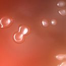
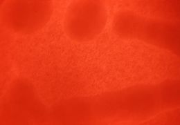
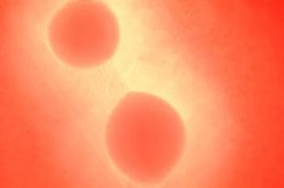
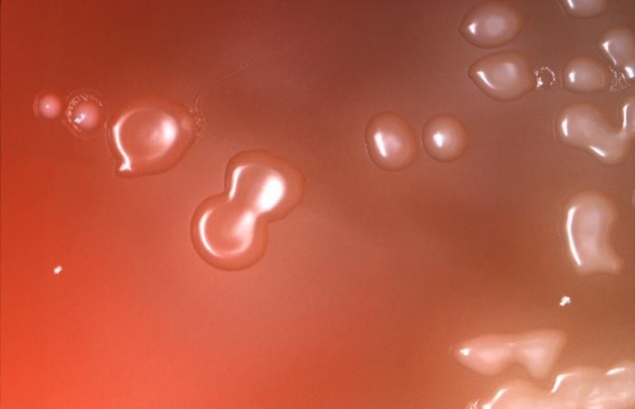
Under an approximate 10X magnification, this image depicts the colonial characteristics displayed by Streptococcus pneumoniae bacterial colonies that were grown on primary isolation medium, consisting of trypticase soy agar containing 5% sheeps blood, as well as 5mg of gentamicin/ml. See PHIL 10863 for another view of this culture.Created: 2008
-
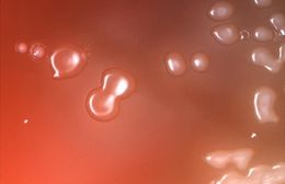
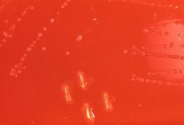
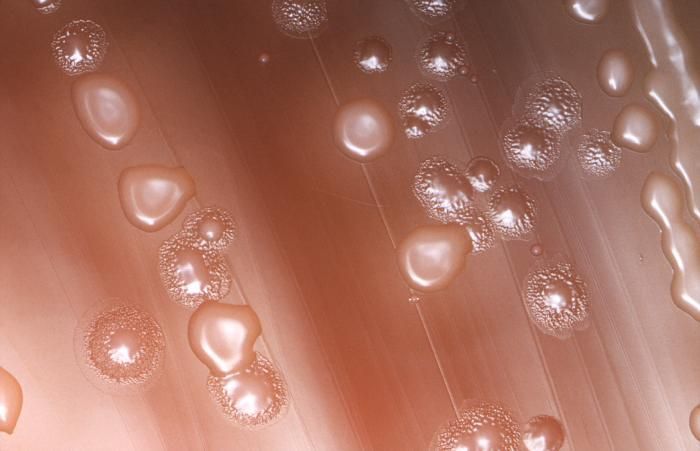
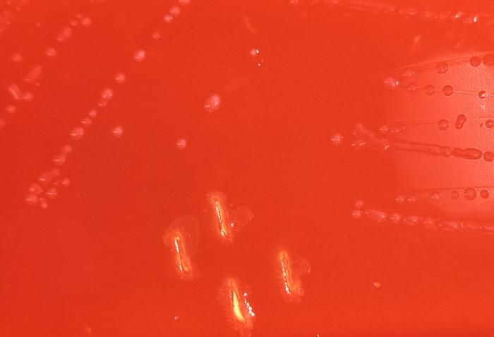
This image depicts the colonial characteristics displayed by Streptococcus pneumoniae bacterial colonies that were grown on primary isolation medium, consisting of trypticase soy agar containing 5% sheeps blood, as well as 5mg of gentamicin/ml. See PHIL 10864 for another view of this culture under a magnification of approximately 10X.Created: 2008
-
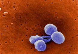
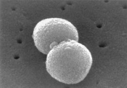
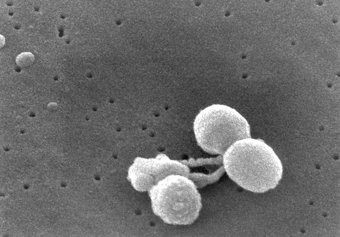
Scanning Electron Micrograph of Streptococcus pneumoniae. See PHIL 265 for a black and white version of this image.Created:
-
Scanning Electron Micrograph of Streptococcus pneumoniae. See PHIL 9996 for a colorized version of this image.Created:
-
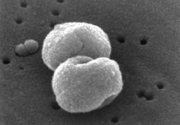
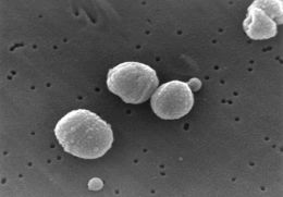
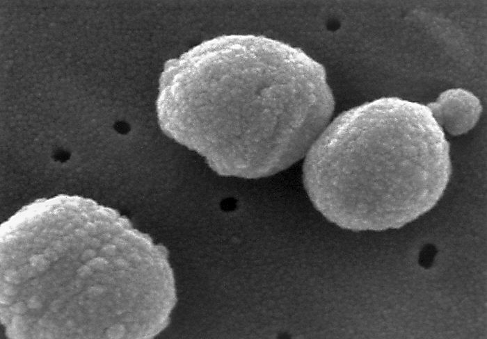
Scanning Electron Micrograph of Streptococcus pneumoniae.Created:
-
Scanning Electron Micrograph of Streptococcus pneumoniae.Created:
-
Scanning Electron Micrograph of Streptococcus pneumoniae.Created:
-
Scanning Electron Micrograph of Streptococcus pneumoniae.Created:
-
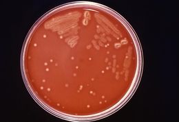
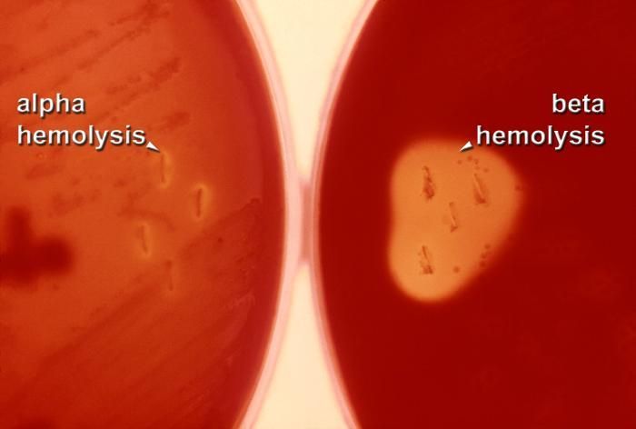
This 1977 photograph depicted two Petri dishes each filled with trypticase soy agar medium containing 5% defibrinated sheep's blood, i.e., blood agar plate (BAP). The plate on the left had been stabbed and streaked with an inoculum containing Streptococcus mitis, alpha-hemolytic bacteria, a member of the Viridans group, while the right plate was stabbed with an inoculum containing Group A Streptococcus pyogenes (GAS), a typical beta-hemolytic bacteria. The inoculation was performed using a wire loop, which had been dipped into a primary culture medium. The BAPs were incubated in a carbon dioxide enriched atmosphere at 35oC for 24 hours. There is no magnification of this image.Created: 1977
-
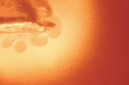
This photograph depicted a colony of a Streptococcus salivarius growing in the Petri dish filled with trypticase soy agar with 5% sheep's blood, (BAP). A loop of diluted culture of S. salivarius was put into the melted agar (50oC) just before the blood was added. The melted agar with blood was allowed to solidify, and then incubated at 35oC for 24 hours in a normal atmosphere. The culture grew subsurface bacterial colonies, one of which is seen here. There were no color changes in the region surrounding the colony, indicating that the red blood cells in the blood agar medium have not been altered in any way, which meant that these bacteria were indeed non-hemolytic in nature.Created: 1977
-
Magnified 100X, this image depicted a Petri dish filled with trypticase soy agar medium containing 5% defibrinated sheep's blood, i.e., blood agar plate (BAP). After having been inoculated by stabbing the surface of the BAP with a non-hemolytic group A Streptococcus pyogenes (GAS) bacteria. The BAP was incubated in a carbon dioxide enriched atmosphere at 35oC for 24 hours. The culture grew bacterial colonies along the stab in the BAP. There are no clear characteristic color changes in the region surrounding the stabbed area of the BAP in which the red blood cells in the blood agar medium have been altered to some extent. This "hemolyzed zone" indicated that these bacteria appear more like alpha- or WZ-alpha colonies in nature, which means that stabbing the BAP with non-hemolytic GAS is not helpful in the identification of the non-hemolytic variants of GAS.Created: 1977
-
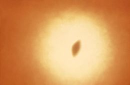
This photograph depicted a subsurface bacterial colony of a non-hemolytic S. pyogenes growing in a Petri dish filled with trypticase soy agar with 5% sheeps blood, (BAP). A loop of diluted non-hemolytic S. pyogenes culture was put into the melted agar (50oC) just before the blood was added to the melted agar, which was then allowed to solidify. It was then incubated at 35oC for 24 hours in a normal atmosphere. There was only a very small color change in the region surrounding the colony indicating that a narrow zone of red blood cells in the medium had been altered, which meant that these bacteria were "narrow-zone"-hemolytic in nature. Among the streptococcal species this hemolytic activity is found only with "non-hemolytic GAS".Created: 1977
-
Magnified 100X, this 1977 photograph depicted a Petri dish filled with trypticase soy agar medium containing 5% defibrinated sheep's blood, i.e., blood agar plate (BAP). After having been inoculated by streaking the surface of the BAP with a non-hemolytic group A Streptococcus pyogenes (GAS) bacteria. The BAP was incubated in a carbon dioxide enriched atmosphere at 35oC for 24 hours, and grew bacterial surface colonies with no characteristic color changes surrounding each colony, or in the stabbed areas. Under examination, no red blood cells in the blood agar medium had been altered, or "hemolyzed", indicating that these bacteria were indeed non-hemolytic in nature.Infection with non-hemolytic GAS can result in a range of symptoms identical to that of typical beta-hemolytic GAS:- No illness- Mild illness (strep throat or a skin infection such as impetigo)- Severe illness (necrotizing faciitis, streptococcal toxic shock syndrome)Created: 1977
-
This 1977 photograph (no magnification) depicted a Petri dish filled with trypticase soy agar medium containing 5% defibrinated sheep's blood, i.e., blood agar plate (BAP) that had been inoculated by streaking and stabbing the surface of the BAP with a non-hemolytic group A Streptococcus pyogenes (GAS) bacteria. The BAP was then incubated in a carbon dioxide enriched atmosphere at 35oC for 24 hours, and grew bacterial surface colonies with no characteristic color changes surrounding each colony, or in the stabbed areas. Under examination, no red blood cells in the blood agar medium had been altered, or "hemolyzed", indicating that these bacteria were indeed non-hemolytic in nature.Infection with non-hemolytic GAS can result in a range of symptoms identical to that of typical beta-hemolytic GAS:- No illness- Mild illness (strep throat or a skin infection such as impetigo)- Severe illness (necrotizing faciitis, streptococcal toxic shock syndrome)Created: 1977
-
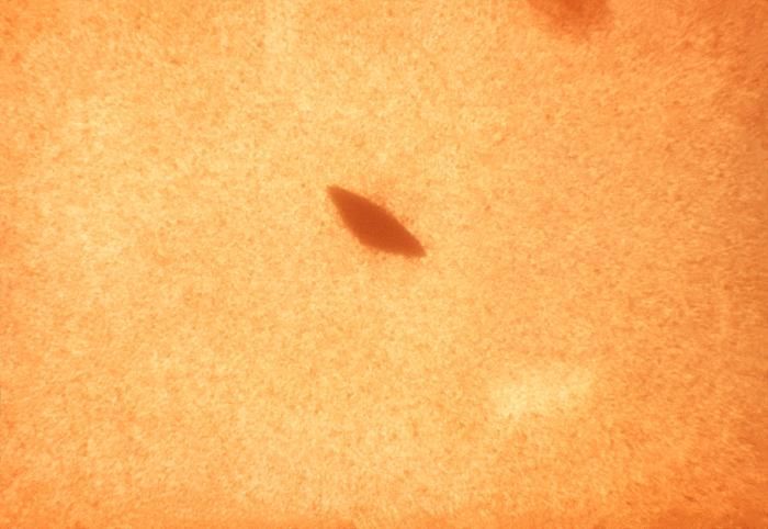
Magnified 100x, this 1977 photograph depicted a Petri dish filled with trypticase soy agar medium containing 5% defibrinated sheep's blood, i.e., blood agar plate (BAP). After having been inoculated with Group A Streptococcus pyogenes (GAS) bacteria using a wire loop stab technique, the BAP was incubated in a carbon dioxide enriched atmosphere at 35oC for 24 hours. The culture grew bacterial colonies along the edge of the stab, a number of which were seen here. The characteristic color changes, i.e., a colorless region around the stabbed area containing colonies of GAS in which the red blood cells in the blood agar medium had been destroyed, or "hemolyzed", indicated that these bacteria were indeed beta-hemolytic in nature.Infection with GAS can result in a range of symptoms:- No illness- Mild illness (strep throat or a skin infection such as impetigo)- Severe illness (necrotizing faciitis, streptococcal toxic shock syndrome)Created: 1977
-
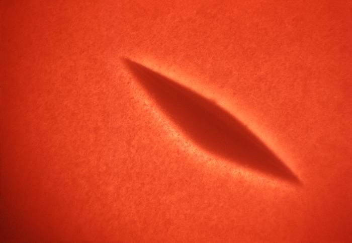
Magnified 100x, this 1977 photograph depicted a Petri dish filled with trypticase soy agar medium containing 5% defibrinated sheep's blood, i.e., blood agar plate (BAP). A loop of diluted culture of Streptococcus pyogenes was put into the melted agar (50oC) just before the blood was added to the melted agar. The melted agar with blood was allowed to solidify, and then incubated at 35oC for 24 hours in a normal atmosphere. The culture grew subsurface bacterial colonies, one of which was seen here. The characteristic color changes, i.e., a colorless region surrounding each colony in which the red blood cells in the blood agar medium had been destroyed, or "hemolyzed", indicated that these bacteria were indeed beta-hemolytic in nature.Infection with GAS can result in a range of symptoms:- No illness- Mild illness (strep throat or a skin infection such as impetigo)- Severe illness (necrotizing faciitis, streptococcal toxic shock syndrome)Created: 1977
-
Magnified 100x, this 1977 photograph depicted a Petri dish filled with trypticase soy agar medium containing 5% defibrinated sheep's blood, i.e., blood agar plate (BAP). After having been inoculated by streaking the surface of the BAP with Group A Streptococcus pyogenes (GAS) bacteria, the dish was incubated in a carbon dioxide enriched atmosphere at 35oC for 24 hours. The culture grew bacterial surface colonies. The characteristic color changes, i.e., a colorless region surrounding each colony in which the red blood cells in the blood agar medium had been destroyed, or "hemolyzed", indicated that these bacteria were indeed beta-hemolytic in nature.Infection with GAS can result in a range of symptoms:- No illness- Mild illness (strep throat or a skin infection such as impetigo)- Severe illness (necrotizing faciitis, streptococcal toxic shock syndrome)Created: 1977
-
This 1977 photograph depicted a Petri dish with Streptococcus pyogenes-inoculated trypticase soy agar containing 5% defibrinated sheep's blood, i.e., blood agar plate (BAP), that had been "streaked", and "stabbed" with a wire loop, which had been dipped into primary culture medium. The BAP was incubated in a normal atmosphere at 35oC for 24 hours. In this case, the culture dish grew colonies of Gram-positive Group A beta-Streptococci (GAS) bacteria. The characteristic color changes, i.e., a clear, colorless region surrounding each colony in which the red blood cells in the blood agar medium had been destroyed, or "hemolyzed", indicated that these bacteria were indeed beta-hemolytic in nature. There is no magnification of this image.Infection with GAS can result in a range of symptoms:- No illness- Mild illness (strep throat or a skin infection such as impetigo)- Severe illness (necrotizing faciitis, streptococcal toxic shock syndrome)Created: 1977
-
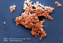
Under a moderate magnification of 2,969X, this scanning electron micrograph (SEM) revealed a number of clusters of Gram-positive, beta-hemolytic Group C Streptococcus sp. bacteria. See PHIL 10590 for a black and white version of this image.Created: 2008
-
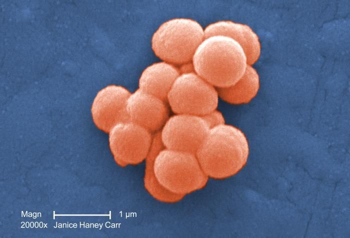
Under a high magnification of 20,000X, this colorized scanning electron micrograph (SEM) revealed a small clustered group of Gram-positive, beta-hemolytic Group C Streptococcus sp. bacteria. See PHIL 10585 for a black and white version of this image.Created: 2008
-
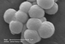
Under a very high magnification of 35,000X, this scanning electron micrograph (SEM) revealed a cluster of Gram-positive, beta-hemolytic Group C Streptococcus sp. bacteria.Created: 2008
-
Under a moderate magnification of 2,969X, this scanning electron micrograph (SEM) revealed a number of clusters of Gram-positive, beta-hemolytic Group C Streptococcus sp. bacteria. See PHIL 10591 for a colorized version of this image.Created: 2008
-
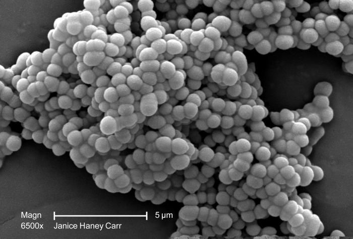
Under a moderately-high magnification of 6,500X, this scanning electron micrograph (SEM) revealed a cluster of Gram-positive, beta-hemolytic Group C Streptococcus sp. bacteria.Created: 2008
-
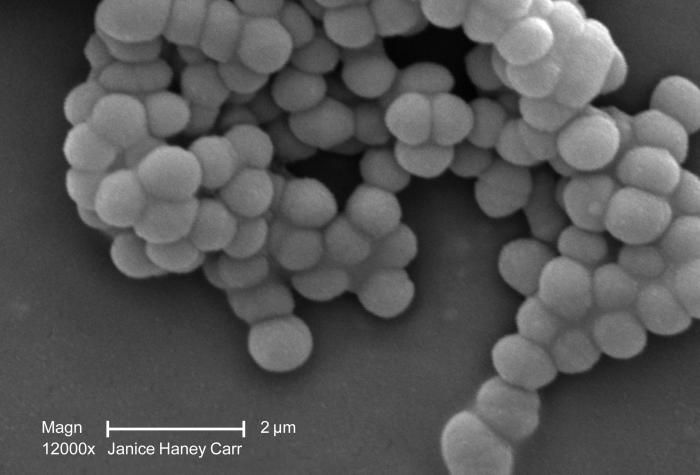
Under a moderately-high magnification of 12,000X, this scanning electron micrograph (SEM) revealed a cluster of Gram-positive, beta-hemolytic Group C Streptococcus sp. bacteria.Created: 2008