-
This scanning electron micrograph (SEM) revealed, a circular lesion on the mucosal microvillous surface of the intestine on the left, which was a result of the tight adhesion produced by the ventral adhesive disk of a Giardia sp. protozoan. The Giardia trophozoite responsible for the lesion on the right, is seen lying on its dorsal surface in this figure, thereby, revealing the circular ventral adhesive disk. Each small circular profile under the protozoan represents the rounded tip of a single microvillous, and it is estimated that 2000 to 3000 microvilli cover the surface of a single intestinal epithelial cell.Created: 1999
-
This scanning electron micrograph (SEM) revealed the presence of circulars lesions that were left on the intestinal surface, as a result of the tight adhesion produced by the ventral adhesive disk of Giardia sp. intestinal protozoa. The dorsal, or upper surfaces of several trophozoites could be seen in this figure, but the protozoan in the center of the photograph is laying upside-down, on its dorsal side, revealing an example of the ventral adhesive disk responsible for the circular lesions.Created: 1999
-
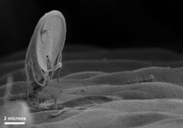
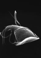
This scanning electron micrograph (SEM) depicted a Giardia sp. intestinal protozoan situated in an upright position on the mucosal surface of the intestine. The ventral adhesive disk, which facilitates adherence to the intestinal surface, can be seen on the underside of the organism.Created: 1999
-
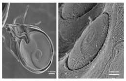
These scanning electron micrographs (SEM) revealed the ultrastructural morphology of a Giardia protozoans ventral adhesive disk on the left, and the circular lesion on the right, which can be left on the intestinal mucosal surface, as a result of the tight adhesion of this disk to the intestines microvillous border.Created: 1999
-
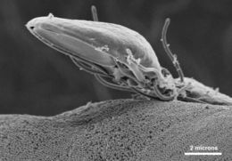
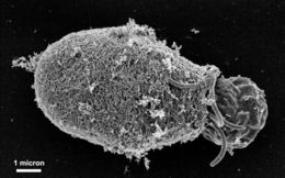
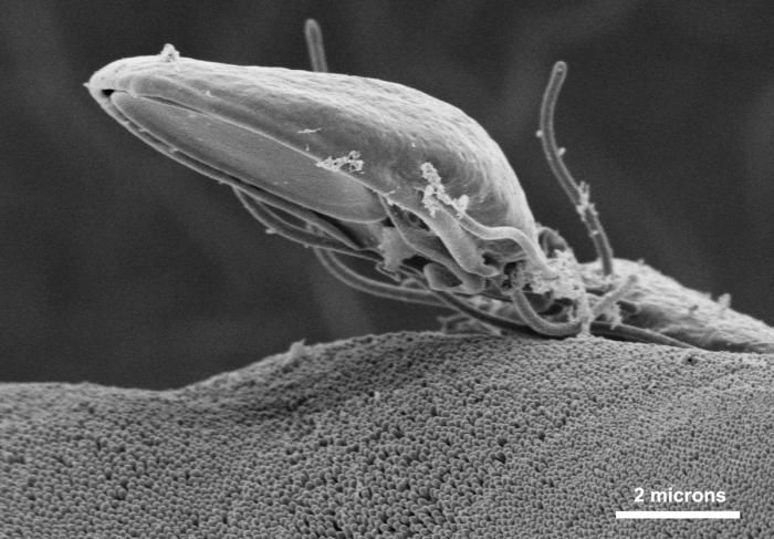
This scanning electron micrograph (SEM) depicted a Giardia specie intestinal protozoan on the microvillous border of intestinal epithelial cells. Each small circular profile under the protozoan represents the rounded tip of a single microvillous, and it is estimated that 2000 to 3000 microvilli cover the surface of a single intestinal epithelial cell. The ventral adhesive disk, which facilitates adherence to the intestinal surface, can be seen on the underside of the organism.Created: 1998
-
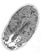
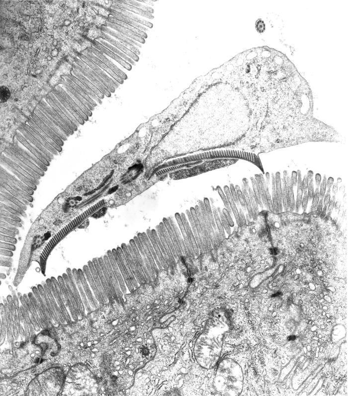
This is a transmission electron micrograph (TEM) of a thin section cut through the ventral adhesive disk (suction-cup-like structure) of a Giardia sp. protozoan, which had adhered itself to the mucosal comb-like microvillous border of an intestinal epithelial cell from a rat ileum. Note the monolayer of microtubules in the organisms ventral adhesive disk.Created: 1996
-
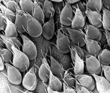
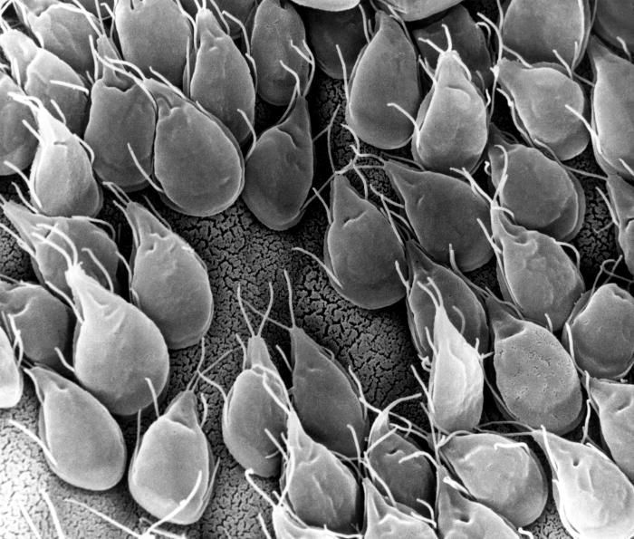
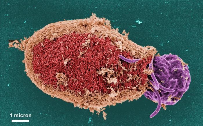
This scanning electron micrograph(SEM) depicted the mucosal surface of the small intestine of a gerbil infested with Giardia sp. protozoa. The intestinal epithelial surface is almost entirely obscured by the attached Giardia trophozoites.Created: 1988
-
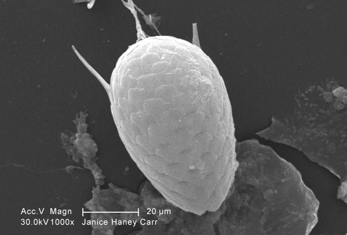
This scanning electron micrograph (SEM) depicted some of the ultrastructural morphologic details of an oblong-shaped Giardia sp. protozoan cyst, revealing the filamentous nature of the cyst wall. Each cyst-wall filament is approximately 7 to 20 nanometers (nm) thick. Note that this cyst was undergoing "excystation", and was captured at a point in the process where a flagellated trophozoite was beginning to emerge from the right side of the cyst.Created: 1999
-
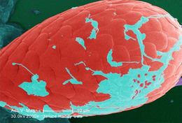
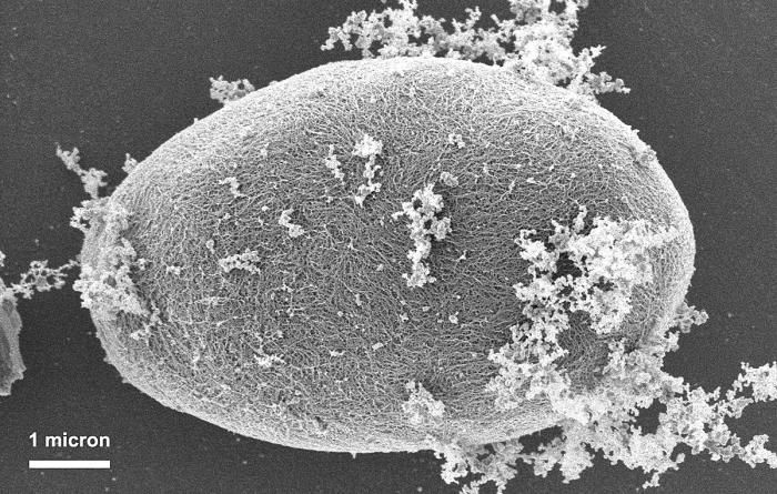
This scanning electron micrograph (SEM) depicted some of the ultrastructural morphologic details of an oblong-shaped Giardia sp. protozoan cyst, revealing the filamentous nature of the cyst wall. Each filament is approximately 7 to 20 nanometers (nm) thick.Created: 1999
-
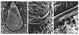
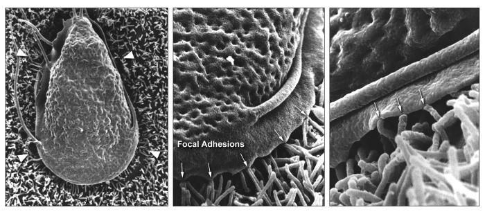
These three scanning electron micrographs (SEM) revealed under successively greater magnifications, some of the ultrastructural morphologic details of a Giardia trophozoite, where it had attached itself to apical microvilli, i.e., numerous small cylindrical structures, some highlighted with white arrowheads in the far left photograph, that covered the mucosal surface of a Caco-2 cultured intestinal epithelial cell. The ventrolateral flange, i.e., ruffle-like structure that surrounds the anterior portion of the protozoan, appeared firmly attached, by way of focal adhesions, to these microvilli.Created: 2009
-
This thin-section transmission electron micrograph (TEM) revealed some of the ultrastructural morphology found within the cyst-stage of a Giardia sp. protozoan. The outer cyst wall is composed of filamentous and membranous portions, and is separated from the cytoplasm of the trophozoites contained within by the peritrophic space. This cyst wall is approximately 0.25 microns thick.Created:
-
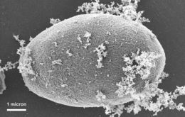
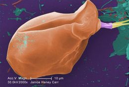
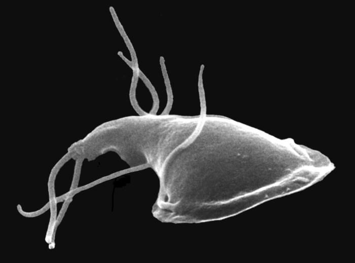
This scanning electron micrograph (SEM) depicted the dorsal (upper) surface of the intestinal protozoan, Giardia. Some of the identifying morphologic characteristics include pairs of thread-like flagella that facilitate motility, and a ventolateral flange that appears as a ruffle around the anterior portion of the organism.Created:
-
This scanning electron micrograph (SEM) depicted the dorsal (upper) surface of a Giardia protozoan that had been isolated from a rats intestine. Some of the identifying morphologic characteristics include pairs of thread-like flagella that facilitate motility, and a ventolateral flange that appears as a ruffle around the anterior portion of the organism. Pairs of flagella seen here include an anterior, posterior-lateral, and caudal pairs.Created: 1982
-
This digitally-colorized scanning electron micrograph (SEM) depicted some of the ultrastructural morphologic details of an oblong-shaped Giardia sp. protozoan cyst, revealing the filamentous nature of the cyst wall. Each cyst-wall filament is approximately 7 to 20 nanometers (nm) thick. Note that this cyst was undergoing "excystation", and was captured at a point in the process where a flagellated trophozoite was beginning to emerge from the right side of the cyst.Created: 1999
-
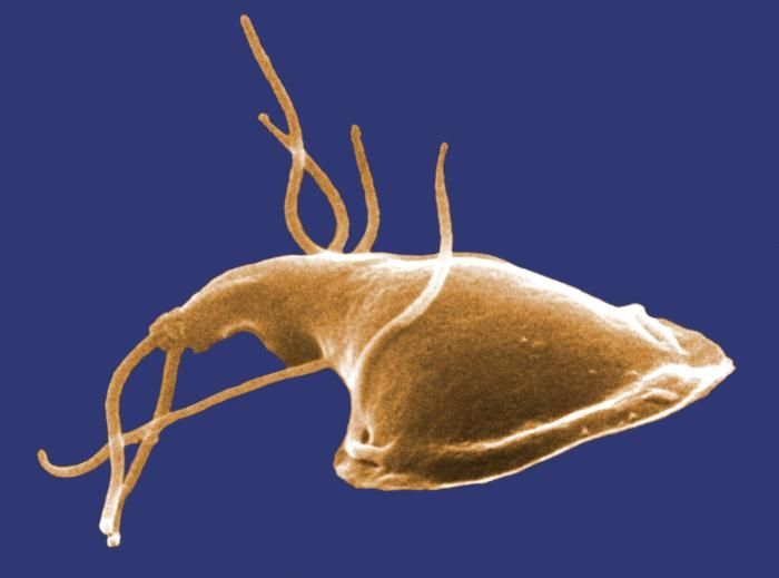
This digitally-colorized scanning electron micrograph (SEM) depicted the dorsal (upper) surface of a Giardia protozoan that had been isolated from a rats intestine. Some of the identifying morphologic characteristics include pairs of thread-like flagella that facilitate motility, and a ventolateral flange that appears as a ruffle around the anterior portion of the organism. Pairs of flagella seen here include an anterior, posterior-lateral, and caudal pairs.Created: 1982
-
Under a moderate magnification of 2000X, this digitally-colorized scanning electron micrograph (SEM) of an untreated water specimen extracted from a wild stream mainly used to control flooding during inclement weather, revealed the presence of unidentified organisms, which included bacteria, protozoa, and algae. In this particular view, a single copepod-like microorganism was seen occupying the field of view. Also, if you look closely towards the upper right corner, youll also notice the small grouping of bacteria, which had become enmeshed in a patch of biofilm.Created: 2009
-
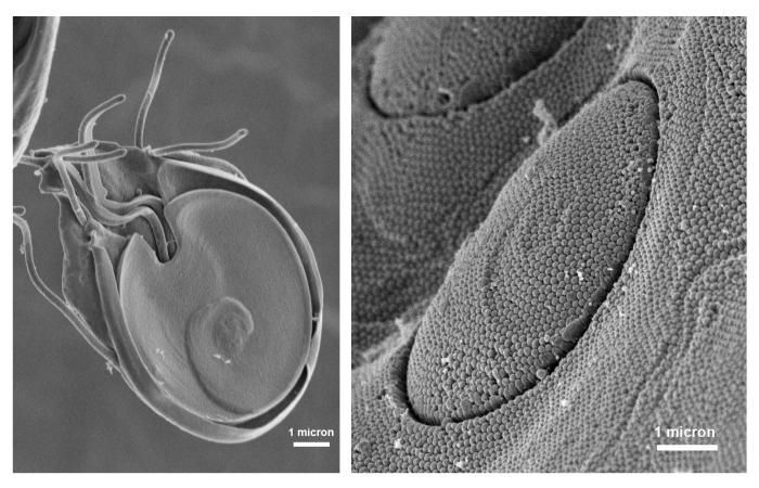
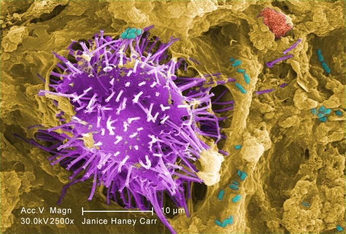
Under a moderately-high magnification of 2500X, this digitally-colorized scanning electron micrograph (SEM) of an untreated water specimen extracted from a wild stream mainly used to control flooding during inclement weather, revealed the presence of unidentified organisms, which included bacteria, protozoa, and algae. In this particular view, a microorganism is featured, the exterior of which is covered by numerous projections imparting an appearance of a sea urchin. This microscopic pin cushion was teathered to its surroundings by a biofilm within which many bacteria, and amoeboid protozoa could be seen enmeshed as well. See PHIL 11781 for a greater magnification of this organisms exterior.Created: 2009
-
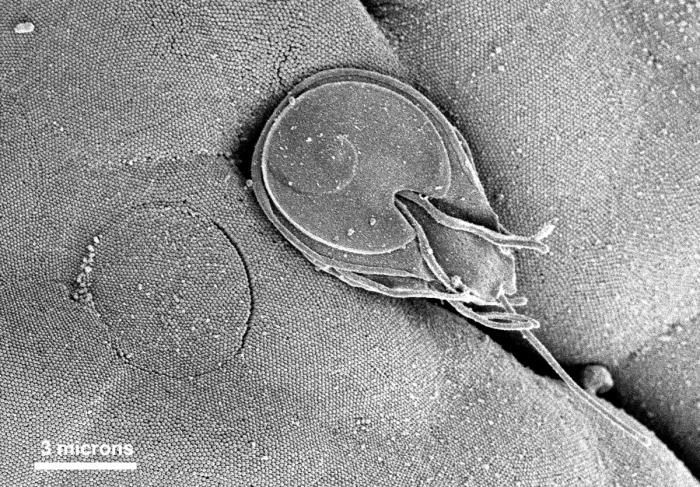
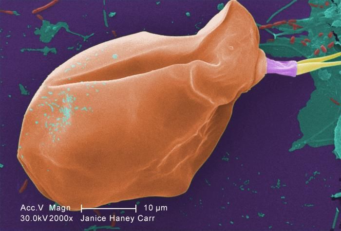
Under a moderate magnification of 2000X, this digitally-colorized scanning electron micrograph (SEM) of an untreated water specimen extracted from a wild stream mainly used to control flooding during inclement weather, revealed the presence of unidentified organisms, which included bacteria, protozoa, and algae. In this particular view, a single copepod-like microorganism was seen occupying the field of view, which seemed to be encased in an outer shell of armour-like plates, or scales. If you look closely, youll also notice the small grouping of bacteria, which had become enmeshed in a patch of biofilm on the dorsal surface of this creature's carapace.Created: 2009
-
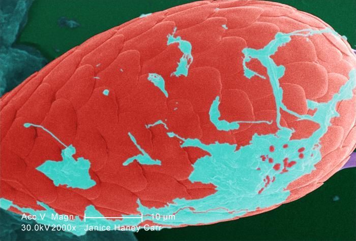
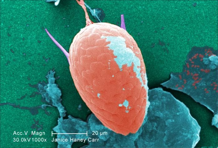
Under a moderate magnification of 1000X, this digitally-colorized scanning electron micrograph (SEM) of an untreated water specimen extracted from a wild stream mainly used to control flooding during inclement weather, revealed the presence of unidentified organisms, which included bacteria, protozoa, and algae. In this particular view, a single copepod-like microorganism was seen occupying the field of view, which seemed to be encased in an outer shell of armour-like plates, or scales. If you look closely, youll also notice the small grouping of bacteria, which had become enmeshed in a patch of biofilm.Created: 2009
-
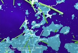
Under a relatively low magnification of 121X, this digitally-colorized scanning electron micrograph (SEM) of an untreated water specimen extracted from a wild stream mainly used to control flooding during inclement weather, revealed the presence of unidentified organisms, which included bacteria, protozoa, and algae. In this particular view, numbers of what appeared to be rod-shaped sections of various sizes were scattered throughout the field of view, which though unconfirmed, may have been vegetative in nature, and possibly algal organisms. There were also patches of biofilm present as well.Created: 2009
-
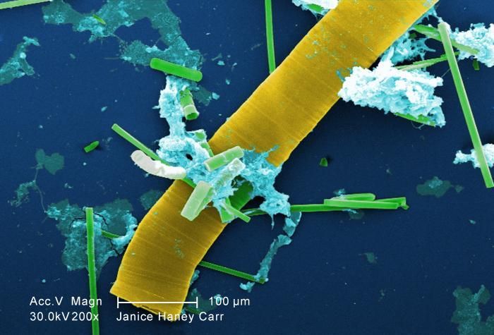
Under a relatively low magnification of 200X, this digitally-colorized scanning electron micrograph (SEM) of an untreated water specimen extracted from a wild stream mainly used to control flooding during inclement weather, revealed the presence of unidentified organisms, which included bacteria, protozoa, and algae. In this particular view, numbers of what appeared to be rod-shaped sections of various sizes were scattered throughout the field of view, which though unconfirmed, may have been vegetative in nature, and possibly algal organisms. There were also patches of biofilm present as well.Created: 2009
-
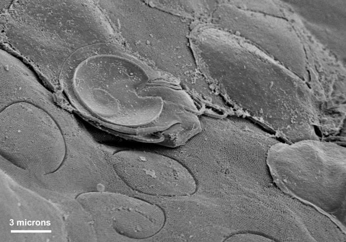
Under a moderate magnification of 1000X, this scanning electron micrograph (SEM) of an untreated water specimen extracted from a wild stream mainly used to control flooding during inclement weather, revealed the presence of unidentified organisms, which included bacteria, protozoa, and algae. In this particular view, a single copepod-like microorganism was seen occupying the field of view, which seemed to be encased in an outer shell of armour-like plates, or scales. If you look closely, youll also notice the small grouping of bacteria, which had become enmeshed in a patch of biofilm. See PHIL 11787 for a colorized version of this image.Created: 2009
-
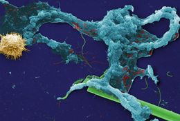
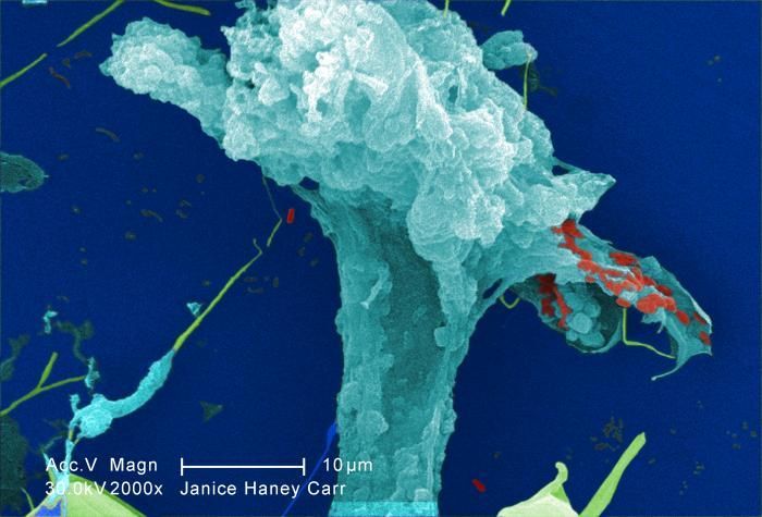
This digitally-colorized scanning electron micrograph (SEM) of an untreated water specimen extracted from a wild stream mainly used to control flooding during inclement weather, revealed the presence of unidentified organisms, which included bacteria, protozoa, and algae. Occupying most of the field of view, an unidentified amorphous mucoidal biofilm was featured, which appeared to have enmeshed numbers of amoeboid organisms, while on the right was a strangely-beautiful microorganism displaying an outer surface studded with numerous projections, making it appear like a microscopic sea urchin.Created: 2009
-
At a magnification of 2000X, this digitally-colorized scanning electron micrograph (SEM) of an untreated water specimen extracted from a wild stream, which is mainly used to control flooding during inclement weather, revealed the presence of unidentified organisms, which included bacteria, protozoa, and algae. In this particular image, an expanding amorphous organic biofilm was featured.Created: 2009