-
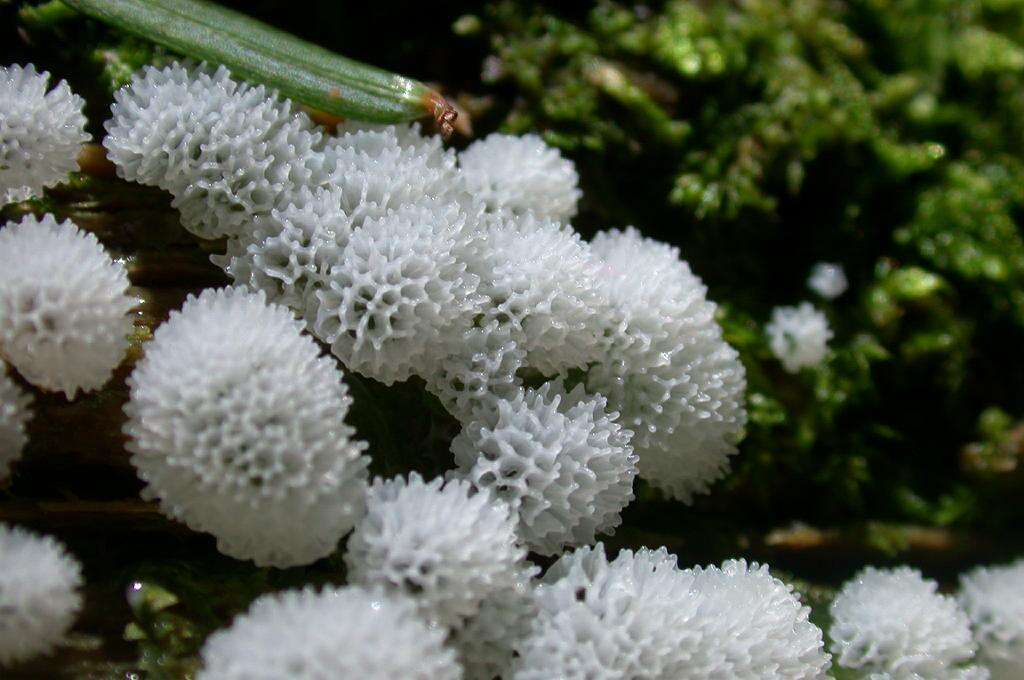
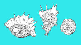
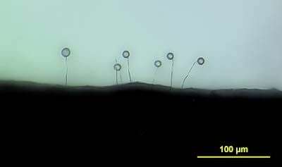
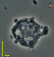
Ceratiomixa sp. (Myxomycota?) Japanese name;Tunohokori immature Fruiting body date:2004.06.13:Susami Town,Wakayama pref.Japan Author;Keisotyo
: English: This image was selected as a
picture of the month on the Japanese language Wikipedia and is considered one of the finest images. Svenska: Denna bild valdes som
månadens bild på japanskspråkiga Wikipedia och anses vara en av de finaste bilderna. 日本語: これは日本語版ウィキペディアにおける
月間新記事賞 今月の一枚に選ばれた画像であり、最も優れた画像の一つであると評価されました。. : Permission is granted to copy, distribute and/or modify this document under the terms of the
GNU Free Documentation License, Version 1.2 or any later version published by the
Free Software Foundation; with no Invariant Sections, no Front-Cover Texts, and no Back-Cover Texts. A copy of the license is included in the section entitled
GNU Free Documentation License.http://www.gnu.org/copyleft/fdl.htmlGFDLGNU Free Documentation Licensetruetrue. : This file is licensed under the
Creative Commons Attribution-Share Alike 3.0 Unported license.:.. This licensing tag was added to this file as part of the GFDL
licensing update.http://creativecommons.org/licenses/by-sa/3.0/CC-BY-SA-3.0Creative Commons Attribution-Share Alike 3.0truetrue.
-
-

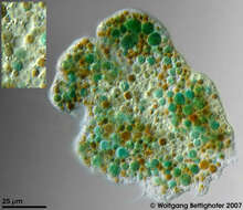
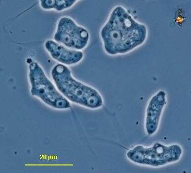
Enteromyxa paludosa Enteromyxa paludosa cell was showing very colorful metabolites and numerous nuclei and contractile vacuoles. In order to deliver depth of focus 50 high resolution frames (Planapo 63/1.4) were processed. The picture inserted shows 4 nuclei and 3 contractile vacuoles in higher magnification. Sample from sphagnum pond situated in the northern alpine region of Austria near Salzburg. Images were taken using Zeiss Universal with Olympus C7070 CCD camera.Image under Creative Commons License V 3.0 (CC BY-NC-SA). Place name: Bogs near Salzburg (Austria) Latitude: 48.068516 Longitude: 12.954134 Die Enteromyxa paludosa -Zelle zeigt sehr farbintensive Metabolite und zahlreiche Kerne und kontraktile Vakuolen. Die ins Bild eingefügten Ausschnittsvergrößerungen zeigen 4 Kerne und 3 kontraktile Vakuolen. Tiefenschärfe durch Multiebenenabbildung aus 50 Bildebenen, manuell gestapelt. Probe aus einem Moor in den nördlichen Kalkalpen von Österreich in der Nähe von Salzburg. Mikrotechnik: Zeiss Universal, Kamera: Olympus C7070. Creative Commons License V 3.0 (CC BY-NC-SA). For permission to use of (high-resolution) images please contact postmaster@protisten.de.
-
-
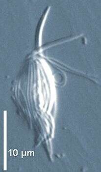
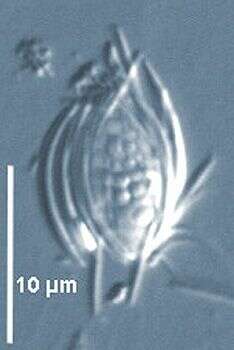
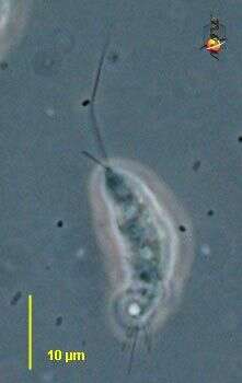
Polymastix is an oxymonad flagellate with and almond or spindle-shaped body with four free anterior flagella arranged in two pairs. Flagella deflected backwards but not adherent to the cell body. Nucleus pointed anteriorly, reduced anterior pelta and slender axostyle generally not protruding posteriorly. Several species occurring in xylophagous insects such as Polymastix melolonthae from coleopterid larvae. Polymastix sp. from the cockroach Parasphaeria boleiriana from Brazil, anterior flagella, body covered with epibiontic Fusiformis bacteria (interference contrast).
-
Polymastix is an oxymonad flagellate with and almond or spindle-shaped body with four free anterior flagella arranged in two pairs. Flagella deflected backwards but not adherent to the cell body. Nucleus pointed anteriorly, reduced anterior pelta and slender axostyle generally not protruding posteriorly. Several species occurring in xylophagous insects such as Polymastix melolonthae from coleopterid larvae. Polymastix sp. from the cockroach Parasphaeria boleiriana from Brazil with epibiontic Fusiformis bacteria (interference contrast).
-
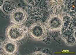
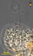
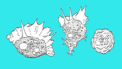
This is an image of multiple fruiting bodies.
-
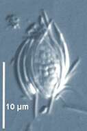
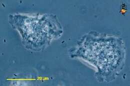
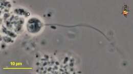
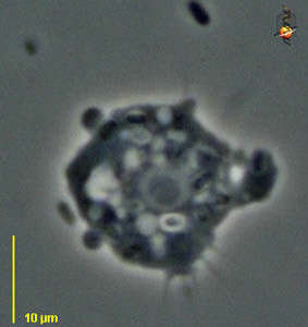
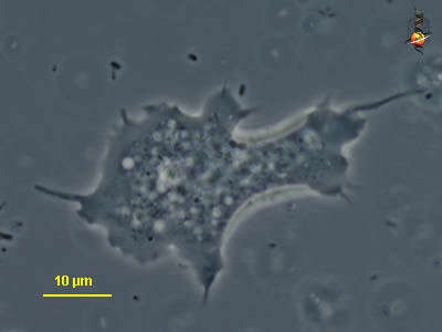
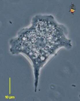
Planoprotostelium (plan-owe-pro-toe-steal-ee-um) is a true myxomycete (or closely related to the true myxomycete), there is a flagellated stage typically with a single flagellum but occasionally a rod or filament may project from the front end of the cell. The flagellate body is very amoeboid and may have uroidal filaments projecting from the back end. The species may also become an amoeba (top right) and may produce a stalk and encyst at the top of that stalk. Contractile vacuoles (white) nuclei with nucleoli (some cells have more than one) are visible. Phase contrast.
-
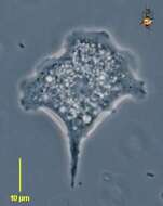
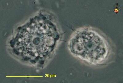
Planoprotostelium (plan-owe-pro-toe-steal-ee-um) is a true myxomycete (or closely related to the true myxomycete), there is a flagellated stage typically with a single flagellum but occasionally a rod or filament may project from the front end of the cell as may be seen here. The flagellate body is very amoeboid and may have uroidal filaments projecting from the back end. Phase contrast.
-
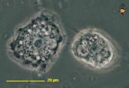
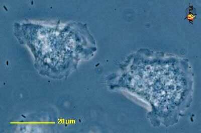
Planoprotostelium (plan-owe-pro-toe-steal-ee-um) is a true myxomycete (or closely related to the true myxomycete), there is a flagellated stage typically with a single flagellum but occasionally a rod or filament may project from the front end of the cell. The flagellate body is very amoeboid and may have uroidal filaments projecting from the back end. The species may also become an amoeba and may produce a stalk and encyst at the top of that stalk. Contractile vacuole (white) and nuclei with nucleoli are visible. Phase contrast.
-
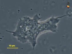
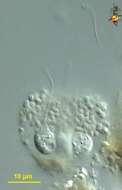
Filamoeba (file-a-me-ba) small free-living amoeba, with filamentous uroid, (bottom). Central nucleus with nucleolus, and with a contractile vacuole being formed by the fusion of several smaller vesicles to the left. Phase contrast.
-
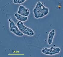
Flamella (flam-ell-a), a naked amoeba. Body form changeable, from an ovoid shape to a fan-shaped form. A more irregular extended form is illustrated here. Hyaline region well developed. Sub-pseudopodia are evident in this image. Phase contrast.
-
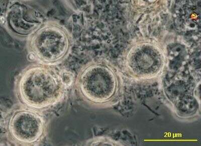
Flamella (flam-ell-a), a naked amoeba. Body form changeable, from an ovoid shape to a fan-shaped form. This image is of the cysts. Phase contrast.
-
Flamella (flam-ell-a), a naked amoeba. Body form changeable, from an ovoid shape to a fan-shaped form. The irregular form is illustrated here. Hyaline region well developed. Phase contrast.
-
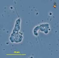
Flamella (flam-ell-a), a naked amoeba. Body form changeable, from an ovoid shape to a fan-shaped form. A more contracted form is illustrated here. Hyaline region well developed. Subpseudopodia, nucleus, and contractile vacuole (to the right) are evident in this image. Phase contrast.
-
Flamella (flam-ell-a), a naked amoeba. Body form changeable, from an ovoid shape to a fan-shaped form - as illustrated here. Hyaline region well developed. Subpseudopodia a may form, but are not evident in this image. Phase contrast.
-
Flamella (flam-ell-a), a naked amoeba. Body form changeable, from an ovoid shape to a fan-shaped form. This image is of the cysts. Phase contrast.
-
-
-

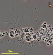
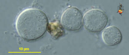
Multicilia marina Cienkowski, 1881. Cells are normally spherical in a culture, about 30-40 microns in diameter, however,some cells attain an oblong or irregular form. The majority of cells possess 20-30 long flagella, 1.5-2 times the cell diameter. The flagella differ from those of other heterotrophic flagellates by very weak movements, resulting in a similarity to heliozoan axopods. The locomotion of M ulticilia is slow, rotatory, without any definitive direction, therefore moving cells normally lack anterior and posterior ends. Cells glide in one direction for a very short distance and may change direction rapidly (including moving backwards). The cell rolls over when changing direction. The temporary anterior flagellum is then stretched and clasped along the substratum, only its apex continues to oscillate. The temporary posterior flagellum shows the same behaviour. Other flagella perform irregular movements without any visible coordination. After changing the salinity, cells become free floating and continue a weak oscillation of the flagella.
-
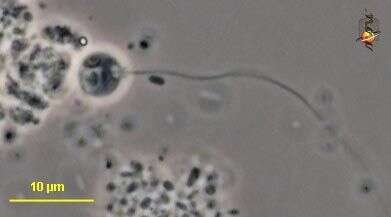
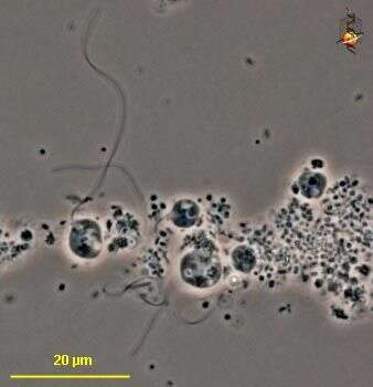
Phalansterium (fah-lan-stear-ee-um) is a free-living flagellate, usually found in mucoid colonies, with a single apical flagellum surrounded with a tight apical collar. Phase contrast.
-
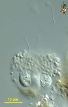
Phalansterium (fah-lan-stear-ee-um) is a free-living flagellate, usually found in mucoid colonies, with a single apical flagellum surrounded with a tight apical collar. Phase contrast.
-
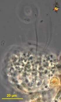
Phalansterium (fah-lan-stear-ee-um) is a free-living flagellate, usually found in mucoid colonies, with a single apical flagellum surrounded with a tight apical collar. Mucus globular. Differential interference contrast.
-
Phalansterium (fah-lan-stear-ee-um) is a free-living flagellate, usually found in mucoid colonies, with a single apical flagellum surrounded with a tight apical collar. Phase contrast.


_Japan.svg)