-
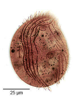
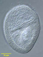
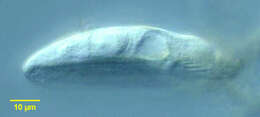
Surface detail of the marine Phyllopharyngeid ciliate, Dysteria brasiliensis Da Cunha, De Faria & Pinto, 1922. This is one of the largest species of this genus (100-130 um).The posterior terminates in a sharp spinous process (slightly out of focus here) not to be confused with the ventral posterior podite by which the cell attaches to the substrate. The podite is angled anteriorly in this image (the the viewer's right).Collected from a commercial saltwater aquarium in Boise, Idaho. March 2004. DIC.
-

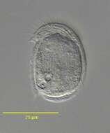
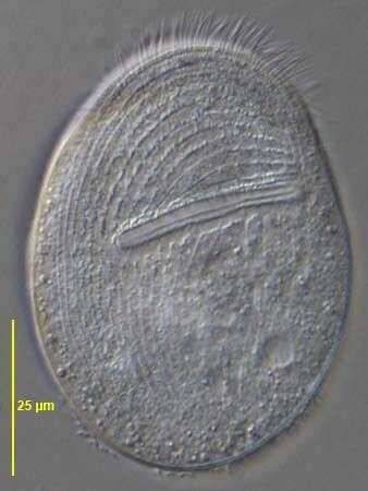
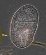
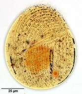
Gastronauta membranaceus (Engelmann in Bütschli,1889), a hypostome ciliate, distinguished by its long transversely oriented cytostome. The cytostome lacks trichites. The body is ovoid in outline and strongly dorsoventrally flattened. Ciliature is restricted to the ventral surface except for two short dorsal kineties anteriorly. A few somatic kineties run uninterrupted to the right of the cytostome arching around the anterior of the cell. Several right somatic kineties are interrupted by the cytostome. The left somatic kineties terminate at the cytostome. A single kinety runs around the circumference of the cytostome. An unciliated bare are overlies the region of the macronucleus posterior to the cytostome. The macronucleus is oblong and heteromerous (i.e. containing areas with markedly differing RNA and DNA contents resulting in irregular staining and optical characteristics). The single micronucleus is quite prominent. Two contractile vacuoles are present, one in the anterior half and one posteriorly. Gastronauta feeds mainly on diatoms. From a freshwater pond near Boise, Idaho. Phase contrast illumination.
-
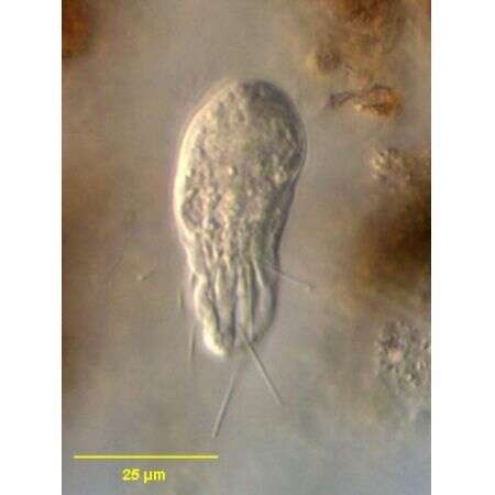
Atopochilodon distichum (Deroux,1976). Collected from a commercial saltwater aquarium in Boise, Idaho. February 2006.DIC.
-
Infraciliature (ventral surface) of the chilodonellid ciliate Pseudochilodonopsis piscatoris (Blochmann, 1895) Foissner, 1979 in early division. Stomatogenesis is of the telokinetal type in which the oral apparatus of the posterior daughter cell (opisthe) derives from fragments of several left sided kineties. As can be seen here, stomatogenesis precedes cytokinesis. Collected from a freshwater pond near Boise, Idaho February 2005.Stained by the silver carbonate technic (see Foissner, W.Europ. J. Protistol.27,313-330;1991). Brightfield.
-
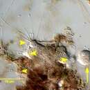
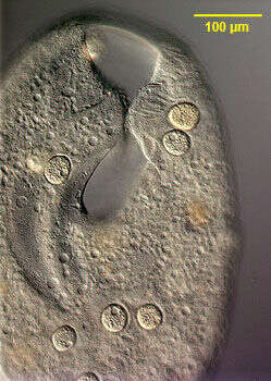
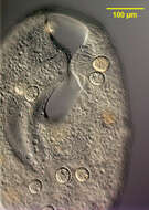
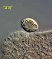
Image of the stalkless parasitic suctorian, Sphaerophrya insolita (Jankowski, 1973) infesting the large colpodid ciliate, Bursaria truncatella (Muller, 1773). The Sphaerophrya cells are ellipsoid and approximately 35 u in diameter. One suctorian can be seen adhering to the right lip of the vestibulum of the host cell. At least 7 others can be seen adhering to the pellicle where they may be mistaken for food vacuoles on cursory examination. Sphaerophrya is thought to have lost its stalk during the transition to a parasitic mode of existence. The cells have capitate tentacles by which they adhere to the pellicle of the host cell. There is a central ellipsoid granular nucleus (the micronuclei have not been characterized). There is a single peripheral contractile vacuole. These individuals were found on B. truncatella collected from a temporary rainwater pool containing decaying grass near Boise, Idaho March 2005. DIC.
-
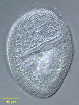
Gastronauta membranaceus (Engelmann in Bütschli,1889), a hypostome ciliate, distinguished by its long transversely oriented cytostome. The cytostome lacks trichites. The body is ovoid in outline and strongly dorsoventrally flattened. Ciliature is restricted to the ventral surface except for two short dorsal kineties anteriorly. A few somatic kineties run uninterrupted to the right of the cytostome arching around the anterior of the cell. Several right somatic kineties are interrupted by the cytostome. The left somatic kineties terminate at the cytostome. A single kinety runs around the circumference of the cytostome. An unciliated bare are overlies the region of the macronucleus posterior to the cytostome. The macronucleus is oblong and heteromerous (i.e. containing areas with markedly differing RNA and DNA contents resulting in irregular staining and optical characteristics). The single micronucleus is quite prominent. Two contractile vacuoles are present, one in the anterior half and one posteriorly. Gastronauta feeds mainly on diatoms. From a freshwater pond near Boise, Idaho. Phase contrast illumination.
-
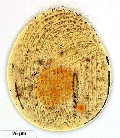
Ventral surface of the marine phyllopharyngiid ciliate, Coeloperix sleighi (Gong and Song,2004).The cell is broadly ovoid in outline and strongly dorsoventrally flattened. The dorsum is slightly convex and the ventral surface flattened. Ciliature is restricted to the ventral surface. The preoral and postoral kineties are separated by a transverse suture and the preoral kineties are transversely oriented. The postoral kineties are continuous , lacking the central bare gap seen in Chlamydodon. There is a peripheral cross-striated band (CSB) similar to that seen in Chlamydodon however the CSB in Coeloperix is interrupted on the right and left sides by two slightly offset gaps. There are 3or 4 short "tentacles" on the posteromedial ventral surface. These are about 5 µm long and are quite difficult to see even with DIC. The anterior ventral cytostome is supported by prominent nematodesmata. There is a central heteromerous macronucleus. There are two contractile vacuoles situated diagonally. They empty through single pores on the ventral surface. Collected from a commercial marine aquarium in Boise, Idaho. pH 7.93. January 2004. DIC.
-
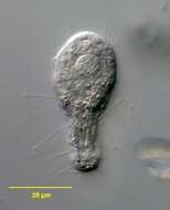
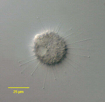
n vivo view of the swarmer of the suctorian, Parapodophrya soliformis (LAUTERBORN,1908) KAHL,1931. The swarmers have numerous capitate tentacles arising randomly from the body posterior to the anterior subapical ciliary wreath. The body has longitudinal irregular pellicular wrinkles. Collected from sapropelic bottom sediments of a stagnant freshwater pond near Boise, Idaho 43°40â 57.20â N 116° 15â 15.44â W . September, 2006.DIC.
-
Detail view stalkless parasitic suctorians, Sphaerophrya insolita (Jankowski, 1973) infesting the large colpodid ciliate, Bursaria truncatella (Muller, 1773). The Sphaerophrya cells are ellipsoid and approximately 35 u in diameter. Two suctorians can be seen on the left side (viewer's right) of the vestibular cleft of the host cell and one on the right. There are several posterior to the cleft. Sphaerophrya is thought to have lost its stalk during the transition to a parasitic mode of existence. The cells have capitate tentacles by which they adhere to the pellicle of the host cell. There is a central ellipsoid granular nucleus (the micronuclei have not been characterized). There is a single peripheral contractile vacuole (seen well in a number of these cells). These individuals were found on B. truncatella collected from a temporary rainwater pool containing decaying grass near Boise, Idaho March 2005. DIC.
-
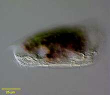
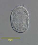
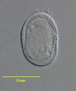
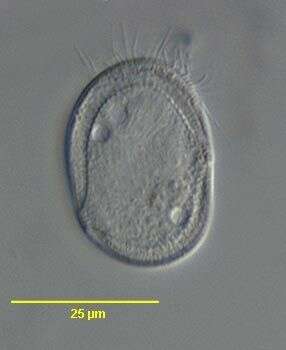
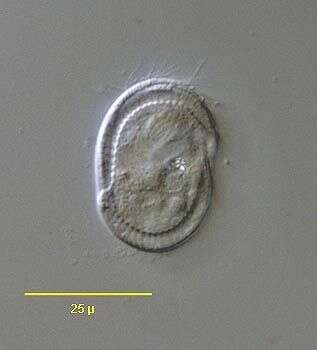
Gastronauta membranaceus (Engelmann in Butschli, 1889). Seen from the side, the body is dorso-ventrally flattened.
-
Ventral surface of the marine phyllopharyngiid ciliate, Coeloperix sleighi (Gong and Song,2004).The cell is broadly ovoid in outline and strongly dorsoventrally flattened. The dorsum is slightly convex and the ventral surface flattened. Ciliature is restricted to the ventral surface. The preoral and postoral kineties are separated by a transverse suture and the preoral kineties are transversely oriented. The postoral kineties are continuous , lacking the central bare gap seen in Chlamydodon. There is a peripheral cross-striated band (CSB) similar to that seen in Chlamydodon however the CSB in Coeloperix is interrupted on the right and left sides by two slightly offset gaps. There are 3or 4 short "tentacles" on the posteromedial ventral surface. These are about 5 µm long and are quite difficult to see even with DIC. The anterior ventral cytostome is supported by prominent nematodesmata. There is a central heteromerous macronucleus. There are two contractile vacuoles situated diagonally. They empty through single pores on the ventral surface. Collected from a commercial marine aquarium in Boise, Idaho. pH 7.93. January 2004. DIC.
-
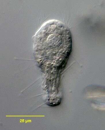
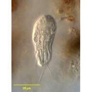
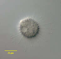
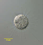
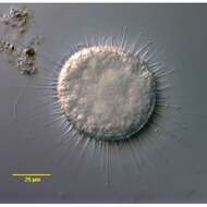
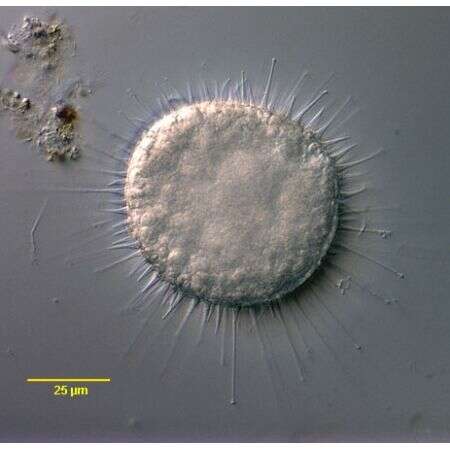
Portrait of the exogenid suctorian, Parapodophrya soliformis (Lauterborn,1908)Kahl,1931.This is a tentative identification.Members of this genus usually have a very fine stalk but sometimes this is absent (as in this example).A firm diagnosis requires identification of the swarmer cell which is elongate, the anterior end wider than the posterior. There is an anterior wreath of cilia in the swarmer.The cell body is roughly spherical with tentacles distributed over the entire surface rather than in fascicles. The tentacles widen at their bases giving the cell a serrated outline. Only the extended tentacles are capitate. When they contract they appear as short wide-based spines Seen best here at 12 o'clock). These features are also typical of this species of Parapodophrya.The single contractile vacuole is seen at 8 o'clock here. The spherical macronucleus (not well seen here) is central. Parapodophrya species are free-living and never parasitic unlike Podophrya.Collected from sapropelic bottom sediments of a freshwater aquaculture tub near Boise, Idaho.December 2005.DIC.
-
Sphaerophrya insolita (Jankowski, 1973) infesting the large colpodid ciliate, Bursaria truncatella (Muller, 1773). The Sphaerophrya cells are ellipsoid and approximately 35 u in diameter. Sphaerophrya is thought to have lost its stalk during the transition to a parasitic mode of existence. The cells have capitate tentacles by which they adhere to the pellicle of the host cell (several of these are visible on the viewer's left). There is a central ellipsoid granular nucleus seen well here (the micronuclei have not been characterized). There is a single peripheral contractile vacuole (seen well in this cell). These individuals were found on B. truncatella collected from a temporary rainwater pool containing decaying grass near Boise, Idaho March 2005. DIC.
-
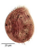
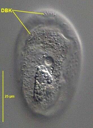
Dorsal view of Gastronauta membranaceus (Engelmann in Bütschli,1889). DBK= dorsal brush kineties. The bipartite dorsal brush is one of the distinguishing characteristics of this species. G. derouxi has a long anterior marginal dorsal brush row of dikinetids. From a freshwater pond near Boise, Idaho. DIC.
-
Optical section of the marine phyllopharyngiid ciliate, Coeloperix sleighi (Gong and Song,2004).The cell is broadly ovoid in outline and strongly dorsoventrally flattened. The dorsum is slightly convex and the ventral surface flattened. Ciliature is restricted to the ventral surface. The preoral and postoral kineties are separated by a transverse suture and the preoral kineties are transversely oriented. The postoral kineties are continuous , lacking the central bare gap seen in Chlamydodon. There is a peripheral cross-striated band (CSB) similar to that seen in Chlamydodon however the CSB in Coeloperix is interrupted on the right and left sides by two slightly offset gaps. There are 3or 4 short "tentacles" on the posteromedial ventral surface. These are about 5 µm long and are quite difficult to see even with DIC. The anterior ventral cytostome is supported by prominent nematodesmata. There is a central heteromerous macronucleus. There are two contractile vacuoles situated diagonally. They empty through single pores on the ventral surface. Collected from a commercial marine aquarium in Boise, Idaho. pH 7.93. January 2004. DIC.
-
Portrait of the exogenid suctorian, Parapodophrya soliformis (Lauterborn,1908)Kahl,1931.This is a tentative identification.Members of this genus usually have a very fine stalk but sometimes this is absent (as in this example).A firm diagnosis requires identification of the swarmer cell which is elongate, the anterior end wider than the posterior. There is an anterior wreath of cilia in the swarmer.The cell body is roughly spherical with tentacles distributed over the entire surface rather than in fascicles. The tentacles widen at their bases giving the cell a serrated outline. Only the extended tentacles are capitate. When they contract they appear as short wide-based spines Seen best here at 10 o'clock). These features are also typical of this species of Parapodophrya.The single contractile vacuole is eccentric (not seen here). The spherical macronucleus (not well seen here) is central. Parapodophrya species are free-living and never parasitic unlike Podophrya.Collected from sapropelic bottom sediments of a freshwater aquaculture tub near Boise, Idaho.December 2005.DIC.
-
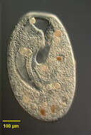
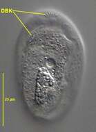
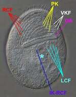
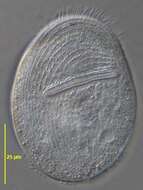
Ventral view (in vivo) of Gastronauta derouxi (FOISSNER & BLATTERER, 1992). OA=oral large transverse oral aperture. RCF=right ventral somatic ciliary field. LCF= left ventral somatic ciliary field. PK=preoral kineties. VKF=vertical kinety fragments.IK-RCF=anteriorly curved innermost kinety of right ciliary field. *= unciliated postoral area between right and left somatic ciliary fields.Collected from an ephemeral puddle on a grass lawn in Boise, Idaho. July 2007.DIC
-
Optical section of the marine phyllopharyngiid ciliate, Coeloperix sleighi (Gong and Song,2004).The cell is broadly ovoid in outline and strongly dorsoventrally flattened. The dorsum is slightly convex and the ventral surface flattened. Ciliature is restricted to the ventral surface. The preoral and postoral kineties are separated by a transverse suture and the preoral kineties are transversely oriented. The postoral kineties are continuous , lacking the central bare gap seen in Chlamydodon. There is a peripheral cross-striated band (CSB) similar to that seen in Chlamydodon however the CSB in Coeloperix is interrupted on the right and left sides by two slightly offset gaps. There are 3or 4 short "tentacles" on the posteromedial ventral surface. These are about 5 ?m long and are quite difficult to see even with DIC. The anterior ventral cytostome is supported by prominent nematodesmata. There is a central heteromerous macronucleus. There are two contractile vacuoles situated diagonally. They empty through single pores on the ventral surface. Collected from a commercial marine aquarium in Boise, Idaho. pH 7.93. January 2004. DIC.
-
Portrait of the exogenid suctorian, Parapodophrya soliformis (Lauterborn,1908)Kahl,1931.This is a tentative identification.Members of this genus usually have a very fine stalk but sometimes this is absent (as in this example).A firm diagnosis requires identification of the swarmer cell which is elongate, the anterior end wider than the posterior. There is an anterior wreath of cilia in the swarmer.The cell body is roughly spherical with tentacles distributed over the entire surface rather than in fascicles. The tentacles widen at their bases giving the cell a serrated outline. Only the extended tentacles are capitate. When they contract they appear as short wide-based spines. These features are also typical of this species of Parapodophrya.There is a single contractile vacuole. The spherical macronucleus (not well seen here) is central. Parapodophrya species are free-living and never parasitic unlike Podophrya.Collected from sapropelic bottom sediments of a freshwater aquaculture tub near Boise, Idaho.December 2005.DIC.
-
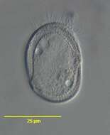
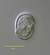
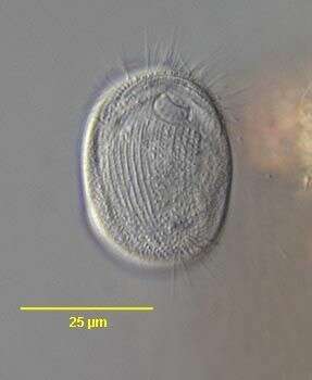
Ventral view (in vivo) of Gastronauta derouxi (FOISSNER & BLATTERER, 1992). Collected from an ephemeral puddle on a grass lawn in Boise, Idaho. July 2007.DIC.
-
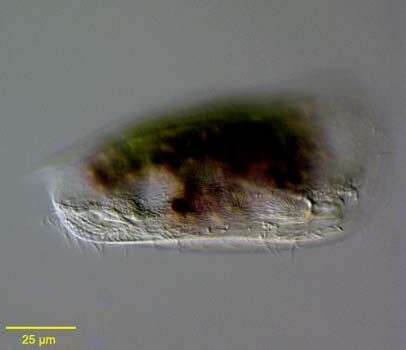
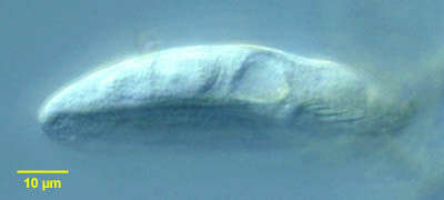
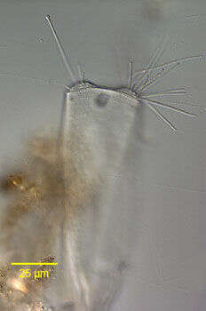
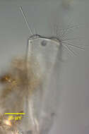
Portrait of the discophryid suctorian, Periacineta buckei (Kent,1882).This periphytonic species has a laterally compressed tectinous lorica which is snug against the cell body.The truncate anterior end of the cell protrudes from the slit-like aperture of the lorica.The lorica tapers posteriorly to a narrow attachment with the substarte. A true stalk is absent. Capitate tentacles occur in two fasicles on either side of the anterior end. There are three contractile vacuoles at the anterior end (seen here).The macronucleus is ellipsoid.Swarmers are elongate and flattened.Collected from a freshwater pond near Boise, Idaho.DIC.
-
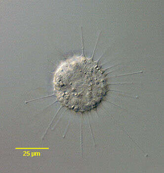
In vivo view of the swarmer of the suctorian, Parapodophrya soliformis (LAUTERBORN,1908) KAHL,1931. The swarmers have numerous capitate tentacles arising randomly from the body posterior to the anterior subapical ciliary wreath. The body has longitudinal irregular pellicular wrinkles. Collected from sapropelic bottom sediments of a stagnant freshwater pond near Boise, Idaho 43°40â 57.20â N 116° 15â 15.44â W . September, 2006.DIC.
-
Ventral infraciliature of Gastronauta derouxi (FOISSNER & BLATTERER, 1992).Collected from an ephemeral puddle on a grass lawn in Boise, Idaho. July 2007. Stained by the silver carbonate technique (Foissner,W. Europ. J. Protistol.27:313-330;1991).Brightfield.
-
Portrait of the discophryid suctorian, Periacineta buckei (Kent,1882).This periphytonic species has a laterally compressed tectinous lorica which is snug against the cell body.The truncate anterior end of the cell protrudes from the slit-like aperture of the lorica.The lorica tapers posteriorly to a narrow attachment with the substarte. A true stalk is absent. Capitate tentacles occur in two fasicles on either side of the anterior end. There are three contractile vacuoles at the anterior end.The macronucleus is ellipsoid.Swarmers are elongate and flattened.Collected from a freshwater pond near Boise, Idaho.DIC.