-
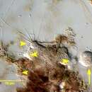
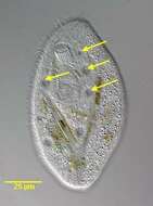
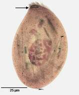
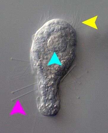
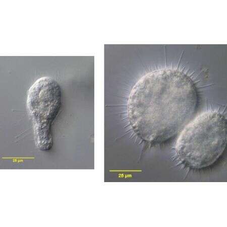
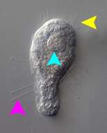
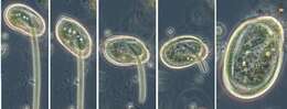
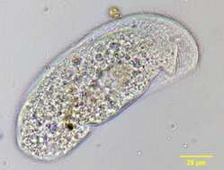
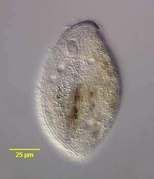
In vivo view of the swarmer of the suctorian, Parapodophrya soliformis (LAUTERBORN,1908) KAHL,1931. The swarmers have numerous capitate tentacles (pink arrowhead) arising randomly from the body posterior to the anterior subapical ciliary wreath (yellow arrowhead). The body has longitudinal irregular pellicular wrinkles. The ellipsoid nucleus is seen in cross-section here (light blue arrowhead).Collected from sapropelic bottom sediments of a stagnant freshwater pond near Boise, Idaho 43°40â 57.20â N 116° 15â 15.44â W . September, 2006.DIC.
-
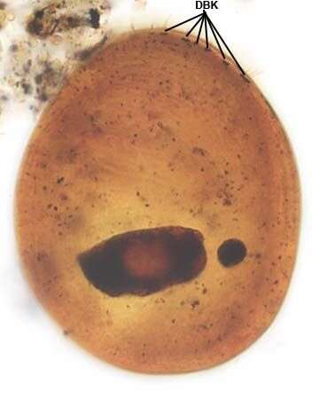
Dorsal infraciliature of Gastronauta derouxi (FOISSNER & BLATTERER, 1992).DBK=dorsal brush dikinetids. Collected from an ephemeral puddle on a grass lawn in Boise, Idaho. July 2007. Stained by the silver carbonate technique (Foissner,W. Europ. J. Protistol.27:313-330;1991).Brightfield.
-
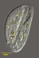
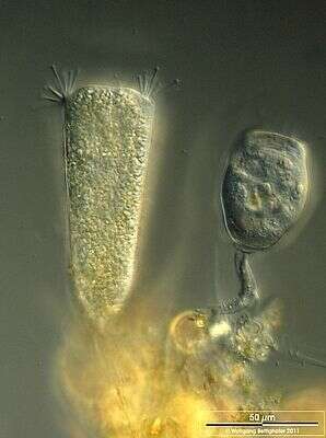
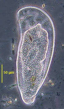
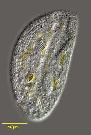
Scale bar indicates 50 µm. Sample from a pond situated in the vicinity of Lake Constance (Bodensee, Southern Germany). The image was built up using several photomicrographic frames with manual stacking technique. Images were taken using Zeiss Universal with Olympus C7070 CCD camera.
-
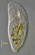
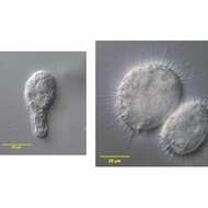
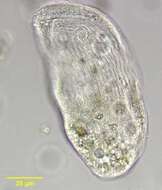
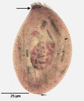
In vivo views of the swarmer (left) and the adult form, recently divided (right) of the suctorian, Parapodophrya soliformis (LAUTERBORN,1908) KAHL,1931. Both collected from sapropelic bottom sediments of the same stagnant freshwater pond near Boise, Idaho 43°40â 57.20â N 116° 15â 15.44â W . September, 2006.DIC.
-
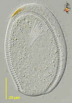
Ventral infraciliature of Gastronauta derouxi (FOISSNER & BLATTERER, 1992).Collected from an ephemeral puddle on a grass lawn in Boise, Idaho. July 2007. Stained by the silver carbonate technique (Foissner,W. Europ. J. Protistol.27:313-330;1991).Brightfield.
-
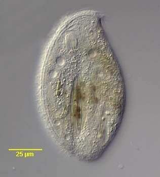
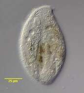
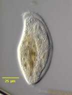
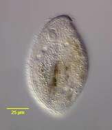
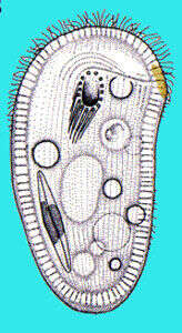
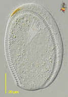
Ventral view of the chillodonellid ciliate, Pseudochilodonopsis polyvacuolata (Foissner and Didier, 1981). The cell is ovoid. The anterior end is drawn to the left as a bluntly pointed rostrum. The ventral surface is flat and the central dorsal surface is arched. There is a flattened narrow circumferential margin. Ciliature is restricted to the ventral surface except for a short dorsal brush. The 7 left somatic kineties are separated from 5 right somatic kineties by an unciliated postoral bare area. The lateral-most 5 left somatic kineties terminate at a right angle to short separate preoral kineties arranged in stair-step fashion from the cytostome to the tip of the rostrum. The medial two left somatic kineties are shorter. There are two short circumoral kineties. The cyrtos opens ventrally. The heteromerous macronucleus is approximately central with one adherent ovoid micronucleus. There are 7-10 contractile vacuoles each with a single ventral excretory pore. The similar species, P. fluviatilis is smaller and has only two contractile vacuoles.Collected from a freshwater stream with abundant pennate diatoms near Boise, Idaho;43° 34' 41.92" N 116° 08' 50.49" W. March 2006. DIC.
-
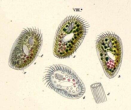
Chlamydodon (clam-ee-doe-don), alga-eating hypostome ciliate. This cell was photographed immediately after dividing. During the division process, the cells do not eat. Food ingested before division is initiated is digested, with the rest that the cytoplasm is very empty and the major organelles can be seen with some clarity: orange zone, ingestion apparatus, macronucleus and railway track. Differential interference contrast.
-
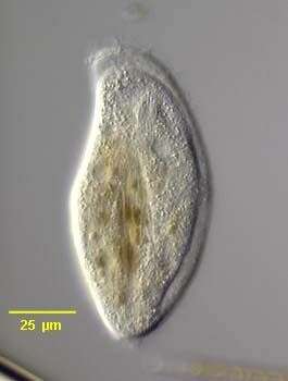
Ventral view of the chillodonellid ciliate, Pseudochilodonopsis polyvacuolata (Foissner and Didier, 1981). The cell is ovoid. The anterior end is drawn to the left as a bluntly pointed rostrum. The ventral surface is flat and the central dorsal surface is arched. There is a flattened narrow circumferential margin. Ciliature is restricted to the ventral surface except for a short dorsal brush. The 7 left somatic kineties are separated from 5 right somatic kineties by an unciliated postoral bare area. The lateral-most 5 left somatic kineties terminate at a right angle to short separate preoral kineties arranged in stair-step fashion from the cytostome to the tip of the rostrum. The medial two left somatic kineties are shorter. There are two short circumoral kineties. The cyrtos opens ventrally. The heteromerous macronucleus is approximately central with one adherent ovoid micronucleus. There are 7-10 contractile vacuoles each with a single ventral excretory pore. Collected from a freshwater stream with abundant pennate diatoms near Boise, Idaho;43° 34' 41.92" N 116° 08' 50.49" W. March 2006. DIC.
-
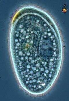
Chlamydodon (clam-ee-doe-don), alga-eating hypostome ciliate, so called because the mouth opens on the ventral surface of the cell. Common in marine habitats. The mouth is used to ingest filamentous algae - and a separate series of images illustrates this process. Phase contrast.
-
Ventral view of the chillodonellid ciliate, Pseudochilodonopsis polyvacuolata (Foissner and Didier, 1981). The cell is ovoid. The anterior end is drawn to the left as a bluntly pointed rostrum. The ventral surface is flat and the central dorsal surface is arched. There is a flattened narrow circumferential margin. Ciliature is restricted to the ventral surface except for a short dorsal brush. The 7 left somatic kineties are separated from 5 right somatic kineties by an unciliated postoral bare area. The lateral-most 5 left somatic kineties terminate at a right angle to short separate preoral kineties arranged in stair-step fashion from the cytostome to the tip of the rostrum. The medial two left somatic kineties are shorter. There are two short circumoral kineties. The cyrtos opens ventrally. The heteromerous macronucleus is approximately central with one adherent ovoid micronucleus. There are 7-10 contractile vacuoles each with a single ventral excretory pore. Collected from a freshwater stream with abundant pennate diatoms near Boise, Idaho;43° 34' 41.92" N 116° 08' 50.49" W. March 2006. DIC.
-
Chlamydodon (clam-ee-doe-don), alga-eating hypostome like many other hypostome ciliates, eats filamentous bacteria - such as filamentous blue green algae. They make contact with the filament, move along up and down until they find an end. They then tip over, pushing the end of the filament into the mouth - a cylindrical structure supported by a palisade of microtubular rods . They then start to suck the filament into the cell. As it hits the posterior margin, the cell is deformed by the stiff filament. The food is stunningly quickly degraded and begins to break and fold so that the cell can pull in a filament very much longer than itself. Yum. Phase contrast.
-

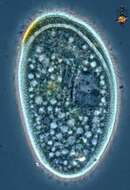
Ventral view of the chillodonellid ciliate, Pseudochilodonopsis polyvacuolata (Foissner and Didier, 1981). The cell is ovoid. The anterior end is drawn to the left as a bluntly pointed rostrum. The ventral surface is flat and the central dorsal surface is arched. There is a flattened narrow circumferential margin. Ciliature is restricted to the ventral surface except for a short dorsal brush. The 7 left somatic kineties are separated from 5 right somatic kineties by an unciliated postoral bare area. The lateral-most 5 left somatic kineties terminate at a right angle to short separate preoral kineties arranged in stair-step fashion from the cytostome to the tip of the rostrum. The medial two left somatic kineties are shorter. There are two short circumoral kineties. The cyrtos opens ventrally. The heteromerous macronucleus is approximately central with one adherent ovoid micronucleus. There are 7-10 contractile vacuoles each with a single ventral excretory pore (arrows). Collected from a freshwater stream with abundant pennate diatoms near Boise, Idaho;43° 34' 41.92" N 116° 08' 50.49" W. March 2006. DIC.
-
-

Ventral view of the chillodonellid ciliate, Pseudochilodonopsis polyvacuolata (Foissner and Didier, 1981). The cell is ovoid. The anterior end is drawn to the left as a bluntly pointed rostrum. The ventral surface is flat and the central dorsal surface is arched. There is a flattened narrow circumferential margin. Ciliature is restricted to the ventral surface except for a short dorsal brush. The 7 left somatic kineties are separated from 5 right somatic kineties by an unciliated postoral bare area. The lateral-most 5 left somatic kineties terminate at a right angle to short separate preoral kineties arranged in stair-step fashion from the cytostome to the tip of the rostrum. The medial two left somatic kineties are shorter. There are two short circumoral kineties. The cyrtos opens ventrally. The heteromerous macronucleus is approximately central with one adherent ovoid micronucleus. There are 7-10 contractile vacuoles each with a single ventral excretory pore. Collected from a freshwater stream with abundant pennate diatoms near Boise, Idaho;43° 34' 41.92" N 116° 08' 50.49" W. March 2006. Stained by the silver carbonate technique (see Foissner, W. Europ. J. Protistol., 27:313-330;1991).Brightfield.
-
Video showing how this ciiate collected from Cedar Swamp around Woods Hole moves around. Really cute guy.
-

Ventral view of the chillodonellid ciliate, Pseudochilodonopsis polyvacuolata (Foissner and Didier, 1981). The cell is ovoid. The anterior end is drawn to the left as a bluntly pointed rostrum. The ventral surface is flat and the central dorsal surface is arched. There is a flattened narrow circumferential margin. Ciliature is restricted to the ventral surface except for a short dorsal brush. The 7 left somatic kineties are separated from 5 right somatic kineties by an unciliated postoral bare area. The lateral-most 5 left somatic kineties terminate at a right angle to short separate preoral kineties arranged in stair-step fashion from the cytostome to the tip of the rostrum (arrows). The medial two left somatic kineties are shorter. There are two short circumoral kineties. The cyrtos opens ventrally. The heteromerous macronucleus is approximately central with one adherent ovoid micronucleus. There are 7-10 contractile vacuoles each with a single ventral excretory pore. Collected from a freshwater stream with abundant pennate diatoms near Boise, Idaho;43° 34' 41.92" N 116° 08' 50.49" W. March 2006. Stained by the silver carbonate technique (see Foissner, W. Europ. J. Protistol., 27:313-330;1991).Brightfield.
-
Originally described by Ehrenberg under the name Chlamidodon mnemosyne.
-
Dorsal view of the chillodonellid ciliate, Pseudochilodonopsis polyvacuolata (Foissner and Didier, 1981). The cell is ovoid. The anterior end is drawn to the left as a bluntly pointed rostrum. The ventral surface is flat and the central dorsal surface is arched. There is a flattened narrow circumferential margin. Ciliature is restricted to the ventral surface except for a short anterior dorsal brush. (arrow). The 7 left somatic kineties are separated from 5 right somatic kineties by an unciliated postoral bare area. The lateral-most 5 left somatic kineties terminate at a right angle to short separate preoral kineties arranged in stair-step fashion from the cytostome to the tip of the rostrum. The medial two left somatic kineties are shorter. There are two short circumoral kineties. The cyrtos opens ventrally. The heteromerous macronucleus is approximately central with one adherent ovoid micronucleus. There are 7-10 contractile vacuoles each with a single ventral excretory pore. Collected from a freshwater stream with abundant pennate diatoms near Boise, Idaho;43° 34' 41.92" N 116° 08' 50.49" W. March 2006. Stained by the silver carbonate technique (see Foissner, W. Europ. J. Protistol., 27:313-330;1991).Brightfield.
-
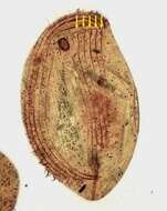
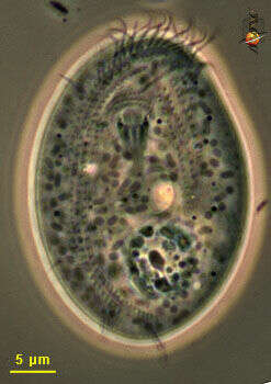
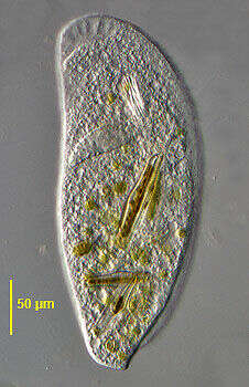
Trithigmostoma, a large hypostome ciliate. The cell is elongate, the right margin curving to meet the relatively straight left margin. The cell is flat ventrally with a slightly domed dorsum. There is a dorsal row of bristles which angles posteriorly toward the left margin (seen clearly here). Protrusible nemadesmata are seen surrounding the cytostome. Multiple small contractile vacuoles are scattered throughout the cytoplasm. From freshwater pond near Boise, Idaho. Brightfield illumination.
-
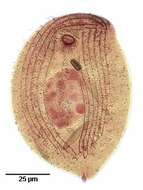
Dorsal view of the large Chlamydodontid ciliate, Trithigmostoma steini (Blochman,1895) Foissner, 1988. The colorless cell is ellipsoid in outline, broader anteriorly than posteriorly. The right side is convex and the left slightly concave. The right side curves anteriorly to meet the left as a definite beak. There is a dorsal hump extending from the level of the cytostome anteriorly and terminating as a lobular projection that extends beyond the posterior end of the ventral side. the ventral side is flat. The somatic ciliature is restricted to the ventral surface except for an oblique "dorsal brush" of cilia at the left side anteriorly. The somatic cilia cover the ventral surface unlike Chilodonella which has a bare postoral area. There are three preoral kineties the longest of which extends obliquely from the cytostome to the beak along the suture between the right and left kineties. The right kineties curve anterior to the cytostome. The left kineties are straight. There are 2 to 4 postoral kineties. The anterior cytostome is supported by very stout nematodesmata which are slightly protrusible. The ellipsoid macronucleus is central (seen here). There are 10-40 small contractile vacuoles (not well seen in this image). T. steini feeds primarily on algae and diatoms. T. steini differs from T. cucullulus, T. srameki and T. bavariensis are generally smaller, have fewer somatic kineties and lack a dorsal hump. Collected from a freshwater pond near Boise Idaho. Phase contrast.
-
Trithigmostoma, a large hypostome ciliate. The cell is elongate, the right margin curving to meet the relatively straight left margin. The cell is flat ventrally with a slightly domed dorsum. There is a dorsal row of bristles which angles posteriorly toward the left margin (seen clearly here). Protrusible nemadesmata are seen surrounding the cytostome. Multiple small contractile vacuoles are scattered throughout the cytoplasm. From freshwater pond near Boise, Idaho. Brightfield illumination.
-
Dorsal view of the large Chlamydodontid ciliate, Trithigmostoma steini (Blochman,1895) Foissner, 1988. The colorless cell is ellipsoid in outline, broader anteriorly than posteriorly. The right side is convex and the left slightly concave. The right side curves anteriorly to meet the left as a definite beak. There is a dorsal hump extending from the level of the cytostome anteriorly and terminating as a lobular projection that extends beyond the posterior end of the ventral side. the ventral side is flat. The somatic ciliature is restricted to the ventral surface except for an oblique "dorsal brush" of cilia at the left side anteriorly. The somatic cilia cover the ventral surface unlike Chilodonella which has a bare postoral area. There are three preoral kineties the longest of which extends obliquely from the cytostome to the beak along the suture between the right and left kineties. The right kineties curve anterior to the cytostome. The left kineties are straight. There are 2 to 4 postoral kineties. The anterior cytostome is supported by very stout nematodesmata which are slightly protrusible. The ellipsoid macronucleus is central (seen here). There are 10-40 small contractile vacuoles. T. steini feeds primarily on algae and diatoms. T. steini differs from T. cucullulus, T. srameki and T. bavariensis are generally smaller, have fewer somatic kineties and lack a dorsal hump. Collected from a freshwater pond near Boise Idaho. DIC.
-
Ventral face, kineties extend to the right and left of the mouth. The mouth is supported by strong microtubular nematodesmata. The granular structure near the rear is the macronucleus. The contractile vacuoles are light structure. the oblong grey organelles are probably mitochondria. Phase contrast microscopy.
-
Ventral view of the large Chlamydodontid ciliate, Trithigmostoma steini (Blochman,1895) Foissner, 1988. The colorless cell is ellipsoid in outline, broader anteriorly than posteriorly. The right side is convex and the left slightly concave. The right side curves anteriorly to meet the left as a definite beak. There is a dorsal hump extending from the level of the cytostome anteriorly and terminating as a lobular projection that extends beyond the posterior end of the ventral side. The ventral side is flat. The somatic ciliature is restricted to the ventral surface except for an oblique "dorsal brush" of cilia at the left side anteriorly. The somatic cilia cover the ventral surface unlike Chilodonella which has a bare postoral area. There are three preoral kineties the longest of which extends obliquely from the cytostome to the beak along the suture between the right and left kineties (seen well here). The right kineties curve anterior to the cytostome. The left kineties are straight. There are 2 to 4 evenly spaced postoral kineties. The anterior cytostome is supported by very stout nematodesmata which are slightly protrusible. The macronucleus is ellipsoid. There are 10-40 small contractile vacuoles (visible in this image). T. steini feeds primarily on algae and diatoms. T. cucullulus usually has <10 contractiloe vacuoles and <23 ventral kineties. T. srameki has ,10 contractile vaucoles and its postoral kineties are more widely spaced than the othe r ventral somatic kineties. Both lack a dorsal hump. Collected from a freshwater pond near Boise, Idaho. DIC.