Optical section
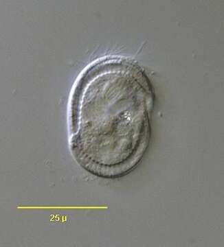
Description:
Optical section of the marine phyllopharyngiid ciliate, Coeloperix sleighi (Gong and Song,2004).The cell is broadly ovoid in outline and strongly dorsoventrally flattened. The dorsum is slightly convex and the ventral surface flattened. Ciliature is restricted to the ventral surface. The preoral and postoral kineties are separated by a transverse suture and the preoral kineties are transversely oriented. The postoral kineties are continuous , lacking the central bare gap seen in Chlamydodon. There is a peripheral cross-striated band (CSB) similar to that seen in Chlamydodon however the CSB in Coeloperix is interrupted on the right and left sides by two slightly offset gaps. There are 3or 4 short "tentacles" on the posteromedial ventral surface. These are about 5 ?m long and are quite difficult to see even with DIC. The anterior ventral cytostome is supported by prominent nematodesmata. There is a central heteromerous macronucleus. There are two contractile vacuoles situated diagonally. They empty through single pores on the ventral surface. Collected from a commercial marine aquarium in Boise, Idaho. pH 7.93. January 2004. DIC.
Included On The Following Pages:
- Life (creatures)
- Cellular (cellular organisms)
- Eukaryota (eukaryotes)
- SAR (Stramenopiles, Alveolates, Rhizaria)
- Alveolata (alveolates)
- Ciliophora (ciliates)
- Intramacronucleata
- Phyllopharyngea
- Phyllopharyngia
- Chlamydodontida
- Lynchellidae
- Coeloperix
- Coeloperix sleighi
This image is not featured in any collections.
Source Information
- license
- cc-by-nc
- author
- Bill Bourland
- provider
- micro*scope
- original
- original media file
- visit source
- partner site
- micro*scope
- ID