-
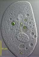
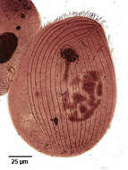
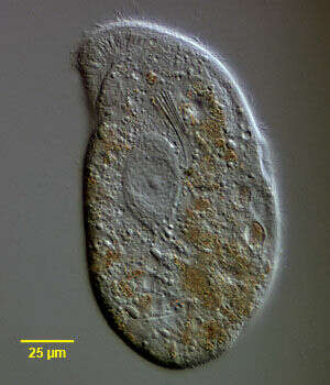
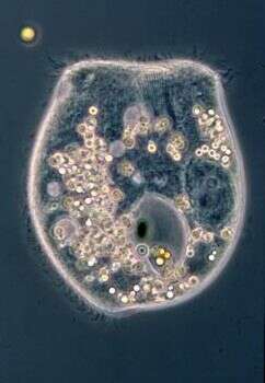
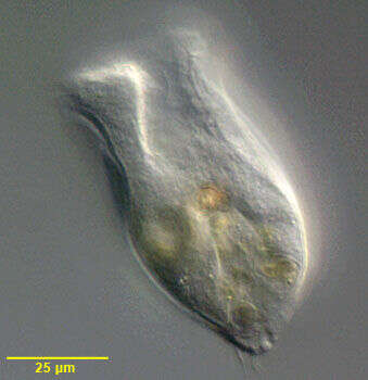
Browsing ciliate, consumes diatoms (frustules are visible inside the cell) using the mouth - upper right. The large central mass is the macronucleus. From coastal debris caught adjacent to the Tvarminne Zoological Station, 3rd April 2012.
-

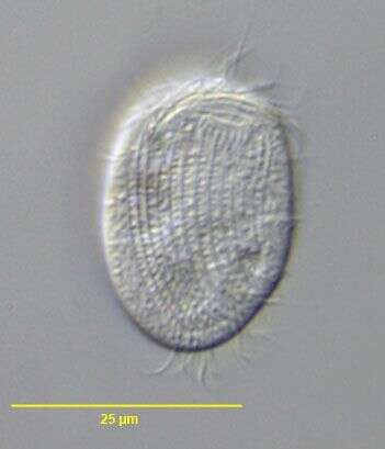
Ventral infraciliature of the large Chlamydodontid ciliate, Trithigmostoma steini (Blochman,1895) Foissner, 1988. The colorless cell is ellipsoid in outline, broader anteriorly than posteriorly. The right side is convex and the left slightly concave. The right side curves anteriorly to meet the left as a definite beak. There is a dorsal hump extending from the level of the cytostome anteriorly and terminating as a lobular projection that extends beyond the posterior end of the ventral side. The ventral side is flat. The somatic ciliature is restricted to the ventral surface except for an oblique "dorsal brush" of cilia at the left side anteriorly (not seen in this view). The somatic cilia cover the ventral surface unlike Chilodonella which has a bare postoral area. There are three preoral kineties the longest of which extends obliquely from the cytostome to the beak along the suture between the right and left kineties (seen well here). The right kineties curve anterior to the cytostome. The left kineties are straight. There are 2 to 4 evenly spaced postoral kineties. The anterior cytostome is supported by very stout nematodesmata which are slightly protrusible. The macronucleus is ellipsoid. There are 10-40 small contractile vacuoles. T. steini feeds primarily on algae and diatoms. T. cucullulus usually has <10 contractiloe vacuoles and <23 ventral kineties. T. srameki has ,10 contractile vaucoles and its postoral kineties are more widely spaced than the othe r ventral somatic kineties. Both lack a dorsal hump. Collected from a freshwater pond near Boise, Idaho. Stained by the silver carbonate technic (see Foissner, W. Europ. J. Protistol., 27:313-330;1991). Brightfield.
-
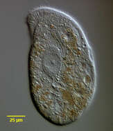
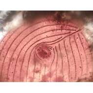
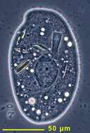
Trithigmostoma, browsing ciliate that will ingest attached bacteria and algae. Image emphasizing mouth with rods and apical teeth, but also showing the frustules of diatoms that have been ingested. Collected with debris from the shore adjacent to Tvarminne Zoological Station, 3rd April 2012.
-
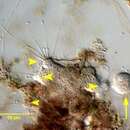
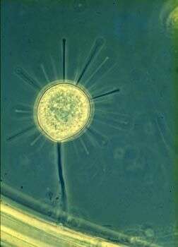
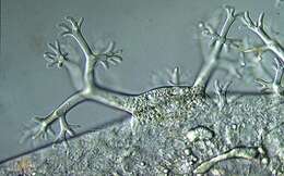
This unusual suctorian lives on the gill plates of the freshwater amphipod, Gammarus pulex.
-
Trithigmostoma, browsing ciliate that will ingest attached bacteria and algae. Image emphasizing mouth with rods and apical teeth, but also showing the frustules of diatoms that have been ingested. Collected with debris from the shore adjacent to Tvarminne Zoological Station, 3rd April 2012.
-
Trophic suctorian cell, each tentacle or arm is a mouth, the expanded termini contain extrusomes that will grab hold of prey - mostly other ciliates.
-
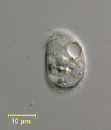
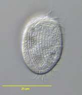
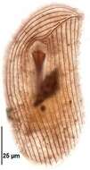
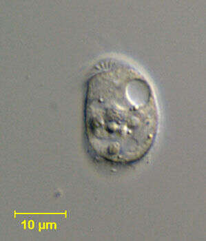
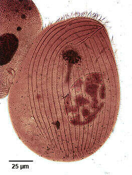
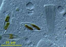
Chamydonella alpestris (Foissner,1979), a small hypostome ciliate. The body is strongly curved dorsally and flattened ventrally as shown in this lateral view. The single round macronucleus is located in the mid-body. There are two contractile vacuoles, one anterior and one posterior. This individual has been consuming diatoms. From freshwater pond near Boise, Idaho. Brightfield illumination.
-
Chamydonella alpestris ( Foissner, 1979), a small hypostome ciliate. The body is strongly curved dorsally and flattened ventrally. Ciliature is limited to the ventral surface except for a small dorsal anterior tuft on the left (seen in this image). Kineties curve anterior to the cytostome on the right. More central kineties terminate at the cytostome. A flattened transverse Y-shaped kinety just anterior to the cytostome is considered distinctive. The circular oral aperture is supported by trichites. The single round macronucleus is located in the mid-body. There are two contractile vacuoles, one anterior and one posterior. From freshwater pond near Boise, Idaho. DIC.
-
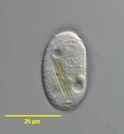
Ventral surface of Chlamydonella alpestris (Foissner, 1979), a small hypostome ciliate. The body is strongly curved dorsally and flattened ventrally. The right side is convex and the left is straight. Ciliature is limited to the ventral surface except for a small dorsal anterior tuft on the left. Kineties curve anterior to the cytostome on the right. About 10 evenly spaced longitudinal kineties extend from the level of the cytostome to the posterior end. A flattened transverse Y-shaped kinety just anterior to the cytostome is considered distinctive. A second short curved kinety lies just anterior to this. The circular oral aperture is supported by trichites. The single round macronucleus is located in the mid-body. There are two contractile vacuoles, one anterior and one posterior. This individual has consumed a diatom and green alga. Collected from freshwater pond near Boise, Idaho October 2003. DIC optics.
-
Ventral surface of Chlamydonella alpestris (Foissner, 1979), a small hypostome ciliate. The body is strongly curved dorsally and flattened ventrally. The right side is convex and the left is straight. Ciliature is limited to the ventral surface except for a small dorsal anterior tuft on the left. Kineties curve anterior to the cytostome on the right. About 10 evenly spaced longitudinal kineties extend from the level of the cytostome to the posterior end. A flattened transverse Y-shaped kinety just anterior to the cytostome is considered distinctive. A second short curved kinety lies just anterior to this. The circular oral aperture is supported by trichites. The single round macronucleus is located in the mid-body. There are two contractile vacuoles, one anterior and one posterior(both seen here). This individual has consumed a diatom and green alga. Collected from freshwater pond near Boise, Idaho October 2003. DIC optics.
-

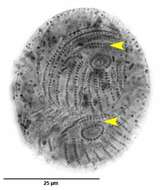
Ventral surface of Chlamydonella alpestris (Foissner, 1979) in mid-division. C. alpestris is a small hypostome ciliate. The body is strongly curved dorsally and flattened ventrally. The right side is convex and the left is straight. The cell shape has been distorted by fixation. Ciliature is limited to the ventral surface except for a small dorsal anterior tuft on the left. Kineties curve anterior to the cytostome on the right. About 10 evenly spaced longitudinal kineties extend from the level of the cytostome to the posterior end. A flattened transverse Y-shaped kinety just anterior to the cytostome is considered distinctive (yellow arrowheads). A second short curved kinety lies just anterior to this. The circular oral aperture is supported by trichites. The single round macronucleus is located in the mid-body (not visible here). There are two contractile vacuoles, one anterior and one posterior. This individual has consumed a diatom and green alga. Collected from freshwater pond near Boise, Idaho October 2003. Silver carbonate stain (see Foissner, W.Europ. J. Protistol.27,313-330;1991). Black and white.Brightfield optics.
-
Ventral surface of Chlamydonella alpestris (Foissner, 1979), a small hypostome ciliate. The body is strongly curved dorsally and flattened ventrally. The right side is convex and the left is straight. Ciliature is limited to the ventral surface except for a small dorsal anterior tuft on the left. Kineties curve anterior to the cytostome on the right. About 10 evenly spaced longitudinal kineties extend from the level of the cytostome to the posterior end. A flattened transverse Y-shaped kinety just anterior to the cytostome is considered distinctive. A second short curved kinety lies just anterior to this. The circular oral aperture is supported by trichites. The single round macronucleus is located in the mid-body. There are two contractile vacuoles, one anterior and one posterior(both seen here). This individual has consumed a diatom and green alga. Collected from freshwater stream near Boise, Idaho March 2007. DIC optics.
-
Ventral view of the chillodonellid ciliate, Trithigmostoma cucullus (Mueller, 1786) Jankowski, 1967.
-
Dorsal view of the chillodonellid ciliate, Trithigmostoma cucullulus (Mueller, 1786) Jankowski, 1967.
-
Ventral infraciliature of the chillodonellid ciliate, Trithigmostoma cucullulus (Mueller, 1786) Jankowski, 1967.
-
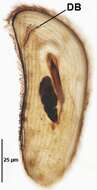
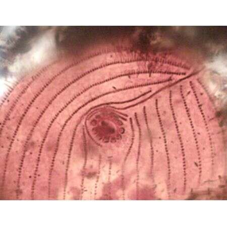
Dorsal brush (DB) of the chillodonellid ciliate, Trithigmostoma cucullulus (Mueller, 1786) Jankowski, 1967. Collected from a freshwater irrigation canal in Boise, Idaho.October 2007.Stained by the protargol A protocol (see Foissner, W. Europ. J. Protistol., 27:313-330;1991).Brightfield.
-
Detail of the ventral infraciliature of the chillodonellid ciliate, Trithigmostoma cucullulus (Mueller, 1786) Jankowski, 1967. Stained by the silver carbonate technique (Foissner,W. Europ. J. Protistol.27:313-330;1991).Brightfield.
-
Ventral infraciliature of the chillodonellid ciliate, Trithigmostoma cucullulus (Mueller, 1786) Jankowski, 1967.Collected from a freshwater irrigation canal in Boise,Idaho,October 2007. Stained by the protargol A protocol (see Foissner, W. Europ. J. Protistol., 27:313-330;1991).Brightfield.
-
Originally described by Ehrenberg under the name Chilodon cucullulus.
-
Originally described by Ehrenberg under the name Chilodon cucullulus.
-
-
-
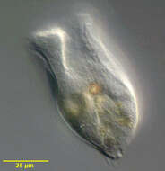
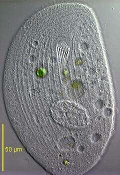
The anterior is to the tiop of the image, an ingestion apparatus lies near the middle of the anterior margin. Phase contrast micrograph.
-
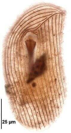
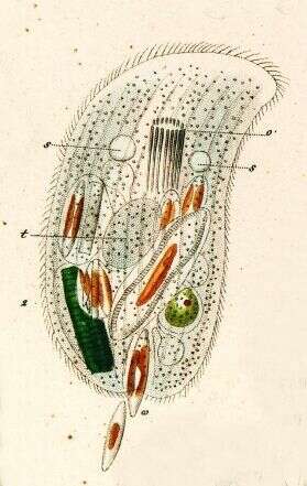
Ventral view of the Phyllopharyngeid ciliate, Phascolodon vorticella (Stein, 1859). The genus is probably monotypic. P. vorticella is easily recognized by its distinctive horse-saddle shape. The cell is dorsoventrally compressed with a deep ventral furrow (seen in this image) that bears 5-6 kineties on the right and 8-10 kineties on the left. There is one transverse file of cilia on the dorsal surface (i.e. the dorsal brush). The right kineties curve around the flared anterior end. The posterior tapers to a blunt point. At the anterior end of the ventral furrow is a slit-like cytostome with a prominent dorsally directed cytopharyngeal basket of nematodesmata. The coarsely granular macronucleus is located in the posterior ½. There are two contractile vacuoles, one on the right anterior side of the ventral furrow and the other on the left posterior side of it. P. vorticella is planktonic and feeds mainly on algae and cyanobacteria. Collected from freshwater pond near Boise, Idaho December 2004. DIC optics.