-
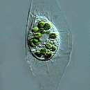
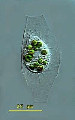
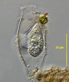
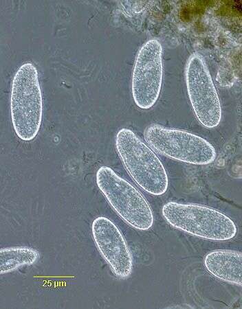
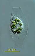
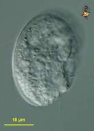
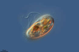
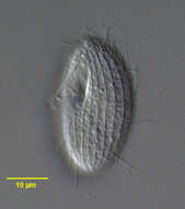
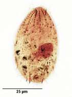
Calyptotricha (kah-lip-toe-trike-a) pleuronemoides is an ovoid to pyriform ciliate. The ciliate forms a transparent lorica. The lorica is tube-like and has apertures at both ends. The middle the tube can have parallel sides or a central bulbous region in which the ciliate is housed. The undulating membrane of the oral aperture stretches down the right side of the body to form a pouch in the posterior body half. Extrusomes are present. There is a conspicuous caudal cilium. Contractile vacuole in posterior body region. The macronucleus is spherical with attached micronuclei. Several endosymbiotic algae are visible and the conspicuous caudal cilium. Ciliate measuring 28 microns, lorica 64 microns. This specimen was collected in freshwater ponds near Konstanz, Germany. Differencial interference contrast.
-
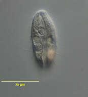
Portrait (ventral surface) of the pleuronematine scuticociliate, Cristigera phoenix (Penard, 1922). Collected from a freshwater aquaculture pond near Boise, Idaho November 2004. DIC.
-
-
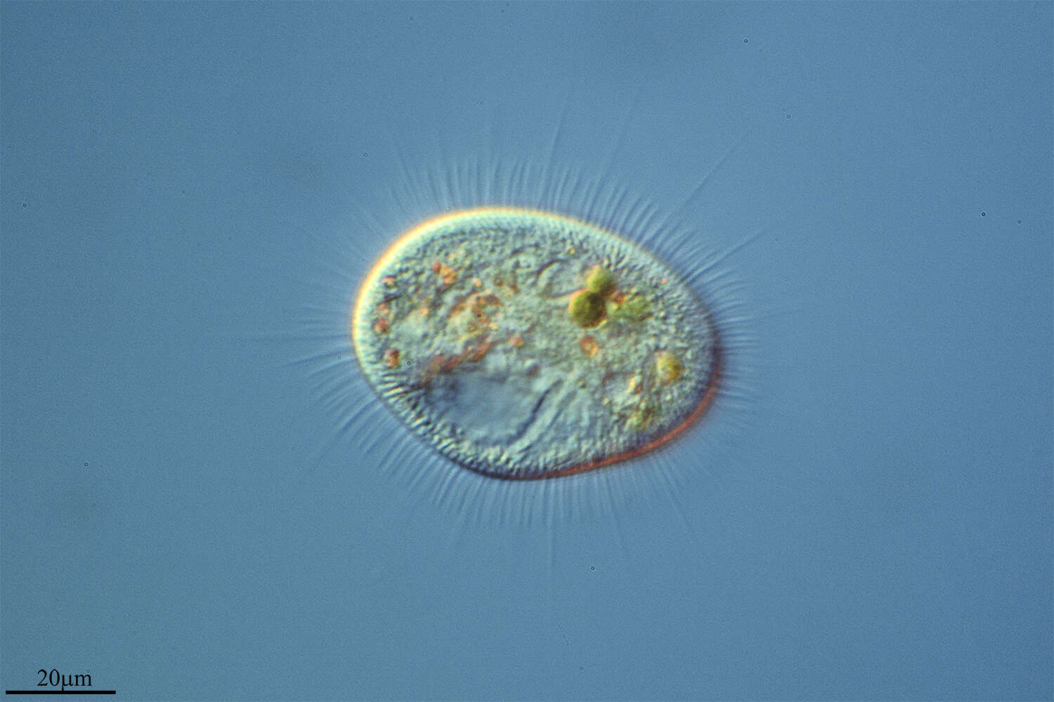
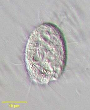
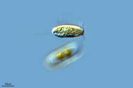
Cinetochilum (sigh-neat-owe-kai-lum) is a small bacterivorous ciliate, common and widely distributed. Differential interference contrast. Material from Nymph Creek and Nymph Lake, thermal sites within Yellowstone National Park, photograph by Kathy Sheehan and David Patterson.
-
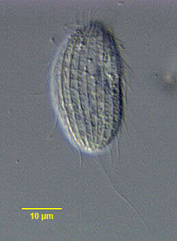
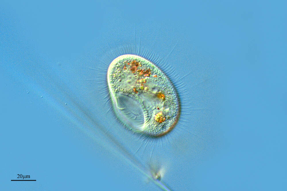
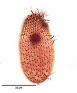
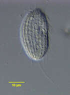
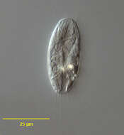
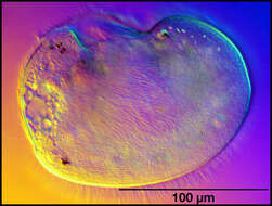
Portrait (dorsal surface) of the cinetochilid scuticociliate ciliate, Sathrophilus muscorum (Kahl, 1931) Corliss, 1960. The ellipsoid cell is dorsoventrally flattened. The left side is slightly convex and the right side more straightened. The approximately triangular cytostome is at the junction of the first and middle 1/3. There are three obliquely oriented adoral membranelles. M1 is longest, M2 intermediate in length and M3 quite short. There is a short, slightly curved paraoral membrane bordering the right margin of the peristome. The oral apparatus is quite similar to that of Cinetochilum margaritaceum. The central spherical macronucleus is usually single but may be in as many as four parts. There are 12-17 longitudinal somatic kineties that run between prominent pellicular ridges. There is a single long caudal cilium that inserts on the dorsal surface (seen here). There is a short preoral and longer postoral suture. The slit-like cytoproct is in the postoral suture. There are inconspicuous wedge shaped peripheral extrusomes. Its ellipsoid shape and pellicular characteristics differentiate S. muscorum from the similar Cinetochilum and Platynematum both of which have more truncate notched posterior margins. Collected from sapropelic bottom sediments of a freshwater aquaculture tub near Boise, Idaho. DIC.
-
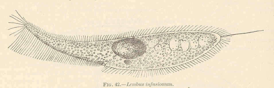
Lembus infusionum.
-
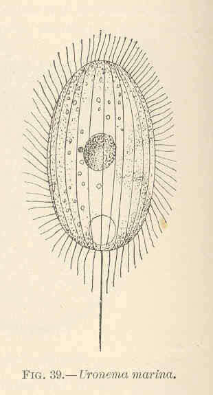
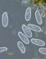
Uronema marina.
-
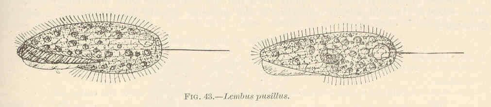
Lembus pusillus.
-
The species now known as Peniculistoma mytili was first described as Conchopthirus mytili by William De Morgan in 1925 in an article in the Journal of the Marine Biological Association (vol 13, 600-660).
-
Canencia, Madrid, Spain
-
Peniscola, Valencia, Spain
-
Logroo, La Rioja, Espaa
-
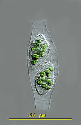
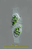
Calyptotricha (kah-lip-toe-trike-a) pleuronemoides is an ovoid to pyriform ciliate. The ciliate forms a transparent lorica. The lorica is tube-like and has apertures at both ends. The middle the tube can have parallel sides or a central bulbous region in which the ciliate is housed. The undulating membrane of the oral aperture stretches down the right side of the body to form a pouch in the posterior body half. Extrusomes are present. There is a conspicuous caudal cilium. Contractile vacuole in posterior body region. The macronucleus is spherical with attached micronuclei. This image taken shortly after cell division when there are two specimens in the lorica. Lorica measuring 68 microns. This specimen was collected in freshwater ponds near Konstanz, Germany. Differential interference contrast.
-
Portrait (ventral surface) of the pleuronematine scuticociliate, Cristigera phoenix (Penard, 1922). Collected from a freshwater aquaculture pond near Boise, Idaho November 2004. DIC.
-
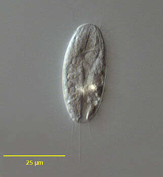
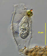
Dexiotricha tranquilla (Kahl,1926). DIC.
-
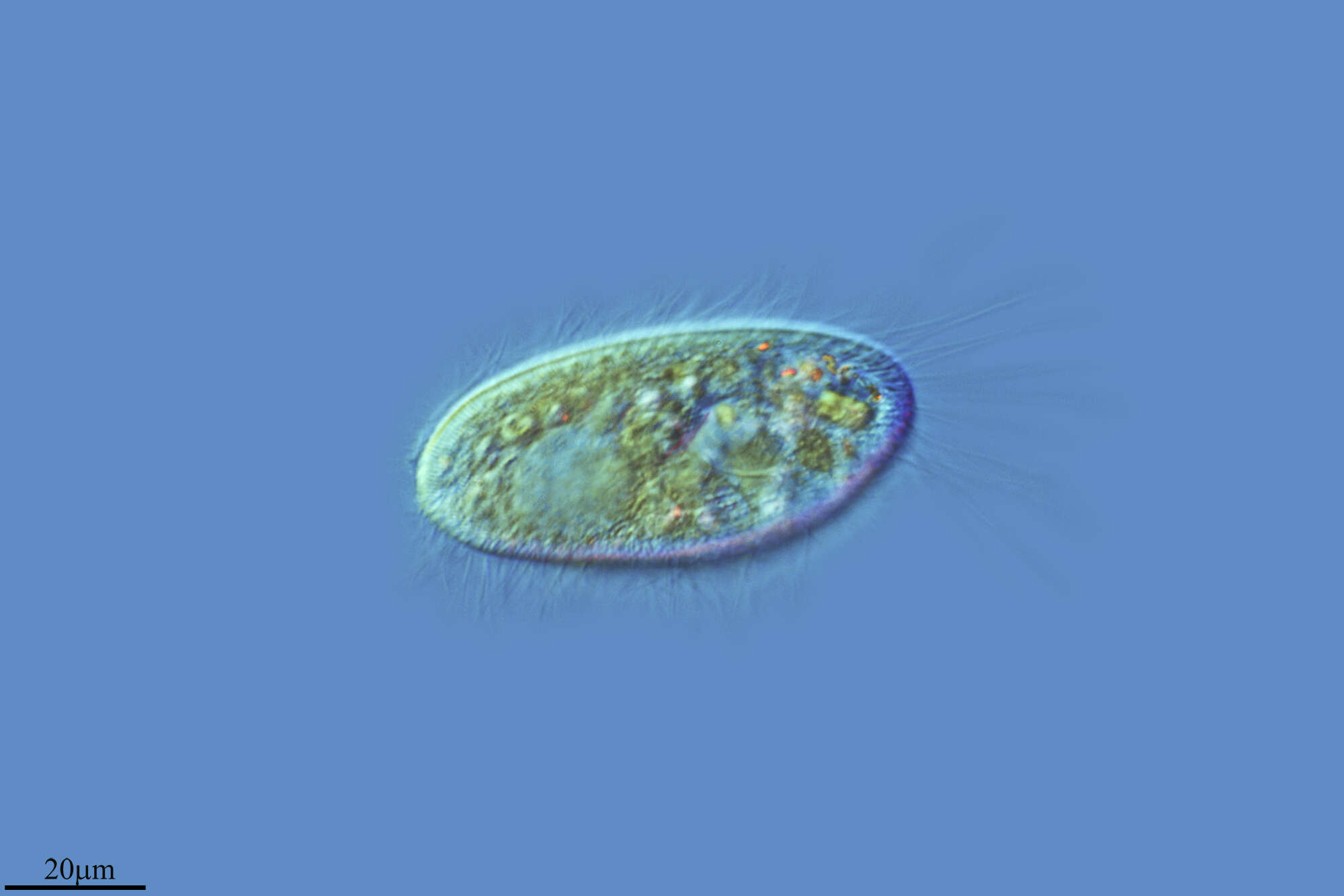
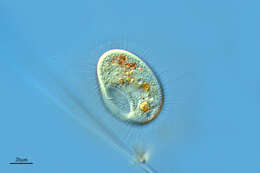
Cinetochilum margaritaceum (Ehrenberg, 1831) Perty, 1849, small rounded dorsoventrally flattened hymenostome ciliate. Cilia are located in shallow ventral furrows. Prominent oral aperture with small membranelles is seen on the organism's right posteriorly. Usually with long caudal cilia. Nucleus is central. Common. From freshwater pond near Boise, Idaho. Oblique illumination
-
Portrait (ventral surface) of the cinetochilid scuticociliate ciliate, Sathrophilus muscorum (Kahl, 1931) Corliss, 1960. The ellipsoid cell is dorsoventrally flattened. The left side is slightly convex and the right side more straightened. The approximately triangular cytostome is at the junction of the first and middle 1/3. There are three obliquely oriented adoral membranelles. M1 is longest, M2 intermediate in length and M3 quite short. There is a short, slightly curved paraoral membrane bordering the right margin of the peristome. The oral apparatus is quite similar to that of Cinetochilum margaritaceum. The central spherical macronucleus is usually single but may be in as many as four parts. There are 12-17 longitudinal somatic kineties that run between prominent pellicular ridges. There is a single long caudal cilium that inserts on the dorsal surface. There is a short preoral and longer postoral suture. The slit-like cytoproct is in the postoral suture. There are inconspicuous wedge shaped peripheral extrusomes. Its ellipsoid shape and pellicular characteristics differentiate S. muscorum from the similar Cinetochilum and Platynematum both of which have more truncate notched posterior margins. Collected from sapropelic bottom sediments of a freshwater aquaculture tub near Boise, Idaho. DIC.
-
Image of Peniculistoma mytili (endocommensal of the blue mussel Mytilis edulis) using DIC optics. Image by G. A. Antipa.
-
Galende, Castille and Leon, Spain
-
Talveila, Castille and Leon, Spain
-
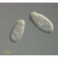
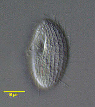
Portrait of the loricate pleuronematid ciliate, Calyptotricha pleuronemoides (Phillips, 1882). The transparent lorica of this species is open at both ends and dilated in the center where the cell resides. The cell is bluntly pointed anteriorly and broadly rounded posteriorly. The peristome is about 3/4 cell length. There is a prominent undulating membrane on the right margin of the peristome curving around its posterior end to form a shallow pouch (seen well here). There are three inconspicuous adoral membranelles. The longitudinal somatic kineties are uniformly distributed. There is a preoral and postoral suture. There is a single long caudal cilium. There is a single posterior contractile vacuole. The spherical macronucleus is centrally located. C. lanuginosa is similar in appearance of the cell except that it has two long anterior apical cilia and a cylindrical lorica with parallel sides. Collected from a freshwater dredge pond near Idaho City, Idaho June 2003. DIC.
-
Stained by the silver carbonate technique (see Foissner, W. Europ. J. Protistol., 27:313-330;1991).Brightfield.
-
Dexiotricha tranquilla (Kahl,1926). Phase contrast.
-
Dorsal view of the silverline system of the hymenostome ciliate, Cinetochilum margaritaceum (Ehrenberg, 1831) Pert, 1849.Stained by the dry silver nitrate technic (see Foissner, W. Europ. J. Protistol.27, 313-330; 1991). Collected from a freshwater pond near Boise, Idaho. Brightfield. Black and white.