Ventral surface in vivo
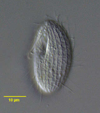
Description :
Portrait (ventral surface) of the cinetochilid scuticociliate ciliate, Sathrophilus muscorum (Kahl, 1931) Corliss, 1960. The ellipsoid cell is dorsoventrally flattened. The left side is slightly convex and the right side more straightened. The approximately triangular cytostome is at the junction of the first and middle 1/3. There are three obliquely oriented adoral membranelles. M1 is longest, M2 intermediate in length and M3 quite short. There is a short, slightly curved paraoral membrane bordering the right margin of the peristome. The oral apparatus is quite similar to that of Cinetochilum margaritaceum. The central spherical macronucleus is usually single but may be in as many as four parts. There are 12-17 longitudinal somatic kineties that run between prominent pellicular ridges. There is a single long caudal cilium that inserts on the dorsal surface. There is a short preoral and longer postoral suture. The slit-like cytoproct is in the postoral suture. There are inconspicuous wedge shaped peripheral extrusomes. Its ellipsoid shape and pellicular characteristics differentiate S. muscorum from the similar Cinetochilum and Platynematum both of which have more truncate notched posterior margins. Collected from sapropelic bottom sediments of a freshwater aquaculture tub near Boise, Idaho. DIC.
Inclus dans les pages suivantes :
- Life
- Cellular (Organismes cellulaires)
- Eukaryota (eucaryotes)
- SAR (Stramenopiles, Alveolates, Rhizaria)
- Alveolata
- Ciliophora
- Intramacronucleata
- Oligohymenophorea
- Scuticociliatia
- Philasterida
- Loxocephalidae
- Kariphilus
- Kariphilus muscorum
Cette image ne figure dans aucune collection.
Informations sur la provenance
- licence
- cc-by-nc
- auteur
- William Bourland
- fournisseur
- micro*scope
- original
- fichier de média d’origine
- visiter la source
- site partenaire
- micro*scope
- ID