nimet breadcrumb-navigoinnissa
Diatome of kieselwiere (klas Bacillariophyceae) is eensellige wiere wat gewoonlik 'n geelbruin tot groengeel kleur het. Die wier se kieselagtige selwand bestaan uit twee helftes wat op mekaar pas soos 'n deksel op 'n pildosie. As die selinhoud van die diatoom doodgaan, sink die diatoomskelet na die meer- of seebodem. Deur die eeue heen het daar op hierdie manier 'n baie dik laag diatoomgrond op die seebodem gevorm. Tans word meer as 10 000 soorte diatome onderskei, wat nie net in die see en in varswater voorkom nie, maar ook in vogtige plekke op land.
Die sowat 10 000 diatome kan van alle ander eensellige wiere onderskei word op grond van hul tweedelige selwand. Die boonste doppie van die selwand pas soos 'n deksel op die ietwat kleiner onderste doppie. Die twee helftes van die selwand word deur 'n bandvormige ring, die gordel, bymekaar gehou. Die harde, kieselagtige wand van die diatoom bevat geen kalkbestanddele soos dikwels met die harde dele van ander organismes die geval is nie, maar wel silikonverbindings, soos kieselsure en silikaat.
Daar word twee ordes diatome onderskei, naamlik die ronde Centrales en die ellips- of bootvormige Pennales. Die Centrales het dikwels 'n growwe oppervlak met fyn groefies. Daarbenewens het die meeste soorte Centrales lang hare of stekelhare, wat die dryfvermoë van die sel verbeter. Die Pennales het 'n gladde selwand met 'n veervormige patroon van porieë. In die sitoplasma van die sel is korrel- of plaatvormige kleurstofdraers (chromatofore) waarin daar chlorofiel en ook geel en bruin pigmente (karoteen, karotenoïed en xantofil) voorkom. Die chromatofore gee aan die diatoom 'n kenmerkende bruingeel tot groengeel kleur.
Diatome word oral op aarde aangetref. Die meeste soorte lewe in die see of in varswater, maar ook in 'n verskeidenheid van vogtige woonplekke op land, soos byvoorbeeld in boomstamme of onder klippe. Die meeste Centrales lewe in die see, waar hulle 'n belangrike bestanddeel van die plantaardige plankton vorm en 'n groot rol in suurstofproduksie speel. Daarenteen is die Pennales hoofsaaklik varswaterbewoners. Hulle kom gewoonlik voor in kettingvormige of vertakte kolonies wat aan die een of ander bodem vasgeheg is. Die selle word aan mekaar vasgeheg met behulp van 'n klewerige stof wat deur die sitoplasma afgeskei word.
Die diatome plant gewoonlik voort deur middel van seldeling. Met die seldeling erf albei dogterselle een van die doppies van die moedersel se tweedelige wand en bou dan self die ander helfte om daarby aan te pas. Die doppies wat van die moedersel verkry word, vorm altyd die deksel van die nuwe dogtersel. Die onderste deel word altyd self deur die dogtersel gevorm. Dit lei daartoe dat die dogtersel wat die grootste doppie (die moederseldeksel) erf, altyd so groot soos die moedersel word, terwyl die ander dogtersel baie kleiner is. Hierdie proses van seldeling duur 'n paar generasies voort, en met elke seldeling ontstaan 'n klein en 'n groot nakomeling. Later is die kleinste selle só klein dat hulle nie meer lewensvatbaar is nie. In sulke gevalle vorm die diatoom spore (ouksospore).
By die Centrales deel die selkern 'n paar keer; die protoplasma wring hom uit die sel na buite en neem normale afmetinge aan. Hierná word twee volledige nuwe doppies gevorm.
In die geval van die Pennales word geslagselle gevorm. Hierdie geslagselle verlaat die oorspronklike doppies en versmelt met mekaar. Die sigote hou aan met hulle groei totdat hulle volwassenheid bereik het en kort daarna deel hulle en vorm nuwe doppies. In ongunstige omstandighede vorm die selle russpore, wat weer sal ontwikkel as die sel se lewensomstandighede verbeter.
Die diatome speel nie net 'n belangrike rol in suurstofvoorsiening nie, maar ook in voedselverskaffing vir planktondiertjies, soos vislarwes en mikroklein krefies.
Wanneer die selinhoud van 'n diatoom doodgaan, of die ou doppies verlaat, sink die diatoomskelet na die bodem van die see of die meer. As gevolg hiervan het daar deur die eeue heen 'n dik laag diatoomgrond op die seebodem gevorm. Hierdie diatoomgrond, wat dikwels kieselguhr genoem word, is van groot belang vir die nywerheid. Nadat Alfred Nobel dinamiet uitgevind het, het hy diatoomgrond gebruik om die gevaarlike nitrogliserien te absorbeer. Dit het die waarskynlikheid van onbeheerde ontploffings baie verminder. Diatoomgrond is vroeër baie gebruik as 'n poleermiddel in tandepasta.
Diatome of kieselwiere (klas Bacillariophyceae) is eensellige wiere wat gewoonlik 'n geelbruin tot groengeel kleur het. Die wier se kieselagtige selwand bestaan uit twee helftes wat op mekaar pas soos 'n deksel op 'n pildosie. As die selinhoud van die diatoom doodgaan, sink die diatoomskelet na die meer- of seebodem. Deur die eeue heen het daar op hierdie manier 'n baie dik laag diatoomgrond op die seebodem gevorm. Tans word meer as 10 000 soorte diatome onderskei, wat nie net in die see en in varswater voorkom nie, maar ook in vogtige plekke op land.
Les diatomees (taxón Diatomea, Diatomeae o Bacillariophyceae sensu llato), ye un grupu d'algues unicelulares que constitúi un de los tipos más comunes de fitoplancton. Contién anguaño unes 20.000 especies vives que son importantes productores dientro de la cadena alimenticia.[3] Munches diatomees son unicelulares, anque dalgunes d'elles pueden esistir como colonies en forma de filamentos o cintes (y.g. Fragillaria), abanicos (y.g. Meridion), zigzags (y.g. Tabellaria) o colonies estrellaes (y.g. Asterionella). Una carauterística especial d'esti tipu d'algues ye que se topen arrodiaes por una paré celular única fecha de xil opalino (dióxidu de siliciu hidratao) llamada frústula. Estes frústules amuesen una amplia variedá na so forma, pero xeneralmente consisten en dos partes asimétriques con una división ente elles, carauterística que-y da nome al grupu. La evidencia fósil suxer que les diatomees aniciáronse mientres o dempués del periodu Xurásicu inferior, anque los primeros restos corpóreos son del Paleógeno. Les comunidaes de diatomees son una ferramienta usada recurrentemente pa la vixilancia de les condiciones medioambientales, de la calidá de l'agua y nel estudiu de los cambeos climáticos.
Les diatomees son organismos fotosintetizadores que formen parte del plancton (fitoplancton). Anguaño conócense más de 200 xéneros de diatomees, 20.000 especies vives y envalórase qu'hai alrodíu de 100.000 especies extintes.[4][5] Como colonizadores, les diatomees destremanse por atopase en cualesquier tipu d'ambiente yá seya marín, dulceacuícola, terrestre o tamién sobre superficies húmedes. Otres atópense n'ambientes onde esisten condiciones estremes de temperatura o salín y d'igual forma atopamosles interautuando con otros organismos como ye'l casu coles cianofícees filamentoses onde esiste un epifitismo per parte de les diatomees. La mayoría son peláxiques (viven n'agües llibres), dalgunes son bentóniques (sobre'l fondu marín), ya inclusive otres viven so condiciones de mugor atmosférico. Son especialmente importantes nos océanos, onde se calcula qu'apurren hasta un 45 % del total de la producción primaria oceánica.[6] La distribución espacial del fitoplancton marín ye acutada tantu horizontal como verticalmente. Les diatomees viven en tolos océanos dende los polos hasta los trópicos; les rexones polar y subpolar contienen relativamente poques especies en contraste cola biota templada.[7] Anque les rexones tropicales esiben la mayor cantidá d'especies, les mayores poblaciones de diatomees alcuentrense ente les rexones polar y templao.
Les diatomees pertenecen a un gran grupu llamáu Heterokontophyta, que inclúi especies tantu autótrofes (y.g algues pardes) como heterótrofes (y.g. oomicetos). Los cloroplastos mariellu-marrones de les diatomees son típicos de los heteroncontos, con cuatro membranes, clorofiles a, c1 y c2 y pigmentos tales como β-caroteno, fucoxantina, diatoxantina y diadinoxantina. Les reserves d'alimentu almacénense como carbohidratos o aceites, qu'amás de sirvir de reserva, contribúin a la so flotabilidá.
Los sos individuos usualmente escarecen de flaxelu, pero tán presentes en gametos y usualmente presenten una estructura heteroconta, sacante en qu'escarecen de vellosidaes (mastigonemas) carauterísticos d'otros grupos. Munches diatomees nun tienen movimientu, anque delles otres tienen movimientu flaxeláu. Por cuenta de la so relativamente pesada paré celular afondien con facilidá, les formes planctónices n'agües abiertes polo xeneral dependen de la turbulencia oceánica producida pol vientu nes capes cimeres pa caltenese suspendíes nes agües superficiales allumaes pol Sol. Delles especies regulen viviegamente la so flotabilidá colos lípidos intracelulares pa faer frente al fundimientu.
A pesar de ser xeneralmente microscópiques, delles especies de diatomees pueden algamar los 2 milímetros de llargor. Les diatomees tán conteníes dientro d'una única paré celular de silicatu (frústula) compuesta de dos valves separaes. El xil biogénica de la que la paré celular componse ye sintetizada intracelularmente por polimerización de monómeros d'ácidu silícico. Esti material ye depués secretado escontra l'esterior de la célula onde participa na conformanza de la paré celular. Les cáscares de les diatomees se superponen una a otra como los dos metaes d'una placa de Petri (esto ye, como una caxa y la so tapadoria). El frústulo apaez delicadamente afatáu con relieves que formen dibuxos variaos y perfeutamente simétricos. Dando llugar a dos tipos de diatomees, les que tienen simetría radial y les de simetría llateral. Los frústulos de les diatomees posense por gravedá cuando ye dixerida o muerre la célula, dando orixe a roques sedimentaries como les diatomites y moronites.
Les diatomees alternen ente la reproducción asexual por división celular, que ye la más frecuente, y la reproducción sexual.[8] Na reproducción por división celular, cada célula fía recibe una de les frústules de la célula padre y utilizala como frústula mayor (o epiteca), reconstruyendo una frústula menor (o hipoteca). De resultes d'esti procesu, la célula que recibió la frústula menor resulta nuna diatomea de menor tamañu que la orixinal. Per otra parte, la frústula nun pudi crecer. D'esta forma, na primer xeneración el 50% de les diatomees son de menor tamañu que la orixinal y vanse faciendo cada vez más pequeñes en cada xeneración. Reparóse sicasí, que delles especies pueden estremase ensin causar un amenorgamientu nel tamañu de célula.[9] El procesu sigue hasta que les célules algamen una tercer parte del so tamañu máximu.[10] Dempués d'un determináu númberu de xeneraciones, les diatomees reproducir de forma sexual, produciendo gametos ensin frústules que se funden formando una auxospora. Esti mecanismu ayuda a restablecer el tamañu orixinal de les diatomees porque'l cigotu crez muncho primero de producir una nueva frústula.
Les célules vexetatives de les diatomees son diploides (2N) y la meiosis xenera gametos machos y fema qu'entós se funden pa formar el cigotu. El cigotu lliberase de la so cubierta y crez na forma d'una célula esférica cubierta por una membrana orgánica (auxospora). Cuando la auxospora algama la so midida máxima (la de diatomea inicial), forma nel so interior a una diatomea que da empiezu a una nueva xeneración. Tamién pueden formase espores como respuesta a condiciones medioambientales desfavorables, produciéndose la guañada cuándo les condiciones ameyoren.[11]
Les diatomees son mayoritariamente non móviles, anque la espelma de delles especies puede ser flaxeláu, con movimientu de normal llindáu al deslizamiento.[12] Nes diatomees céntriques, los gametos machos son pequeños y tienen un flaxelu, ente que los gametos fema son grandes ya inmóviles (oogamia). Otra manera, nes diatomees pennades dambos gametos escarecen de flaxelos (isogamia).[10] Haise documentáu qu'una especie del grupu de les diatomees pennades ensin rafe ye anisógama y, por tanto, considerase una etapa transicional ente les diatomees céntriques y les diatomees pennades con rafe.[13]
Les diatomees, Diatomeae (Dumortier, 1821) o Bacillariophyceae (Haeckel, 1878), son clasificaes según la distribución de los sos poros y ornamentación. Si les frústules tienen una simetría radial o trímera nomense diatomees centraes, ente que si tienen una simetría billateral y forma allargada nomenanse pennadas. El primer tipu ye parafiléticu con respectu al segundu. Delles diatomees pennades tamién presenten una fisura a lo llargo de la exa llonxitudinal, denominada rafe, que ta implicada nos movimientos realizaos pola diatomea. Al grupu Bolidophyceae, apocayá descubiertu, considérase-y una clase estreme. La clasificación más recién considera cuatro grupos de diatomees:[14]
Diatomea
Coscinodiscophyceae (P): Centrales radiales.
Mediophyceae (P) : Centrales polares.
Fragilariophyceae (P): Pennales sin rafe.
Bacillariophyceae: Pennales con rafe.
La tierra diatomea ye un material constituyíu poles frústules de diatomees fosilizaes, aplicáu como fertilizante ya inseuticida en tierres pa cultivu, al ser un productu natural, ye inocuo y nun presenta riesgos pa la salú o contaminación. La tierra diatomea aprove micronutrientes al suelu que son de gran importancia pa la crecedera de les plantes, pudiendo amontar la fertilidá del suelu, actuando sinérgicamente con calciu y magnesiu, amás amenorga la lixiviación de fósforu, nitróxenu y potasiu y favorez la so absorción nes plantes. La tierra diatomea tamién actúa como reconstituyente en tierres contaminaes por metales pesaos o hidrocarburos, amás neutraliza la tosicidá del aluminiu en suelos acedos y amenorga l'absorción de fierro y manganesu.[15]
Les propiedaes d'esos materiales, formaos por partícules microscópiques, entrevesgaes y bien regulares en tamañu, facenlos curiosos pa diversos usos, como la fabricación de la dinamita, onde la nitroglicerina ye enfiñida, amenorgando la probabilidá d'una esplosión accidental.
El polvu de diatomees emplégase como inseuticida n'animales y plantes. Actúa deshidratando a inseutos hasta matalos, amás curtia y fura el exoesqueleto, mancándolos y esaniciándolos de forma progresiva y efeutiva. Les frústules de les diatomees son d'orixe natural, polo cual son inocues p'animales y plantes. A diferencia d'inseuticides tóxicos, el polvu de diatomees nun puede ingresar nos texíos animales por cuenta del so tamañu.[16]
Les diatomees pertenecen a les microalges oleaxinoses por cuenta de que presenten fracciones lipídices del 25 % (condiciones normales) al 45 % (condiciones de estrés), cultivables en fotobioreautores (FBR). La producción de biodiésel a partir de diatomees dase por mediu de transesterificación del aceite preveniente de les microalgas. La producción de biodiésel basase na producción y captación de biomasa de diatomees, que ye deshidratada y sometida a ultrasoníos por que llibere los sos componentes, darréu los lípidos son dixebraos de carbohidratos y proteínes. L'aceite llográu ye sometíu a transesterificación alcalina, aceda o enzimática pa producir glicerol y biodiésel.
Los aceites provenientes de diatomees son principalmente triglicéridos, que xeneren amiestos de ésteres d'arriendo al convertise en biocombustible. El cracking térmicu o pirólisis, ye un procesu alternu a la transesterificación que tresforma triglicéridos n'otros compuestos orgánicos simples.[17]
Determinóse que les diatomees tienen la capacidá de producir ácidos grasos poliinsaturados (AGPI) n'altes concentraciones, como por casu la producción de diatomees del xéneru Nitzschia d'ácidu eicosapentanoico (GUO). Nitzschia tien ventayes como la resistencia a temperatures d'hasta -6 °C y ambientes altamente salobres, amás el so aceite algama'l 50 % del pesu secu de la biomasa.[18]
Les comunidaes de diatomees pueden utilizase pa la determinación de les condiciones ambientales tantu de presente como del pasáu y del cambéu climáticu. Tamién pueden utilizase pa determinar la calidá de l'agua.
La demostración de la presencia de Diatomees dientro de los cuévanos cardiacos o nel migollu óseu de cadabres ye utilizada en melicina forense como fuerte niciu de muerte por sumersión (afogamientu).[19]
Skeltomema potamos
Cocconeis krammerii
Melosira sp.
Cyclotella meneghiniana
Triceratium dubium
Aulacodiscus
Amphoraovalis
Phaeodactylum tricornutum
Surirela sublinearis
Cymbella sp.
Licmophora sp.
Les diatomees (taxón Diatomea, Diatomeae o Bacillariophyceae sensu llato), ye un grupu d'algues unicelulares que constitúi un de los tipos más comunes de fitoplancton. Contién anguaño unes 20.000 especies vives que son importantes productores dientro de la cadena alimenticia. Munches diatomees son unicelulares, anque dalgunes d'elles pueden esistir como colonies en forma de filamentos o cintes (y.g. Fragillaria), abanicos (y.g. Meridion), zigzags (y.g. Tabellaria) o colonies estrellaes (y.g. Asterionella). Una carauterística especial d'esti tipu d'algues ye que se topen arrodiaes por una paré celular única fecha de xil opalino (dióxidu de siliciu hidratao) llamada frústula. Estes frústules amuesen una amplia variedá na so forma, pero xeneralmente consisten en dos partes asimétriques con una división ente elles, carauterística que-y da nome al grupu. La evidencia fósil suxer que les diatomees aniciáronse mientres o dempués del periodu Xurásicu inferior, anque los primeros restos corpóreos son del Paleógeno. Les comunidaes de diatomees son una ferramienta usada recurrentemente pa la vixilancia de les condiciones medioambientales, de la calidá de l'agua y nel estudiu de los cambeos climáticos.
Diatom yosunlar (lat. Bacillariophyta). Bu tipə tək, nadir hallarda qonur rəngli (piqmenti - fukoksantin) mikroskopik (0,75 mkm – 2 mm) kolonial orqanizmlər daxildir. Diatom yosunlar planktonda geniş, daha az miqdarda dəniz və şorsu hövzələrinin bentosunda, bəziləri torpaqda məskunlaşırlar; müasir diatom yosunlar qütb dairəsinə yaxın və əsasən arktik hövzələrdə yayılmışdır. Hüceyrə ikitaylı silisium qabıq içərisində yerləşir, qabıq iki elementdən ibarətdir; bunlardan kiçiyi qapaq kimi həmişə digər tayı örtür. Sonuncunun içərisində dairəvi lövhə (tayın özülü) və kəmərcik (yan tərəflər) ayrılır.
Lövhənin formasına görə diatom yosunlar tipi iki sinifə ayrılır: pennatlar və sentriklər. Pennat diatom yosunlar ikitərəfli simmetriyalı olub, formaları uzunsov oval (yumurtavari), iynəşəkilli olur, adətən, qabığın ortasında yarıqşəkilli dəlik (tikiş xətti) yerləşir. Sentrik diatom yosunlar radial simmetriya ilə səciyyələnir, formaca dairəvi, üçbucaq, ulduz şəkillidir. Bütün diatomeaların kəmərcik tərəfdən görünüşü çubuqşəkillidir (uzunsov-düzbucaqlı) ki, bu da tipin ikinci adını – Bacillariophyta (lat. bacillus - çubuq) təyin etmişdir. Qabığın taylarında çoxsaylı hamar və ya qabarıq nazik haşiyəli iri və xırda dəliklər vardır.
Diatom yosunlar 300-ə qədər cinsi və 1200-dən çox müasir və fossil növləri məlumdur. Cinsi və qeyri-cinsi yolla artırlar. Süxur əmələgətirən orqanizmlərdir; bitkilər məhv olduqdan sonra dibə çökərək (çox vaxt bir-birindən ayrılmış tayalar şəklində) silisium lillərini, qazıntı halında isə silisium süxurlarını (diatomitlər, trepellər, opokalar və b.) əmələ gətirirlər. Diatom yosunlar - sentriklər təbaşirdə (yura tapıntıları haqqında məlumatlar mübahisəlidir), pennatlar isə paleogendə meydana gəlmişdir; orta paleogenin şirinsu hövzələrində yaşamağa uyğunlaşmışdır, buna qədər isə təmamilə dənizlərdə məskunlaşmışlar. Cavan çöküntülərin (neogendən başlayaraq) zonal bölgülərinin əsaslandırılmasında diatom yosunlar böyük əhəmiyyəti vardır. Fatsiya göstəricisi (göl, çay və b.) kimi diatom yosunlar olduqca əhəmiyyətlidir. Təbaşir - müasir.
Xəzərdə 292 növü qeyd edilmişdir, onlardan planktonda 165 növ, bentosda isə 129 növü yaşayır. Chaetoceros, Thalassiosira,Coscinodiscus və Rhizosolenia cinsləri Xəzərin planktonunda və fitoplanktonunda mühüm rol oynayır.
Geologiya terminlərinin izahlı lüğəti. — Bakı: Nafta-Press, 2006. — Səhifələrin sayı: 679.
Diatom yosunlar (lat. Bacillariophyta). Bu tipə tək, nadir hallarda qonur rəngli (piqmenti - fukoksantin) mikroskopik (0,75 mkm – 2 mm) kolonial orqanizmlər daxildir. Diatom yosunlar planktonda geniş, daha az miqdarda dəniz və şorsu hövzələrinin bentosunda, bəziləri torpaqda məskunlaşırlar; müasir diatom yosunlar qütb dairəsinə yaxın və əsasən arktik hövzələrdə yayılmışdır. Hüceyrə ikitaylı silisium qabıq içərisində yerləşir, qabıq iki elementdən ibarətdir; bunlardan kiçiyi qapaq kimi həmişə digər tayı örtür. Sonuncunun içərisində dairəvi lövhə (tayın özülü) və kəmərcik (yan tərəflər) ayrılır.
Lövhənin formasına görə diatom yosunlar tipi iki sinifə ayrılır: pennatlar və sentriklər. Pennat diatom yosunlar ikitərəfli simmetriyalı olub, formaları uzunsov oval (yumurtavari), iynəşəkilli olur, adətən, qabığın ortasında yarıqşəkilli dəlik (tikiş xətti) yerləşir. Sentrik diatom yosunlar radial simmetriya ilə səciyyələnir, formaca dairəvi, üçbucaq, ulduz şəkillidir. Bütün diatomeaların kəmərcik tərəfdən görünüşü çubuqşəkillidir (uzunsov-düzbucaqlı) ki, bu da tipin ikinci adını – Bacillariophyta (lat. bacillus - çubuq) təyin etmişdir. Qabığın taylarında çoxsaylı hamar və ya qabarıq nazik haşiyəli iri və xırda dəliklər vardır.
Diatom yosunlar 300-ə qədər cinsi və 1200-dən çox müasir və fossil növləri məlumdur. Cinsi və qeyri-cinsi yolla artırlar. Süxur əmələgətirən orqanizmlərdir; bitkilər məhv olduqdan sonra dibə çökərək (çox vaxt bir-birindən ayrılmış tayalar şəklində) silisium lillərini, qazıntı halında isə silisium süxurlarını (diatomitlər, trepellər, opokalar və b.) əmələ gətirirlər. Diatom yosunlar - sentriklər təbaşirdə (yura tapıntıları haqqında məlumatlar mübahisəlidir), pennatlar isə paleogendə meydana gəlmişdir; orta paleogenin şirinsu hövzələrində yaşamağa uyğunlaşmışdır, buna qədər isə təmamilə dənizlərdə məskunlaşmışlar. Cavan çöküntülərin (neogendən başlayaraq) zonal bölgülərinin əsaslandırılmasında diatom yosunlar böyük əhəmiyyəti vardır. Fatsiya göstəricisi (göl, çay və b.) kimi diatom yosunlar olduqca əhəmiyyətlidir. Təbaşir - müasir.
Xəzərdə 292 növü qeyd edilmişdir, onlardan planktonda 165 növ, bentosda isə 129 növü yaşayır. Chaetoceros, Thalassiosira,Coscinodiscus və Rhizosolenia cinsləri Xəzərin planktonunda və fitoplanktonunda mühüm rol oynayır.
Les diatomees (Bacillariophyceae) són una classe d'algues unicel·lulars microscòpiques (encara que n'existeixen algunes que formen colònies), que s'enquadra dintre del fílum Heterokontophyta, superfílum Chromista, regne protoctist, domini Eukarya. El nom científic de la classe és Bacillariophyceae (o Diatomeae) i es relaciona filogenèticament amb la classe Chrysophyceae i d'altres del grup Chromista.
Les diatomees són organismes fotosintetitzadors que viuen en aigua dolça o marina i constitueixen una part molt important del fitoplàncton.
Un dels trets característics de les cèl·lules de diatomees és la presència d'una coberta de sílice (diòxid de silici hidratat), anomenat frústul. Els frústuls mostren una gran diversitat de formes, algunes de molt belles i ornamentades, i generalment consten de dues parts asimètriques o valves amb una divisió entre si, d'aquí el nom del grup. Moltes espècies apareixen formant encadenaments o altres agregats ordenats. L'evidència fòssil suggereix que es van originar durant o abans del període Juràssic primerenc.
Actualment, es coneixen més de 200 gèneres vivents de diatomees i s'estima que hi ha al voltant de 100.000 espècies extintes (%[1] %[2]). Com a colonitzadores, les diatomees es distingeixen per trobar-se en qualsevol tipus d'ambient, ja sigui marí o d'aigua dolça. També es troben en ambients on existeixen condicions extremes de temperatura o salinitat i, de la mateixa forma, les trobem interaccionant amb altres organismes com és el cas de cianofícies filamentoses, en què existeix un epifitisme per part de les diatomees.
La majoria són pelàgiques (viuen en aigües lliures), encara que algunes són bentòniques (sobre el fons marí), o fins i tot poden viure sobre superfícies humides. Són especialment importants en els oceans, on es calcula que proporcionen fins a un 45% del total de la producció primària oceànica (%[3]). Encara que són generalment microscòpiques, algunes espècies de diatomees poden arribar fins a 2 mil·límetres de longitud. Els frústuls de les diatomees se sedimenten per gravetat quan és digerida o mor la cèl·lula, i donen origen a roques sedimentàries, com les diatomites i moronites.
Les propietats d'aquests materials, formats per partícules microscòpiques, intricades i molt regulars en grandària, els han fets atractius per a diversos usos, com la fabricació de la dinamita, en què la nitroglicerina és embeguda, reduint la probabilitat d'una explosió accidental.
Els tàxons més rics en lípids es fan servir per desenvolupar el biocombustible d'alga.
Amphora ovalis
Biddulphia pulchella
Cocconeis krammerii
Cyclotella meneghiniana
Phaeodactylum tricornutum
Skeletonema potamos
Surirella sublinearis
Triceratium dubium
Diatomees. Microscopi òptic. 300X. Mètode de la gota penjant
.jpg)
Phaeodactylum tricornutum és una alga diatomea i és l'única espècie del gènere Phaeodactylum. Aquesta alga adopta diferents tipus morfològics, fet que la fa útil per a estudiar alteracions en la forma cel·lular, que poden ser estimulats per canvis en les condicions ambientals. A més a més, pot créixer en absència de silicona, fet que proporciona noves eines en el camp de la fabricació de nanoinstruments de silicona.
Les diatomees (Bacillariophyceae) són una classe d'algues unicel·lulars microscòpiques (encara que n'existeixen algunes que formen colònies), que s'enquadra dintre del fílum Heterokontophyta, superfílum Chromista, regne protoctist, domini Eukarya. El nom científic de la classe és Bacillariophyceae (o Diatomeae) i es relaciona filogenèticament amb la classe Chrysophyceae i d'altres del grup Chromista.
Les diatomees són organismes fotosintetitzadors que viuen en aigua dolça o marina i constitueixen una part molt important del fitoplàncton.
Un dels trets característics de les cèl·lules de diatomees és la presència d'una coberta de sílice (diòxid de silici hidratat), anomenat frústul. Els frústuls mostren una gran diversitat de formes, algunes de molt belles i ornamentades, i generalment consten de dues parts asimètriques o valves amb una divisió entre si, d'aquí el nom del grup. Moltes espècies apareixen formant encadenaments o altres agregats ordenats. L'evidència fòssil suggereix que es van originar durant o abans del període Juràssic primerenc.
Rozsivky (Diatomeae, syn Bacillariophyceae) jsou velkou skupinou jednobuněčných fotosyntetizujících organismů s dvojdílnou křemičitou schránkou, tradičně řazenou mezi hnědé řasy.
Rozsivky jsou jednobuněčné řasy s dvojdílnou křemitou schránkou. Skupinu tvoří 285 rodů obsahujících 10 až 12 tisíc druhů (Round et al. 1990). Jejich fotosyntetickými barvivy jsou chlorofyly a a c, fukoxanthin. Chloroplasty mají čtyři obalné membrány a tylakoidy jsou uspořádány v trojicích. Zásobními látkami jsou chrysolaminaran, olej a volutin.
Schránka rozsivek se nazývá frustula. Je tvořena polymerizovaným oxidem křemičitým, který je proti korozi chráněn vrstvou kyselého polysacharidu diatotepinu. Schránku si buňka vytváří aktivním vychytáváním kyseliny křemičité z prostředí rychlostí až 18 molekul za sekundu. Frustula se skládá ze dvou částí (jako krabice s víkem či Petriho miska) – epithéky a hypothéky, z nichž každá má svou plochu (valvu) a boční stěnu (pleuru či cingulum; viz obrázek). Během nepohlavního rozmnožování získá každá z dceřiných buněk jednu část mateřské frustuly a druhou, vždy tu menší, si dotvoří. V případě nedostatku křemíku v prostředí se nepohlavní rozmnožování zastavuje. Podle tvaru frustuly se rozsivky dělí na dvě hlavní skupiny: centrické, radiálně souměrné a penátní, dvoustranně souměrné.
Při nepohlavním rozmnožování probíhá rovina dělení rovnoběžně s rovinou valv. Dělení frustuly viz výše. Při zmenšení frustuly pod určitou mez dochází ke smrti buňky či k pohlavnímu rozmnožení.
Centrické rozsivky se pohlavně rozmnožují oogamicky. Z jedné buňky vznikne oogonium s oosférou z další pak antheridium, v němž vzniknou čtyři spermatozoidy s jedním bičíkem. Po splynutí spermatozoidu s oosférou vzniká auxospora, velká buňka s polysacharidickou stěnou, na které se časem začne usazovat stěna křemičitá.
Penátní rozsivky se pohlavně rozmnožují izogamicky nebo anizogamicky. Dvě rozsivky se k sobě přiblíží a vytvoří společný slizový obal. Následně v každé z buněk proběhne meióza a jedna ze vzniklých gamet améboidním pohybem pronikne do druhé frustuly, splyne s jednou z gamet v ní za vzniku auxospory (viz výše).
Rozsivky jsou velmi významnými primárními producenty. Podle některých pramenů připadá na mořské rozsivky 20–25 % roční celkové primární produkce Země. Podle některých pramenů se výrazně, spolu se sinicemi, podílely na vzniku kyslíkaté atmosféry na Zemi. Jsou dominantní skupinou mořského planktonu, zvláště v temperátních a chladných mořích. Významně jsou zastoupeny také ve sladkovodním planktonu a bentosu. Mohou žít také přisedle na pevných podkladech ve vodě, jako jsou kameny, dřevo, rostliny či živočichové. Některé rozsivky žijí i v půdě.
Nejstarší rozsivky pocházejí z období spodní křídy. Evolučně starší jsou rozsivky centrické. Záznamy o nejmladších penátních jsou z období před 70 mil. let. V rámci hnědých řas jsou monofyletickou skupinou.
Schránky odumřelých rozsivek tvoří horninu diatomit (křemelinu), který se těží (v ČR například u Borovan u Českých Budějovic) a využívá se jako filtrační či sorpční materiál. V některých oblastech se rozsivky podílely na vzniku ložisek ropy. Vzhledem ke specifickým nárokům některých rozsivek se tyto využívají jako indikační organismy pro určení kvality vody, a to i zpětně v archeologických vykopávkách.
Moderní taxonomové rozsivky oddělují zcela od rostlin a řadí je do samostatné, páté říše živých organismů, Chromista. Zdůvodňuje se to tím, že svými vlastnostmi leží na rozhraní mezi rostlinami a živočichy. V této říši tvoří část jedné ze dvou podříší (Chromobiota), do níž jsou zařazeny jako samostatná třída kmene (oddělení) Ochrophyta.
Třetí možné dělení je řadí do oddělení Heterokontophyta a spolu s ním do čtvrté říše, Protista.
Třída Bacillariophyceae se v současných systémech dělí obvykle na dvě velké podtřídy, a to Eunotiophycidae a Bacillariophycidae.
Rozsivky (Diatomeae, syn Bacillariophyceae) jsou velkou skupinou jednobuněčných fotosyntetizujících organismů s dvojdílnou křemičitou schránkou, tradičně řazenou mezi hnědé řasy.
Underrækken Diatomeae kiselalger – tidligere rækken Bacillariophyta – også kaldet diatoméer, hører til planteplanktonet og er levende organismer. De indeholder klorofyl, et grønt stof, der bruges i omdannelsen af solens lys til energi. Algerne har to skaller dannet af kisel, der sidder sammen som låget sidder på en madkasse. Skallerne har små huller, som algen optager næring og udskiller affaldsstoffer igennem. Nogle af disse organismer har små børster og pigge som de bruger til at holde sig svævende i vandet med.
Disse alger blev før i tiden kun inddelt i to grupper: de centriske, der hovedsagelig lever i de frie vandmasser, og de pennate, som især er bundlevende. Algerne er brune eller brungule, de pennate alger kan af og til farve havbunden helt gulbrun, hvis de forekommer i store mængder, f.eks. på de vanddækkede lavvandede, områder i Vadehavet.
Die Kieselalgen oder Diatomeen (Bacillariophyta) bilden ein Taxon von Photosynthese betreibenden Protisten (Protista) und werden in die Gruppe der Stramenopilen (Stramenopiles) eingeordnet.
Oft wird die Gruppe mit dem synonymen Namen Diatomea Dumortier bezeichnet, alternativ sind auch die synonymen Namen Fragilariophyceae, Diatomophyceae in Verwendung. Einige Autoren nennen die Kieselalgen Bacillariophyceae, sie ordnen sie also als Klasse in die, dann als Phylum aufgefassten photosynthetischen Vertreter der Stramenopiles (Ochrophyta) ein, diese Auffassung wird etwa in den Datenbanken DiatomBase[1] und WoRMS vertreten. Diese Verwendung ist allerdings missverständlich, da andere Taxonomen eine enger abgegrenzte Klasse Bacillariophyceae, als eine von drei Klassen innerhalb der Diatomeen aufführen. Bei Verwendung dieses Namens ist also die jeweilige Auffassung zu kontrollieren, da es sonst zu Missverständnissen kommt.
Man unterscheidet heute rund 6000 Arten. Es wird jedoch angenommen, dass insgesamt bis zu 100.000 Arten existieren.[2] Eine Arbeitsstelle für Diatomeen-Forschung (mit umfangreicher Sammlung und Online-Katalog), begründet von Friedrich Hustedt, befindet sich im Alfred-Wegener-Institut.[3]
Ihren deutschen Trivialnamen verdanken die Kieselalgen der Zellenhülle (Frustel), die überwiegend aus Siliziumdioxid (Summenformel: SiO2) besteht, dem Anhydrid der Kieselsäure (vereinfachte Summenformel: SiO2 · n H2O). Die Kieselsäure wird jedoch im deutschen Sprachraum oft fälschlich mit ihrem Anhydrid gleichgesetzt. Das Siliziumdioxid gewinnt der Organismus aus der Monokieselsäure Si(OH)4.
Die Frustel ist schachtelförmig und besteht aus zwei schalenförmigen Teilen unterschiedlicher Größe, von denen die eine („Epitheka“) mit ihrer Öffnung über die Öffnung der anderen („Hypotheka“) greift. Die Schalen sind in charakteristischen Mustern strukturiert. Aufgrund der Schalengeometrie werden zwei Typen von Kieselalgen unterschieden: Zentrische Kieselalgen (Centrales) haben zumeist runde, bisweilen auch dreieckige Schalen, während pennate Kieselalgen (Pennales) stab- oder schiffchenförmige, mitunter auch bogen- oder S-förmig gekrümmte Gehäuse ausbilden. Die Schalen werden mittels spezieller Peptide angelegt, die als Silaffine bezeichnet werden. Die Silaffine ermöglichen die Ausfällung des Siliziumdioxids in kleine globuläre Siliziumdioxidaggregate, den sogenannten „Nanospheren“. Diese haben einen Durchmesser von 30 bis 50 nm und bilden in ihrer Gesamtheit die eigentlichen Schalen aus.[4][5] Viele pennate Kieselalgen können auf einer festen Unterlage mit Hilfe einer Raphe kriechen. Die Geschwindigkeit beträgt bis zu 20 μm/s.
Kieselalgen sind einzellig und fast stets unbegeißelt. Nur bei einigen Arten besitzen die männlichen Gameten eine Geißel, eine nach vorn gerichtete Flimmergeißel. Die durch sekundäre Endosymbiose mit einer Rotalge entstandenen Plastiden[6] sind braun gefärbt, da das Xanthophyll Fucoxanthin die Farbe der Chlorophylle (Chlorophyll a und c) überdeckt. Als Reservestoff dient Chrysolaminarin.
In der Regel sind Kieselalgen mikroskopisch klein. Zum Beispiel erreicht die Achnanthes eine Länge von 40 Mikrometer. Einige Arten können jedoch bis zu 2 Millimeter lang werden.
Die Diatomeen sind diploid und vermehren sich hauptsächlich ungeschlechtlich durch Zellteilung. Die Tochterzellen erhalten jeweils einen Schalenteil und bilden den anderen Teil neu; hiervon leitet sich auch die Bezeichnung „Diatomee“ (altgriechisch διατέμνειν (diatemnein) = spalten) ab. Der neue Schalenteil ist stets die kleinere Hypotheka, so dass im Generationenverlauf die Zellgröße fast aller Nachkommen fortlaufend schwindet, nur die Tochterzelllinie der Ausgangs-Epitheka behält die ursprüngliche maximale Größe bei. Wird eine Minimalgröße unterschritten, stirbt das Individuum. Bevor eine Minimalgröße erreicht wird, können jedoch Sexualvorgänge stattfinden. Aus den Zellen bilden sich durch Meiose haploide Gameten. Bei zentrischen Kieselalgen wurde Oogamie nachgewiesen: Die Gameten werden frei, nach Verschmelzen eines weiblichen mit einem männlichen Gameten bildet sich aus der Zygote unter Größenwachstum eine Dauerform, eine sogenannte Auxospore. Bei pennaten Kieselalgen wurde Konjugation beobachtet: Zwei Partner legen sich aneinander und bilden eine gemeinsame Cytoplasmabrücke („Konjugationskanal“), in die jeweils ein haploider Kern und ein Chloroplast der beiden Partner einwandern. Aus der so gebildeten Zygote bildet sich eine Auxospore, in der die Kernverschmelzung (Karyogamie) stattfindet. Aus den Auxosporen der zentrischen und pennaten Kieselalgen wird jeweils eine größere neue Kieselalge mit einer neuen zweiteiligen Schale gebildet.
Kieselalgen kommen hauptsächlich im Meer und in Süßgewässern planktisch oder benthisch vor, oder sie sind auf Steinen oder Wasserpflanzen (Epiphyten) angesiedelt. Manche Arten brauchen reines und kaum verschmutztes Wasser und sind aus diesem Grunde auch Zeigerorganismen für unbelastete Gewässer. Andere Arten wiederum, die im engl. auch als agricultural guild bezeichnet werden, sind typisch für Gewässer, die durch landwirtschaftliche Einträge, bspw. durch Überdüngung, besonders belastet sind. Zu diesen werden u. a. Navicula radiosa, Melosira varians, Nitzschia palea, Diatoma vulgare oder Amphora perpusilla gezählt.[7] Auch terrestrische Arten finden sich unter den Diatomeen; diese besiedeln Böden, in tropischen Gebieten auch Blätter von Bäumen.
Die Kieselalgen wurden traditionell in die radiärsymmetrischen Centrales und die bilateralsymmetrischen Pennales gegliedert. Die Centrales sind jedoch paraphyletisch, eine stabile Systematik auf molekulargenetischer Grundlage hat sich noch nicht etabliert. Nach der taxonomischen Datenbank Diatombase[1] existiert derzeit keine allgemein akzeptierte Gliederung der Kieselalgen in Subtaxa.
Eine weit verbreitete, aber nicht von allen Taxonomen anerkannte Gliederung könnte so aussehen[8], die höhere Gliederung wurde in vergleichbarer Form in die weithin verwendete Klassifikation der Eukaryoten durch Sina Adl und Kollegen übernommen.[9]
Die Kieselalgen sind Hauptbestandteil des Meeresphytoplanktons und sind die Haupt-Primärproduzenten organischer Stoffe, bilden also einen wesentlichen Teil der Basis der Nahrungspyramide. Als Sauerstoff produzierende (oxygene) Phototrophe erzeugen sie auch einen großen Teil des Sauerstoffs in der Erdatmosphäre.
Aus der relativen Arten-Zusammensetzung der Kieselalgenpopulation eines Gewässers kann recht exakt dessen Trophiegrad abgeleitet werden (Diatomeenindex), sowie weitere Gewässerparameter wie pH-Wert, Salinität, Saprobie etc. Diese Verfahren können auch auf Sedimente oder auf Öllagerstätten angewandt werden und geben dann Aufschluss über die ehemals herrschenden Lebensbedingungen.
Zur Identifizierung der Arten wird eine Aufwuchsprobe mit Kieselalgen mit Schwefelsäure, Wasserstoffperoxid, Kaliumdichromat oder einem anderen Oxidationsmittel behandelt und so werden alle organischen Bestandteile der Probe aufgelöst. Es bleiben nur noch die reinen Siliziumdioxid-Schalen übrig. Diese werden in einem Einschlussmedium mit hohem optischem Brechungsindex (z. B. Naphrax™) eingebettet und lichtmikroskopisch mit dem Phasenkontrast-Verfahren bei ca. 1000-facher Vergrößerung identifiziert.
Sterben die Zellen, sinken sie auf den Grund des Gewässers ab, die organischen Bestandteile werden abgebaut und die Siliziumdioxid-Schalen bilden eine Ablagerung, die sogenannte Kieselgur (Diatomeenerde). Dieser Prozess ist, insbesondere im Marinen, erst unterhalb der CCD (Calcit-Kompensationstiefe) effizient genug, um große Vorkommen zu bilden. Die entstehende Kieselgur wird in Technik und Medizin angewendet. Diatomeenschalen finden unter anderem Verwendung als Filter, zur Herstellung von Dynamit, in Zahnpasta als Putzkörper, als giftfreies Insektenbekämpfungsmittel in Form von Ungezieferpuder, sowie als reflektierendes Material in der Farbe, die für Fahrbahnmarkierungen im Straßenbau verwendet wird. Außerdem finden Kieselalgen in der forensischen Medizin Verwendung (Diatomeennachweis). Ihre Aussagekraft für den Nachweis eines Ertrinkungstodes wird jedoch kontrovers diskutiert.[10][11]
Insbesondere im 19. Jahrhundert dienten Diatomeen zur Anfertigung ästhetischer mikroskopischer Präparate, deren gemeinsame Betrachtung in Salons ein beliebter Zeitvertreib in höheren gesellschaftlichen Kreisen war. Zur höchsten Perfektion in der handwerklich extrem schwierigen Anfertigung solcher Präparate hat es Johann Diedrich Möller aus Wedel bei Hamburg gebracht.
Marine Diatomeen können zu Vergiftungen bei Mensch und Tier führen, da einige Arten, insbesondere Pseudo-nitzschia, Nitzschia oder Amphora Domoinsäure produzieren.[12] In filtirierende Meerwasser-Organismen, wie z. B. Muscheln, oder in sich von diesen Tieren ernährende Arten, wie z. B. Fischen, können sich diese Diatomeen akkumulieren. Kommt es zum Verzehr solcher mit Domoinsäure angereicherten Organismen durch den Menschen, treten Vergiftungserscheinungen auf, die als Amnesic Shellfish Poisoning (ASP) bezeichnet werden. Als Symptome treten insbesondere Gedächtnisverlust, Übelkeit, Krämpfe, Durchfall, Kopfschmerz und Atembeschwerden auf.[13]
Die Kieselalgen oder Diatomeen (Bacillariophyta) bilden ein Taxon von Photosynthese betreibenden Protisten (Protista) und werden in die Gruppe der Stramenopilen (Stramenopiles) eingeordnet.
Oft wird die Gruppe mit dem synonymen Namen Diatomea Dumortier bezeichnet, alternativ sind auch die synonymen Namen Fragilariophyceae, Diatomophyceae in Verwendung. Einige Autoren nennen die Kieselalgen Bacillariophyceae, sie ordnen sie also als Klasse in die, dann als Phylum aufgefassten photosynthetischen Vertreter der Stramenopiles (Ochrophyta) ein, diese Auffassung wird etwa in den Datenbanken DiatomBase und WoRMS vertreten. Diese Verwendung ist allerdings missverständlich, da andere Taxonomen eine enger abgegrenzte Klasse Bacillariophyceae, als eine von drei Klassen innerhalb der Diatomeen aufführen. Bei Verwendung dieses Namens ist also die jeweilige Auffassung zu kontrollieren, da es sonst zu Missverständnissen kommt.
Man unterscheidet heute rund 6000 Arten. Es wird jedoch angenommen, dass insgesamt bis zu 100.000 Arten existieren. Eine Arbeitsstelle für Diatomeen-Forschung (mit umfangreicher Sammlung und Online-Katalog), begründet von Friedrich Hustedt, befindet sich im Alfred-Wegener-Institut.
Ang mga diatomea (di-tom-os 'gupitin sa kalahati', mula sa diá, 'sa pamamagitan ng' o 'hiwalay' at ang ugat ng tém-n-ō, 'Pinutol ko') ay isang pangunahing pangkat ng algae, partikular na microalgae, na matatagpuan sa mga karagatan, mga daluyan ng tubig at mga soils ng mundo. Ang mga numero ng buhay na diatoms sa trillions: bumubuo sila ng halos 20 porsiyento ng oksiheno na ginawa sa planeta bawat taon, tumagal ng higit sa 6.7 bilyon metrikong toneladang silikon bawat taon mula sa tubig kung saan sila nakatira, at nag-ambag ng halos kalahati ng organiko na materyal na natagpuan sa mga karagatan.
![]() Ang lathalaing ito ay isang usbong. Makatutulong ka sa Wikipedia sa nito.
Ang lathalaing ito ay isang usbong. Makatutulong ka sa Wikipedia sa nito.
Diatoms (diá-tom-os 'cut in hauf', frae diá, 'throu' or 'apairt'; an the ruit o tém-n-ō, 'A cut'.) [10] are a major group o algae,[11] specifically microalgae, foond in the oceans, watterweys an siles o the warld.
Diatom suvoʻtlar (yun. diatomos -teng ikkiga boʻlingan) — kremniyli suvoʻtlar (Bacillariophyta); suv-oʻtlar boʻlimiga mansub. 20 mingga yaqin turi bor. D. s.ning mikroskopik (0,75— 1500 mkm), bir hujayrali, yakka yoki kolonial formalari mavjud. D. s. hujayralari kremniyli ikki pallali qattiq qobiq bilan oʻralgan. Qobiq devorida tashqi muhit bilan moddalar almashinuvi sodir boʻladigan tirqishlar bor. D. s.ning hujayralarida bir yoki bir nechta yadrochali yadro ustida va bir yoki bir nechta sariq-qoʻngʻir xromatoforalar va b. qismlar boʻladi. D. s. boʻlinib koʻpayadi: har bir qiz hujayra ona qobigʻining bir pallasini oladi, boshqasi esa qaytadan oʻsadi, bunda eski yarim palla oʻz chekkasi bilan yangi yarim pallani tutib turadi. D. s koʻpayish meʼyoriga qarab sekin-asta maydalashadi. Maydalashgan hujayralardan ikkitasi bir-biriga yaqinlashib, pallalari ochiladi va ular oʻrtasida konʼyugatsiyaga oʻxshash jarayon sodir boʻladi yoki anizogamiya (baʼzan ooga-miya) orqali jinsiy koʻpayish sodir boʻladi. Zigotasi autosporaga aylanib oʻsadi. Bunday zigota oʻsib, dastlabki hujayralarga nisbatan bir necha marta yirik hujayrani hosil qiladi. Baʼzi turlari tinim davrida spora hosil qiladi. D. s. diploidli, ularning gametalari esa gaploidli. D. s. suv-oʻtlarning tabiatda keng tarqalgan guruhi boʻlib, chuchuk suv va dengizlarda, ayniqsa dengiz tubidagi loyqada, suv oʻsimliklari va toshlar, nam va b. joylarda oʻsadi. Hayvonlarga oziq boʻladi.
Diatom suvoʻtlar (yun. diatomos -teng ikkiga boʻlingan) — kremniyli suvoʻtlar (Bacillariophyta); suv-oʻtlar boʻlimiga mansub. 20 mingga yaqin turi bor. D. s.ning mikroskopik (0,75— 1500 mkm), bir hujayrali, yakka yoki kolonial formalari mavjud. D. s. hujayralari kremniyli ikki pallali qattiq qobiq bilan oʻralgan. Qobiq devorida tashqi muhit bilan moddalar almashinuvi sodir boʻladigan tirqishlar bor. D. s.ning hujayralarida bir yoki bir nechta yadrochali yadro ustida va bir yoki bir nechta sariq-qoʻngʻir xromatoforalar va b. qismlar boʻladi. D. s. boʻlinib koʻpayadi: har bir qiz hujayra ona qobigʻining bir pallasini oladi, boshqasi esa qaytadan oʻsadi, bunda eski yarim palla oʻz chekkasi bilan yangi yarim pallani tutib turadi. D. s koʻpayish meʼyoriga qarab sekin-asta maydalashadi. Maydalashgan hujayralardan ikkitasi bir-biriga yaqinlashib, pallalari ochiladi va ular oʻrtasida konʼyugatsiyaga oʻxshash jarayon sodir boʻladi yoki anizogamiya (baʼzan ooga-miya) orqali jinsiy koʻpayish sodir boʻladi. Zigotasi autosporaga aylanib oʻsadi. Bunday zigota oʻsib, dastlabki hujayralarga nisbatan bir necha marta yirik hujayrani hosil qiladi. Baʼzi turlari tinim davrida spora hosil qiladi. D. s. diploidli, ularning gametalari esa gaploidli. D. s. suv-oʻtlarning tabiatda keng tarqalgan guruhi boʻlib, chuchuk suv va dengizlarda, ayniqsa dengiz tubidagi loyqada, suv oʻsimliklari va toshlar, nam va b. joylarda oʻsadi. Hayvonlarga oziq boʻladi.
Diatoms (diá-tom-os 'cut in hauf', frae diá, 'throu' or 'apairt'; an the ruit o tém-n-ō, 'A cut'.) are a major group o algae, specifically microalgae, foond in the oceans, watterweys an siles o the warld.
Diatomeen (Bacillariophyceae of Bacillariophyta) san algen faan di stam Ochrophyta. Daalang san amanbi 6.000 slacher bekäänd, man diar san wel son 100.000 slacher noch goorei fünjen wurden.
Diatomeen (Bacillariophyceae of Bacillariophyta) san algen faan di stam Ochrophyta. Daalang san amanbi 6.000 slacher bekäänd, man diar san wel son 100.000 slacher noch goorei fünjen wurden.
Dijatomeje (lat. Diatomeae, Bacillariophyceae) su alge kremenjašice iz carstva Protisti. Ćelijska membrana im je inkrustrirana (prožeta) kremenom ili silicij dioksidom , a označava se kao frustulum. Ovi jednoćelijski organizmi sastavljeni su od dvije valve (poklopca): gornje (veće) i donje, koje su međusobno bočno vezane pleurom.[5]
Kremene alge se razmnožavaju vegetativno i spolno. Vegetativnog se odvija tako da se valve razmaknu, protoplazma se podijeli, a zatim i jedro, u procesu mitoze. Obje novonatale ćelije dobiju po jednu roditeljsku valvu, koja postaje gornja ili veća valva, dok manju valvu same obnove. Ovim putem postepeno dolazi do smanjivanja veličine ćelije. Kada to dode do veličine kod koje se ne može dalje dijeliti, nastupa posebni način dijeljenja tzv. auksosporulacija i spolni proces. Auksosporulacija rezultira stvaranjem zigot u vidu velike auksospore koja zatim luči veći oklop. Zapaženo je i spolno razmnožavanje muškim spolnim ćelijama koje imaju po jedan bič. U nepovoljnim uvjetima, stvaraju trajne spore sa tvrdom silificiranom ljušturicom koja kod raznih vrsta ima karakterističe nastavke. Trajne spore padnu na morsko dno gdje mogu preživjeti nepovoljnnu sezonu i uvjete i aktivirati se kada nastupe povoljne okolnosti.
Diatomeje se hrane autotrofno. Sadrže pigmente hlorofil a i c, karotenoidi i ksantofil (fukoksantin). Proizvod fotosinteze je hrizolaminarin, polimer glukoze.
Procjenjuje se da danas na Zemlji ima oko 100 miliona vrsta algi kremenjašica, koje se razvrstavaju u 250 rodova.
Staništa su im razna: na kopnu, u stajaćim i tekućim vodama, u slobodnoj vodi (plankton) ili na dnu (bentos). Mogu i obrastati potopljene predmete različitog porijekla. Općenito naseljavaju hladnija mora, a u toplijim su ćešće zastupljene u hladnijem dijelu godine.
Prema strukturi ljušturice dijele se na
Uginuli organizmi, sa ljušturama padaju na dno, gdje se tokom miliona godina na stvara dijatomejska zemlja. Alge kremenjašice upotrebljavaju se za proizvodnju izolacijskih materijala, izradu paste za zube i brušenje metala.
.jpg)
Dijatomeje (lat. Diatomeae, Bacillariophyceae) su alge kremenjašice iz carstva Protisti. Ćelijska membrana im je inkrustrirana (prožeta) kremenom ili silicij dioksidom , a označava se kao frustulum. Ovi jednoćelijski organizmi sastavljeni su od dvije valve (poklopca): gornje (veće) i donje, koje su međusobno bočno vezane pleurom.
Diatomeo esas familio de algi feofices, od algi bruna.
Τα διάτομα (ομοταξία: Βακιλλαριοφύκη (Bacillariophyceae)) αποτελούν μια πολύ σημαντική ομάδα φυτοπλαγκτικών οργανισμών και είναι άφθονα τόσο στα θαλάσσια οικοσυστήματα όσο και στα εσωτερικά νερά. Πρόκειται για μονοκύτταρα φύκη με κυτταρικό τοίχωμα που εμφανίστηκαν την Ιουρασική περίοδο, πριν από περίπου 185 εκατομμύρια χρόνια. Ο αριθμός των ειδών που ανήκουν στα διάτομα δεν είναι απόλυτα γνωστός, αλλά πιστεύεται ότι σήμερα υπάρχουν περίπου 100.000 είδη, τα μισά από τα οποία είναι θαλάσσια. Είναι μονοκύτταροι ευκαρυωτικοί οργανισμοί, αν και πολλά από αυτά σχηματίζουν αποικίες σε μορφή αλυσίδας ή σε αστερόμορφους και άλλους σχηματισμούς. Ιδιαίτερο χαρακτηριστικό της ομάδας είναι το κυτταρικό τοίχωμα, με βασικό του συστατικό το διοξείδιο του πυριτίου. Οι κοινωνίες των διατόμων συχνά χρησιμοποιούνται ως βιολογικοί δείκτες σε προγράμματα παρακολούθησης της οικολογικής κατάστασης των υδάτων.
Το μέγεθος των οργανισμών κυμαίνεται από 2 έως 200 μm. Τα είδη της ομοταξίας έχουν χρώμα από κίτρινο έως καφέ και οφείλεται στην ύπαρξη των κίτρινων – φαιών καροτινοειδών χρωστικών τους. Οι φωτοσυνθετικές χρωστικές των διατόμων γενικά είναι οι χλωροφύλλες a, c1 και c2, οι β- και ε-καροτίνες και από τις ξανθοφύλλες η φουκοξανθίνη (που δίνει στα διάτομα το χαρακτηριστικό τους χρώμα), η διατοξανθίνη και η διαδινοξανθίνη.
Το χαρακτηριστικό της ομοταξίας είναι το ανάγλυφο κυτταρικό τους τοίχωμα ή θήκη, όπως συχνά ονομάζεται, με κύριο συστατικό του το διοξείδιο του πυριτίου (SiO2). Αποτελείται από μια εσωτερική συνεχή μεμβράνη από πηκτινικές ουσίες και εξωτερικά από δύο κελυφοειδείς θυρίδες οι οποίες εφαρμόζουν μεταξύ τους και η μία είναι ελαφρώς μεγαλύτερη από την άλλη. Η θήκη δεν είναι απλή στη μορφή αλλά διάτρητη και μπορεί να φέρει περίπλοκες δομές όπως ραβδώσεις, αγκάθια, πόρους και προεξοχές. Η θήκη των διατόμων είναι διαφανής και διαπερατή από το φως, ώστε οι οργανισμοί να μπορούν να δεσμεύουν την ηλιακή ενέργεια απαραίτητη για τη φωτοσύνθεση, ενώ οι πόροι επιτρέπουν τη διέλευση των διαλυμένων στο νερό αερίων και των θρεπτικών συστατικών μέσα και έξω από τα κύτταρα. Η περίπλοκη δομή του τοιχώματος των διατόμων αποτελεί σημαντικό διαγνωστικό χαρακτηριστικό για την ταξινόμησή τους σε γένη και είδη.
Τα διάτομα είναι άφθονα στις εύκρατες και πολικές περιοχές, τόσο στα παράκτια όσο και στα ωκεάνια νερά. Γενικά είναι κοινά σε νερά πλούσια σε θρεπτικά συστατικά. Η ομάδα των διατόμων περιλαμβάνει είδη της θάλασσας και των εσωτερικών υδάτων (λίμνες και ποτάμια). Τα περισσότερα διάτομα είναι πλαγκτικά, δηλαδή προσαρμοσμένα να ζουν σε αιώρηση μέσα στο νερό, αλλά πολλά είδη διαθέτουν προσαρμογές και εξαρτήματα που τους επιτρέπουν να προσκολλώνται σε σκληρές επιφάνειες όπως βράχους και σημαδούρες.
Τα διάτομα είναι σημαντικοί πρωτογενείς παραγωγοί στα υδάτινα οικοσυστήματα. Συγκεκριμένα υπολογίζεται ότι το 20 – 25% της συνολικής δέσμευσης του διοξειδίου του άνθρακα πραγματοποιείται από αυτά, ενώ θεωρούνται οι σημαντικότεροι μη προσκολλημένοι πρωτογενείς παραγωγοί της ανοικτής θάλασσας στις εύκρατες και πολικές περιοχές. Λίγα μόνο είδη είναι άχρωμα και ζουν προσκολλημένα πάνω στις επιφάνειες των φυκών ως ετερότροφοι οργανισμοί.
Διάτομα όπως το Biddulphia tridens αποτελούν βασικό παράγοντα που οξυγονώνει την γήινη ατμόσφαιρα ενώ παράλληλα αποτελούν τροφή για πολλά θαλάσσια πλάσματα. Οι βιολόγοι χρησιμοποιούν τα διάτομα όπως το Triceratium castelliferum ως βιοδείκτες για την ποιότητα του νερού μια και είναι ιδιαίτερα ευαίσθητα στις αλλαγές του περιβάλλοντος.
Τα διάτομα συμμετέχουν συχνά σε φαινόμενα «άνθισης του νερού» (ή φυτοπλαγκτικής «άνθισης») - ο αντίστοιχος αγγλικός όρος είναι «water bloom», δηλαδή φαινόμενα κατά τα οποία ορισμένοι φυτοπλαγκτικοί οργανισμοί αυξάνονται ανά περιόδους σε αφθονία και βιομάζα, εξ’ αιτίας της επικράτησης ευνοϊκών συνθηκών στο νερό (διαθεσιμότητα θρεπτικών συστατικών και άλλοι παράγοντες) και το νερό χρωματίζεται ανάλογα με τις φωτοσυνθετικές χρωστικές των οργανισμών που επικρατούν. Τα διάτομα εμφανίζουν φαινόμενα άνθισης τόσο σε λίμνες όσο και σε παράκτιες αλλά και ωκεάνιες περιοχές (για παράδειγμα στον Ατλαντικό ωκεανό) κατά την άνοιξη.
Κάτω από δυσμενείς συνθήκες τα διάτομα έχουν την ικανότητα να σχηματίζουν ανθεκτικές δομές γνωστές ως έμμονα σπόρια, τα οποία διατηρούνται αδρανή όσο οι συνθήκες είναι δυσμενείς, ενώ όταν αυτές γίνουν ξανά ευνοϊκές προκύπτουν πάλι διάτομα από τα σπόρια.
Στο βυθό των θαλασσών σε διάφορες ωκεάνιες περιοχές οι θήκες των νεκρών διατόμων καθιζάνουν, συσσωρεύονται και σχηματίζουν μεγάλες αποθέσεις, γνωστές ως γη των διατόμων. Τα ιζήματα αυτά έχουν πολλές εμπορικές εφαρμογές, όπως κατασκευή φίλτρων, ηχομονωτικών και θερμομονωτικών υλικών, καθαριστικών, αποσμητικών, λειαντικών και πολλών άλλων υλικών.
Η ομοταξία των διατόμων χωρίζεται σε δύο τάξεις:
Μια ακόμη διαφορά ανάμεσα στις δύο κλάσεις αφορά στην εγγενή αναπαραγωγή των οργανισμών: τα κεντρικά διάτομα παράγουν αρσενικούς γαμέτες που φέρουν μαστίγιο και κινούνται, ενώ ο θηλυκός γαμέτης δε φέρει μαστίγιο. Αντίθετα τα πτεροειδή διάτομα παράγουν γαμέτες που δε φέρουν μαστίγια.
Ο πιο κοινός τρόπος αναπαραγωγής για τα διάτομα είναι η αγενής αναπαραγωγή με κυτταρική διαίρεση. Κατά τη διαδικασία αυτή το κάθε νέο κύτταρο φέρει μία θυρίδα από το αρχικό κύτταρο και εκκρίνει μια δεύτερη, μικρότερη θυρίδα. Επαναλαμβανόμενες κυτταρικές διαιρέσεις κατά τις περιόδους ταχείας αναπαραγωγής (γνωστή ως φάση ακμής) με ευνοϊκές συνθήκες στο περιβάλλον (θερμοκρασία, διαθεσιμότητα θρεπτικών κ.α.) οδηγούν σε σταδιακά μικρότερο μέγεθος των διατόμων, κυρίως επειδή το διαλυμένο στο νερό πυρίτιο εξαντλείται κατά την κατασκευή των νέων κυτταρικών τοιχωμάτων, αλλά και επειδή τα κελύφη τους δεν έχουν τη δυνατότητα να μαγαλώσουν. Όταν τα κύτταρα φτάσουν σε ένα ελάχιστο κρίσιμο μέγεθος σχηματίζονται τα αυξοσπόρια (στάδια ήπιας ανακαμπτικής ανάπτυξης), τα οποία σταδιακά αυξάνουν το μέγεθός τους και ανακτάται το κανονικό μέγεθος, ή λαμβάνει χώρα εγγενής αναπαραγωγή, σε συνδυασμό με την επικράτηση ορισμένων απαραίτητων ευνοϊκών συνθηκών στο περιβάλλον, όσον αφορά τη θερμοκρασία, τα θρεπτικά, τα ιχνοστοιχεία και άλλους παράγοντες. Με τη συγχώνευση των δύο γαμετών αναπτύσσεται ένα αυξοσπόριο και το μέγεθος αποκαθίσταται όπως περιγράφηκε προηγουμένως.
Η σημαντικότερη και πιο ισχυρή τοξίνη που παράγουν πολλά διάτομα είναι το Δομοϊκό Οξύ (Domoic Acid, DA). Το δομοϊκό οξύ είναι μια ισχυρή νευροτοξίνη φυκών που παράγεται με φυσικό τρόπο από αρκετά Διάτομα. Συγκεκριμένα, παράγεται από 12 είδη του γένους Pseudo-nitzschia καθώς επίσης και από τα είδη: Amphora coffeaeformis και Nitzschia navis-varingica. Το DA είναι ένα υδατοδιαλυτό, πολικό αμινοξύ που συνδέεται δομικά με το καϊνικό οξύ. Δρα αγωνιστικά με το γλουταμινικό οξύ και έχει εξωτοξική δράση στο Κεντρικό Νευρικό Σύστημα αλλά και στα όργανα που είναι πλούσια σε υποδοχείς του γλουταμινικού στα σπονδυλωτά. Η τοξίνη αυτή είναι υπεύθυνη για μια ασθένεια που προκαλεί στους ανθρώπους, γνωστή ως αμνησιακή δηλητηρίαση οστρακοειδών (Amnesic Shellfish Poisoning, ASP). Αυτή η ασθένεια ταυτοποιήθηκε για πρώτη φορά το 1987, όταν 143 άνθρωποι αρρώστησαν και τέσσερις πέθαναν έπειτα από την κατανάλωση μολυσμένων με δομοϊκό οξύ μυδιών, τα οποία είχαν συλλεχθεί από ειδικές εγκαταστάσεις καλλιέργειάς τους στην ανατολική ακτή του Prince Edward Island στον Καναδά. Αν και έχουν αναγνωριστεί πολλές πηγές του DA και σε Μακροφύκη, τη σημαντικότερη απειλή στην ανθρώπινη υγεία προβάλλουν τα τοξικά είδη των διατόμων εξαιτίας της συσσώρευσης του DA στο σύστημα φιλτραρίσματος της τροφής που διαθέτουν κάποιοι θαλάσσιοι οργανισμοί. Τα κλινικά συμπτώματα της ASP στους ανθρώπους περιλαμβάνουν γαστρεντερική διαταραχή, σύγχυση, αποπροσανατολισμό, επιληπτικές κρίσεις, μόνιμη απώλεια της βραχύχρονης μνήμης και σε πιο ακραίες περιπτώσεις θάνατο.
Οι πληθυσμιακές εκρήξεις των τοξικών διατόμων είναι ένα παγκόσμιο πρόβλημα και φαίνεται να παρουσιάζουν αύξηση τόσο στην συχνότητα όσο και στην τοξικότητα απειλώντας την ανθρώπινη υγεία και την ασφάλεια των θαλασσινών τροφίμων. Εξαιτίας αυτού, έχουν εγκαθιδρυθεί πολλά προγράμματα έρευνας και ελέγχου στην δυτική ακτή των Η.Π.Α. με σκοπό τον εντοπισμό του DA σε οστρακοειδή και παράκτια νερά. Στην Ελλάδα γίνονται κάποια παρόμοια προγράμματα, μικρότερης όμως κλίμακας, κυρίως για ακαδημαϊκούς σκοπούς. Την περίοδο Αυγούστου-Οκτωβρίου 2008, έξι στελέχη του είδους Pseudo-nitzschia pseudodelicatissima απομονώθηκαν από τον Θερμαϊκό κόλπο. Τα στελέχη καλλιεργήθηκαν και υποβλήθηκαν σε ελέγχους για να διαπιστωθεί εάν παρήγαγαν DA. Τελικά εντοπίστηκε η παραγωγή του και στα έξι στελέχη και η μελέτη αυτή επιβεβαίωσε την παραγωγή του δομοϊκού οξέος από το P. pseudodelicatissima. Στη λήψη μέτρων για τον έλεγχο της ανθρώπινης έκθεσης στο DA, ηγετικό ρόλο παίζει ο Καναδάς και ακολουθούν οι: Νορβηγία, Σκωτία, Γαλλία, Ιρλανδία, Πορτογαλία, Ισπανία, Ιαπωνία, Αυστραλία και Νέα Ζηλανδία.
Σε πολλές περιοχές έχουν εδραιωθεί προγράμματα ελέγχου της τοξίνης και η λήψη μέτρων και εκτέλεσή τους, έχει συνδράμει στην αποφυγή νέου –καταγεγραμμένου- περιστατικού από αυτό του 1987. Αν και τα προγράμματα αυτά δείχνουν να είναι αποτελεσματικά στο να αποτρέπουν δηλητηριάσεις, είναι πιθανό να υπάρχουν επιπτώσεις από την επαναλαμβανόμενη μακρόχρονη έκθεση σε μικρή ποσότητα DA που δεν έχουν ακόμα διερευνηθεί. Έχει π.χ. διαπιστωθεί ένα σύνδρομο χρόνιας τοξικότητας του DA σε θαλάσσιους λέοντες οι οποίοι χρησιμοποιούνται ως πειραματικό είδος για την ανίχνευση των πιθανών κινδύνων σε ανθρώπους από την τοξίνη. Επιπλέον, βρίσκεται σε εξέλιξη η δημιουργία ενός σπονδυλωτού μοντέλου που θα επιτρέπει την αξιολόγηση των επιπτώσεων από την επαναλαμβανόμενη μακρόχρονη έκθεση σε χαμηλά επίπεδα DA.
Όπως αναφέρθηκε, ο μεγαλύτερος κίνδυνος έκθεσης σε DA για τους ανθρώπους και τη θαλάσσια πανίδα είναι η διατροφική κατανάλωση από μολυσμένα με τοξίνη θαλασσινά, όπως οστρακοειδή και κάποια είδη ψαριών. Επιπλέον, έχει διαπιστωθεί ότι και οι βενθικοί οργανισμοί στους οποίους συσσωρεύεται το δομοϊκό οξύ μπορούν να δράσουν σημαντικά ως τοξικοί φορείς στην πελαγική τροφική αλυσίδα. Με σκοπό την ενημέρωση των καταναλωτών έχουν διαμορφωθεί πίνακες που αναφέρουν τα είδη των σπονδυλωτών ζώων που μπορεί να είναι φορείς DA εξαιτίας της διατροφής τους. Κάποια ενδεικτικά παραδείγματα τέτοιων οργανισμών είναι: Balaenoptera musculus (μπλε φάλαινα), γένος Delphinus (κοινό δελφίνι), Engraulis mordax (γαύρος), Sardinops sagax (σαρδέλα), Psettichthys melanostictus (γλώσσα) κ.ά.
Μετά το επεισόδιο του 1987 πολυάριθμες έρευνες τοξικότητας διενεργήθηκαν με σκοπό να προσδιοριστεί η δραστικότητα του DA σε διάφορα σπονδυλωτά είδη. Τα μέχρι τώρα δεδομένα δείχνουν ότι οι άνθρωποι είναι πιο ευαίσθητοι στο DA και εμφανίζουν δυσμενή συμπτώματα σε αρκετά χαμηλότερα επίπεδα της τοξίνης έναντι των τρωκτικών και των ψαριών.
Προκειμένου να προστατευτούν οι καταναλωτές των θαλασσινών, έπειτα από το περιστατικό του ASP στον Καναδά το 1987, οι αρχές θέσπισαν ένα όριο για το επίπεδο του DA που ανέρχεται στα 20 μg DA/g ιστού οστρακοειδών. Αν τα επίπεδα του DA υπερβαίνουν αυτό το όριο, τότε είναι δυνατόν αυτό να προκαλέσει την απομόνωση της μολυσμένης παραλίας ή περιοχή καλλιέργειας οστρακοειδών. Το παραπάνω όριο έχει υιοθετηθεί πλέον και από άλλες χώρες και εφαρμόζεται υποχρεωτικά στις Η.Π.Α., Ε.Ε., Νέα Ζηλανδία και Αυστραλία για μια ποικιλία οστρακοειδών όπως τα μύδια, χτένια και στρείδια.
Η μεγάλη αύξηση του ανθρώπινου πληθυσμού και η ανάπτυξη των πόλεων συμβάλλουν στην εξάντληση των φυσικών πόρων, στην αύξηση του κόστους παραγωγής τους και συντελούν στο φαινόμενο της κλιματικής αλλαγής. Για να ξεπεραστούν οι δυσκολίες στον εφοδιασμό του πληθυσμού και να μειωθεί το κόστος των πόρων η επιστημονική κοινότητα στράφηκε σε εναλλακτικές πηγές, όπως τα μικροφύκη (που συμπεριλαμβάνουν και τα διάτομα) χρησιμοποιούν διοξείδιο του άνθρακα (CO2) για να παράγουν βιομάζα ή πολύτιμες ενώσεις.
Τα διάτομα έχουν διαδραματίσει καθοριστικό ρόλο στο οικοσύστημα για εκατομμύρια χρόνια ως μία σημαντική ομάδα παραγωγής οξυγόνου στη γη και ως μία από τις σημαντικότερες πηγές βιομάζας στους ωκεανούς. Τα φύκη καλλιεργούνται εδώ και πολλά χρόνια, αλλά μόλις πρόσφατα συνειδητοποιήθηκε ότι ο ωκεανός είναι μία σχετικά αναξιοποίητη και ανεξερεύνητη πηγή βιομάζας. Χάρη στην αποτελεσματική τους ικανότητα για φωτοσύνθεση, μετατρέπουν την φωτεινή ενέργεια σε χημική ενέργεια και σε οργανικά μόρια όπως οι υδατάνθρακες και τα λιπίδια. Μέχρι πριν από κάποια χρόνια τα διάτομα είχαν περιοριστεί σχεδόν αποκλειστικά στην βασική έρευνα με ελάχιστη εκτίμηση των πρακτικών τους χρήσεων πέρα από τις πιο υποτυπώδεις εφαρμογές. Έχουν καταβληθεί προσπάθειες για την καθιέρωσή τους ως αποφασιστικά χρήσιμες εμπορικές και βιομηχανικές εφαρμογές, αλλά και νανοτεχνολογίας.
Οι βιοτεχνολογικές εφαρμογές είναι πολλές και ιδιαίτερα προσοδοφόρες, κάποιες από αυτές είναι:
Βιομηχανική χρήση: λιπάσματα, υδατάνθρακες για παραγωγή αιθανόλης μέσω ζύμωσης, πρωτεΐνες για παραγωγή μεθανίου μέσω αναερόβιας αεριοποίησης και φυσικά έλαια για παραγωγή βιοντίζελ. Το μεγάλο πλεονέκτημα που διαθέτουν τα διάτομα είναι οι διατροφικές τους συνήθειες, απαιτούν διοξείδιο του άνθρακα, νερό, ανόργανα άλατα και φως για να αναπτυχθούν. Επιπλέον, οι χώροι παραγωγής τους μπορούν να βρίσκονται οπουδήποτε, με αποτέλεσμα να μη στερούν καλλιεργήσιμη γη.
Νανοτεχνολογία: σήμερα τα διάτομα χρησιμοποιούνται ως συστατικά φίλτρων σε διαδικασίες καθαρισμού DNA και απορρόφησης βαρέων μετάλλων. Λόγω του ιδιαίτερου κυτταρικού τοιχώματος θα μπορούσαν μελλοντικά να χρησιμοποιηθούν και σε τσιπ υπολογιστών.
Φαρμακευτική και ιατρική χρήση: εμβόλια αντισώματα, ορμόνες, ένζυμα σκόνη ψύλλων, καλλυντικά και συστατικά οδοντόπαστας.
Διατροφική χρήση: βιταμίνες υψηλής ποιότητας, καθώς είναι πλούσια σε ακόρεστα λιπαρά οξέα και αμινοξέα, επίσης λόγω των υδατανθράκων και των λιπιδίων που παράγουν, μπορούν να αξιοποιηθούν ως τρόφιμα αλλά και ως ζωοτροφές.
Με τα τεχνολογικά άλματα των τελευταίων ετών αλλά και με την μεγάλη ανεκτικότητα σε ποικίλα περιβάλλοντα σε συνδυασμό με την οικονομική αποδοτικότητα, τα διάτομα προσφέρουν ευκαιρίες επαγγελματικές τόσο σε επιστημονικό όσο και σε επιχειρηματικό επίπεδο.
Τα διάτομα (ομοταξία: Βακιλλαριοφύκη (Bacillariophyceae)) αποτελούν μια πολύ σημαντική ομάδα φυτοπλαγκτικών οργανισμών και είναι άφθονα τόσο στα θαλάσσια οικοσυστήματα όσο και στα εσωτερικά νερά. Πρόκειται για μονοκύτταρα φύκη με κυτταρικό τοίχωμα που εμφανίστηκαν την Ιουρασική περίοδο, πριν από περίπου 185 εκατομμύρια χρόνια. Ο αριθμός των ειδών που ανήκουν στα διάτομα δεν είναι απόλυτα γνωστός, αλλά πιστεύεται ότι σήμερα υπάρχουν περίπου 100.000 είδη, τα μισά από τα οποία είναι θαλάσσια. Είναι μονοκύτταροι ευκαρυωτικοί οργανισμοί, αν και πολλά από αυτά σχηματίζουν αποικίες σε μορφή αλυσίδας ή σε αστερόμορφους και άλλους σχηματισμούς. Ιδιαίτερο χαρακτηριστικό της ομάδας είναι το κυτταρικό τοίχωμα, με βασικό του συστατικό το διοξείδιο του πυριτίου. Οι κοινωνίες των διατόμων συχνά χρησιμοποιούνται ως βιολογικοί δείκτες σε προγράμματα παρακολούθησης της οικολογικής κατάστασης των υδάτων.
Дијатомеите[1] или силикатните алги (латински: Bacillariophyceae) се голема група на еукариотски алги со космоплитско распространување. Најголем дел од дијатомеите се едноклеточни, но има и такви кои формираат различни типови на колонии. Имињата на групата доаѓаат од морфологијата и градбата на нивните клеточни ѕидови кои се силикатни и се организирани во форма на кутија и поклопец. При микроскопските анализи вообичаено овие два дела се одвојуваат еден од друг па оттука и нивното име: грчки: διά = "низ" + τέμνειν = "да исечеш", односно "исечи на половина".
Дијатомеите претставуваат многу разнообразна група на организми. Се смета дека постојат над 200 дијатомејски родови и над 100000 рецентни видови.[2][3][4][5] Распространети се вo сите типови на слатководни и морски екосистеми како и на влажни почви. Вообичаено го населуваат пелагијалот на стагнантните води како дел од планктонската заедница (биоценоза) или литоралната зона каде формираат биофилмови на површината на седиментот, таканаречени бентосни заедници. Нивното големо значење особено се изразува во океаните каде се смета дека се одговорни за 45% од примарната продукција.
Дијатомеите се вклучени во групата на хетероконтните алги. Оваа голема подгрупа од протистите (Protista) е дефинирана со присуството на два нееднакви по градба флагелуми, поставени на предната и задната страна на клетката. Иако во адултна форма дијатомеите не поседуваат флагелуми овие структури се вообичаено застапени кај машките гамети кај оние дијатомеи кои се размножуваат со оогамен полов процес. Клетите содржат еден или поголем број жолто-кафеави хлоропласти обвиткани со четири мембрани кои се продукт на секундарна ендосимбиоза на хлоропластот (види Ендосимбиотска теорија). Основните хлорофилни пигменти се a и c, додека најзначаен додатен пигмент е фукоксантинот кој е кафеаво обоен. Резервните хранливи материи се депонираат во форма на хризоламинарин, покрај кој можат да се сретнат и масни капки или волутински гранули.
Клетките ги содржат вообичаените органели типични за останатите еукариотски организми. За нив специфична е везикулата за депозиција на силикат во која се врши синтеза на компонентите на силикатниот клеточен ѕид. Оваа органела е типична и за некои од другите хетероконтни алги кои имаат способност за синтеза на силикатни клеточни ѕидови (на пример, некои групи од златните алги Chysophyceae).
Клеточниот ѕид уште наречен и фрустула е типично граден и по својата комплексност и организација единствен во живиот свет. Составен е од два дела наречени теки кои се однесуваат како кутија и поклопец (хипо- и епитека). Секоја од теките е понатаму поделена на лицев дел или валва и страничен дел или плеура. Самата фрустула со сите компоненти е нераздвојливо споена и клетката е заробена во неа, единствениот момент во кој теките се раздвојуваат е при клеточна делба.
Дијатомеите се неподвижни во адултна форма. Способноста за движење е овозможена со специфичната структура наречена рафа присутна кај одредени групи на пенатни дијатомеи (редот Pennales). Движењето со помош на оваа структура (пукнатина на клеточниот ѕид) е лизгачко по површината на супстратот. Планктонските дијатомеи кои живеат слободно во водениот столб во основа се неподвижни и зависат од водените струи, миксијата и ветровите на површината за нивно придвижување. Покрај рафата на површината на клеточниот ѕид се забележуваат и безброј комплексни перфорации (пори, ареоли, ...) кои овозможуваат прекрасен орнаметиран изглед на клеточниот ѕид и енормна морфолошка варијабилност.
Дијатомеите се диплонтски организми. Во целиот тек на нивниот живот поседуваат два комплети на хромозоми кој број се редуцира само при продукција на гамети.
Имајќи в предвид дека теките се нераздвојливи, клетката во својата стаклена (силикат) кутија има ограничен раст. Со акумулација на резервни материи и самиот раст во фрустулата доаѓа зголемување на тургорниот притисок (тургор) со што се врши силен внатрешен притисок кој условува отварање на теките. Митотската делба се одвива докрај по што двете новодобиени клетки заджуваат една од теките на мајката. Онаа ќерка клетка која ја задржала мајкината епитеката (поголемата тека) формира хипотека, додека пак онаа ќерка клетка која ја задржала мајкината хипотека формира сопствена хипотека (мајкината хипотека во случајот претставува епитека). На ваков начин доаѓа до прогресивно намалување на димензиите на клетките во популацијата, односно како како популацијата расте (старее) така популацијата ја намалува својата големина. Овој процес е познат како правилото на MacDonald и Pfitzer. По поголем број на делби најголемиот дел од клетките во популацијата значајно ги намалуваат своите димензии до состојба кога во рамките клетoчниот ѕид нема повеќе простор за нормална функција. Овој момент во животниот циклус, односно оваа големина на клетките се нарекува долен праг на делба по чиешто достигнување клетката мора да премина на процес на полово размножување. Долниот праг на делба е видово специфичен и различен кај различни видови од истиот род.
Половиот процес настапува кога клетката е на ниво на долниот праг. Вегетативна делба и намалување на димензиите под овој праг вообичаено предизвикуваат губење на способноста за полово размножување. Клетките кои се наоѓаат на овој праг на големина можат да преминат во гаметангиуми односно клетки кои ќе продуцираат гамети. Кај дијатомеите се застапени трите основни типови на полово размножување и тоа оогамија, анизогамија и изогамија при што се продуцираат изогамети, хетерогамети (различни по големина) како и јајце клетка и сперматозоиди кај оогамниот процес. Бројот на гаметите исто така може да варира и тоа од 1 до 8. По завршениот полов процес се добива зигот кој кај дијатомеите има типична градба и функција. Зиготот се нарекува ауксоспора и негова најзначајна функција е враќање на првобитната големина на популацијата од конкретниот вид. Ауксоспората е обвиткана со перизониум, еластична, силикатна обвивка составена од повеќе напречни ленти во форма на прстени и овозможува раст на зиготот во должина. Максималната должина која ја достигнува ауксоспората претставува горниот праг на големина и таа е исто така видово специфична. Зиготот е повторно доплоиден со што самиот вид се враќа кон двојниот број на хромозоми по клетка. По завршувањето на зиготната експанзија се формираат првите, ригидни валви под перизониумот. На ваков начин се добиваат првите адултни единки во популацијата кои се наречени иницијални клетки кои вообичаено имаат чуден, некарактеристичен изглед.
Постојат повеќе различни системи за класификацијата на дијатомеите. Нивниот статус како група (Тип, Класа) се менува често во последно време како ресултат на новите сознанија од молекуларната биологија. Историски, најчесто беа класифицирани како Тип Bacillariophyta, но денес претставуваат класа Bacillariohyceae од Типот хетероконтни алги (Heterokontophyta) во кој се вклучени и на пример Кафеавите алги (Phaeophyceae) и Златните алги (Chrysophyceae).
Без разлика на повисоката класификација, дијатомеите секогаш се поделени врз база на симетријата на нивниот клеточен ѕид. Оттука ќе имаме дијатомеи со радијална симетрија, симетрија во однос на точка, кои се наречени центрични дијатомеи (Centrophyceae или Centrales) и дијатомеи со билатерална симетрија, во однос на права, наречени пенатни дијатомеи (Pennatophyceae или Pennales). Дали центричните или пенатните дијатомеи ќе имаат статус на Класа (ceae) или Ред (ales) зависи од повисоките систематски категории (погл. погоре).
Дијатомеите или силикатните алги (латински: Bacillariophyceae) се голема група на еукариотски алги со космоплитско распространување. Најголем дел од дијатомеите се едноклеточни, но има и такви кои формираат различни типови на колонии. Имињата на групата доаѓаат од морфологијата и градбата на нивните клеточни ѕидови кои се силикатни и се организирани во форма на кутија и поклопец. При микроскопските анализи вообичаено овие два дела се одвојуваат еден од друг па оттука и нивното име: грчки: διά = "низ" + τέμνειν = "да исечеш", односно "исечи на половина".
डायटम (Diatoms)[6] सूक्ष्मशैवालों (microalgae) का प्रमुख समूह है जो सबसे आम पादपप्लवक (phytoplankton) भी हैं।
डायटम (Diatoms) सूक्ष्मशैवालों (microalgae) का प्रमुख समूह है जो सबसे आम पादपप्लवक (phytoplankton) भी हैं।
இருகலப்பாசிகள் (இலங்கை வழக்கு: தயற்றம், ஆங்கிலம்: Diatoms) என்னும் சொல் இரண்டு எனப்பொருள் தரும் கிரேக்க மொழிச்சொற்களில் இருந்து உருவானது:: διά (dia, ட'யா) = "through" ("ஊடே")+ τέμνειν (temnein, டெம்னைன்) = "to cut" ("வெட்டு"), அதாவது "பாதியாய் பகுப்பது" ("cut in half" ) பாசிகளிலேயே மிகவும் தனித்தன்மை கொண்டவை. இவற்றின் அமைப்பு சலவைக் கட்டிகளை இட்டுவைக்கும் டப்பிக்களைப் போல, மேலே ஒரு கலமும் கீழே ஒரு கலமுமாக இருக்கும். இதன் கலங்கள் சிலிக்கா செல்களால் அமைந்தவை. ஒவ்வொறு சிற்றினமும் தங்களுக்கே உரிய பல்வேறு வேலைப்பாடுகள் மிகுந்த கல மேற்கூரையைக் கொண்டிருக்கும். இந்த வேலைப்பாடுகளே ஒரு சிற்றினத்திலிருந்து மற்றொன்றைக் கண்டுபிடிக்க வகைப்பாட்டியலில் உதவுகிறது.இருகலப் பாசிகளில் மட்டும் ஒரு லட்சத்துக்கும் மேற்பட்ட சிற்றினங்கள் உள்ளன.
இருகலப்பாசிகளின் இருப்பு அதனை சுற்றியுள்ள சுற்றுப்புறச்சூழலை பொருத்ததாகும். ஒவ்வொரு சிற்றினமும் தங்களுக்குரிய சூழ்நிலைக்கூறுகளுக்குள் (Ecological Niche) மட்டும் வாழும். இருகலப்பாசிகளின் இத்தகைய பண்புகள் இவற்றை மிகவும் சிறந்த உயிர் சுட்டிக்காட்டியாக (Bioindicator) உபயோகிக்க உதவுகின்றது. இருகல பாசிகளின் கல அமைப்பு சிலிகாவாலனது, அவை நைட்ரிக் அமிலத்தையும் எதிர்த்து நிற்க கூடியது. ஒவ்வொரு நீர் நிலையில் உள்ள இருகல பாசியின் வடிவமும் தனி தன்மை உடையது. அது மட்டுமின்றி ஒரே நீர் நிலையில் வெவ்வேறு கால நிலைகளில் வெவ்வேறு வடிவ இருகலப்பாசிகள் காணப்படும்.
இந்தியாவில் பாசிகளை பற்றிய ஆராய்ச்சியை தொடங்கியவர் சென்னையை சேர்ந்த எம்.ஓ.பி. ஐயங்கார். இவர் இந்திய பாசியியல் துறையின் தந்தை எனப் போற்றப்படுபவர். இவர் அனைத்து வகையான பாசிகளைப் பற்றிய ஆராய்ச்சியை தொடங்கினாலும். "இருகலப் பாசிகள்" பற்றிய ஆராய்ச்சியை இவரது மாணவர்கள் தொடங்கி வைத்தனர். இவரை தொடர்ந்து டி.வி. தேசிகாச்சாரி, குசராத்தை சேர்ந்த எச்.பி. காந்தி, மகாராட்டிராவை சேர்ந்த பி.டி. சரோட் மற்றும் என். டி. காமத் என்பவர்கள் இந்தியாவில் காணப்படும் இருகலப் பாசிகளை பற்றிய ஆராய்ச்சியை தொடர்ந்தார்கள்.
ஒருவர் நீரில் மூழ்கி இறக்கும் போது இருகலப் பாசிகள் நுரையீரலில் உள்ள காற்றுப் பைகள் வெடிப்பதன் மூலம் குருதி ஓட்டத்திற் கலந்து உடலின் பல்வேறு திசுக்களை அடைகின்றன. குறிப்பாக, என்பு மச்சையில் இவற்றின் இருப்பை தடயவியல் வல்லுநர்கள் பரிசோதிப்பர். ஒருவரை வேறு ஏதேனும் வழியிற் கொன்று விட்டு நீரிற் தூக்கிப் போட்டிருப்பின், அவரது எலும்பு மச்சையில் இருகலப்பாசி இருக்காது. ஏனென்றால் இருகலப்பாசி என்பு மச்சையை அடைய உயிருள்ள குருதி ஓட்டம் தேவை.
இருகலப்பாசிகள் (இலங்கை வழக்கு: தயற்றம், ஆங்கிலம்: Diatoms) என்னும் சொல் இரண்டு எனப்பொருள் தரும் கிரேக்க மொழிச்சொற்களில் இருந்து உருவானது:: διά (dia, ட'யா) = "through" ("ஊடே")+ τέμνειν (temnein, டெம்னைன்) = "to cut" ("வெட்டு"), அதாவது "பாதியாய் பகுப்பது" ("cut in half" ) பாசிகளிலேயே மிகவும் தனித்தன்மை கொண்டவை. இவற்றின் அமைப்பு சலவைக் கட்டிகளை இட்டுவைக்கும் டப்பிக்களைப் போல, மேலே ஒரு கலமும் கீழே ஒரு கலமுமாக இருக்கும். இதன் கலங்கள் சிலிக்கா செல்களால் அமைந்தவை. ஒவ்வொறு சிற்றினமும் தங்களுக்கே உரிய பல்வேறு வேலைப்பாடுகள் மிகுந்த கல மேற்கூரையைக் கொண்டிருக்கும். இந்த வேலைப்பாடுகளே ஒரு சிற்றினத்திலிருந்து மற்றொன்றைக் கண்டுபிடிக்க வகைப்பாட்டியலில் உதவுகிறது.இருகலப் பாசிகளில் மட்டும் ஒரு லட்சத்துக்கும் மேற்பட்ட சிற்றினங்கள் உள்ளன.
இருகலப்பாசிகளின் இருப்பு அதனை சுற்றியுள்ள சுற்றுப்புறச்சூழலை பொருத்ததாகும். ஒவ்வொரு சிற்றினமும் தங்களுக்குரிய சூழ்நிலைக்கூறுகளுக்குள் (Ecological Niche) மட்டும் வாழும். இருகலப்பாசிகளின் இத்தகைய பண்புகள் இவற்றை மிகவும் சிறந்த உயிர் சுட்டிக்காட்டியாக (Bioindicator) உபயோகிக்க உதவுகின்றது. இருகல பாசிகளின் கல அமைப்பு சிலிகாவாலனது, அவை நைட்ரிக் அமிலத்தையும் எதிர்த்து நிற்க கூடியது. ஒவ்வொரு நீர் நிலையில் உள்ள இருகல பாசியின் வடிவமும் தனி தன்மை உடையது. அது மட்டுமின்றி ஒரே நீர் நிலையில் வெவ்வேறு கால நிலைகளில் வெவ்வேறு வடிவ இருகலப்பாசிகள் காணப்படும்.
ಡಯಾಟಮ್ ಒಂದು ಪ್ರಮುಖ ಯುಕಾರ್ಯೊಟ (ಕೋಶಬೀಜ ಇರುವ ಜೀವ) ಶೈವಲ ಹಾಗೂ ಸಾಮಾನ್ಯ ಸಸ್ಯ ಪ್ಲವಕ ದಲ್ಲಿ ಒಂದು ವಿಧ. ಪ್ರಮುಖವಾಗಿ ಎಲ್ಲಾ ಡಯಾಟಮ್ಗಳು ಏಕಕೋಶ ಜೀವಿಗಳಾಗಿದ್ದರೂ, ತಾಮ್ಡೆಯಲಲ್ಲಿ ಕಂಡುಬರುತ್ತವೆ. ತಾಮ್ಡಯ ಆಕಾರವು ಎಳೆ ಅಥವಾ ಪಟ್ಟಿ (ಉ.ಹ. Fragillaria), ಬೀಸಣಿಗೆ (Meridion), ವಕ್ರವಾದ (Tabellaria), ಅಥವಾ ನಕ್ಷತ್ರಾಕಾರದಲ್ಲಿ (Asterionella) ಕಂಡುಬರುತ್ತದೆ. ಈ ಸಸ್ಯಗಳು ದ್ಯುತಿ ಸಂಶ್ಲೇಷಣೆಯಿಂದ ಆಹಾರವನ್ನು ತಯಾರಿಸುತ್ತವೆ. ಡಯಾಟಮ್ ಕೋಶಬಿತ್ತಿಯು ಎರಡು ಕವಾಟಗಳಿಂದ ಮಾಡಲ್ಪಟ್ಟಿದೆ. ಕವಾಟಗಳು ಸಿಲಿಕಾದಿಂದ ಕೂಡಿದ್ದು, ಒಂದು ಇನ್ನೊಂದನ್ನು ಮುಚ್ಚಿರುತ್ತದೆ. ಇವು ಡಬ್ಬಿಯಂತೆ ಕಾಣುತ್ತವೆ.ಈ ಕವಾಟಗಳಿಗೆ ಪ್ರೊಸ್ಥುಲ್ ಗಳೆಂದು ಕರೆಯುತ್ತಾರೆ. ಡಯಾಟಮ್ ಗಳು ಪ್ರಪಂಚದ ಎಲ್ಲಾ ಭಾಗಗಳಲ್ಲಿಯೂ ಕಂಡುಬರುತ್ತವೆ. ಸಿಹಿನೀರು ಮತ್ತು ಸಾಗರ ವ್ಯೆವಸ್ಥೆಗಳೆರಡರಲ್ಲೂ ಜೀವಿಸುತ್ತವೆ. ಡಯಾಟಮ್ ಗಳು ಜಲ ಆಹಾರ ಸರಪಣಿಯ ಅತಿ ಮುಖ್ಯ ಕೊಂಡಿಗಳು.
ඩයටම් බැසිලාරියෝෆීටා වංශයට අයත් වේ. කරදිය ප්රාථමික නිපැයුම් ප්රජාවේ ප්රධාන ස්ථානයක් ගනී. 7/10ක් කරදිය වේ. මිරිදිය, කරදිය මතුපිට ස්ථරවල ඒක සෛලීය ප්රභා ස්වයංපෝෂී වේ. සෛල බිත්තිය සෙලියුලෝස්, පෙක්ටීන් හා ප්රධාන වශයෙන් සිලිකන් ඇත. දර්ශීය ජීවියා පිනියුලේරියා වේ.
මොවුන්ගේ විශේෂිත ම ලක්ෂණය සෛල වටා ඇති අලංකාර පාරදෘෂ්ය සිලිකන් කවචය යි. එක මත එක පිහිටි දෙපියනකින් යුතු ය. කවචයේ ඇති තන්තු ආධාරයෙන් චලනය විය හැක. ක්ලෝරොෆීල් A, B, හා කැරොටිනොයිඩ ඇත. ප්රභාසංස්ලේෂක වර්ණය ෆියුකොසැන්තීන් වේ. සංචිත ආකාරය ක්රිසොලැමිනරීන් වේ.
අලිංගිකව ද්විඛණ්ඩනයෙන් ප්රජනනය කරන අතර ලිංගිකව ඌනන විභාජනයෙන් කශිකා සහිත සචල පුංජන්මාණුවක් හා අචල ඩිම්බ සාදයි. පුං ජන්මාණුවට තනි කශිකාවක් ඇත.
ඩයටම් බැසිලාරියෝෆීටා වංශයට අයත් වේ. කරදිය ප්රාථමික නිපැයුම් ප්රජාවේ ප්රධාන ස්ථානයක් ගනී. 7/10ක් කරදිය වේ. මිරිදිය, කරදිය මතුපිට ස්ථරවල ඒක සෛලීය ප්රභා ස්වයංපෝෂී වේ. සෛල බිත්තිය සෙලියුලෝස්, පෙක්ටීන් හා ප්රධාන වශයෙන් සිලිකන් ඇත. දර්ශීය ජීවියා පිනියුලේරියා වේ.
A diatom (Neo-Latin diatoma)[a] is any member of a large group comprising several genera of algae, specifically microalgae, found in the oceans, waterways and soils of the world. Living diatoms make up a significant portion of the Earth's biomass: they generate about 20 to 50 percent of the oxygen produced on the planet each year,[10][11] take in over 6.7 billion tonnes of silicon each year from the waters in which they live,[12] and constitute nearly half of the organic material found in the oceans. The shells of dead diatoms can reach as much as a half-mile (800 m) deep on the ocean floor, and the entire Amazon basin is fertilized annually by 27 million tons of diatom shell dust transported by transatlantic winds from the African Sahara, much of it from the Bodélé Depression, which was once made up of a system of fresh-water lakes.[13][14]
Diatoms are unicellular organisms: they occur either as solitary cells or in colonies, which can take the shape of ribbons, fans, zigzags, or stars. Individual cells range in size from 2 to 200 micrometers.[15] In the presence of adequate nutrients and sunlight, an assemblage of living diatoms doubles approximately every 24 hours by asexual multiple fission; the maximum life span of individual cells is about six days.[16] Diatoms have two distinct shapes: a few (centric diatoms) are radially symmetric, while most (pennate diatoms) are broadly bilaterally symmetric. A unique feature of diatom anatomy is that they are surrounded by a cell wall made of silica (hydrated silicon dioxide), called a frustule.[17] These frustules have structural coloration due to their photonic nanostructure, prompting them to be described as "jewels of the sea" and "living opals". Movement in diatoms primarily occurs passively as a result of both ocean currents and wind-induced water turbulence; however, male gametes of centric diatoms have flagella, permitting active movement to seek female gametes. Similar to plants, diatoms convert light energy to chemical energy by photosynthesis, but their chloroplasts were acquired in different ways.[18]
Unusually for autotrophic organisms, diatoms possess a urea cycle, a feature that they share with animals, although this cycle is used to different metabolic ends in diatoms. The family Rhopalodiaceae also possess a cyanobacterial endosymbiont called a spheroid body. This endosymbiont has lost its photosynthetic properties, but has kept its ability to perform nitrogen fixation, allowing the diatom to fix atmospheric nitrogen.[19] Other diatoms in symbiosis with nitrogen-fixing cyanobacteria are among the genera Hemiaulus, Rhizosolenia and Chaetoceros.[20]
Dinotoms are diatoms that have become endosymbionts inside dinoflagellates. Research on the dinoflagellates Durinskia baltica and Glenodinium foliaceum have shown that the endosymbiont event happened so recently, evolutionarily speaking, that their organelles and genome are still intact with minimum to no gene loss. The main difference between these and free living diatoms is that they have lost their cell wall of silica, making them the only known shell-less diatoms.[21]
The study of diatoms is a branch of phycology. Diatoms are classified as eukaryotes, organisms with a nuclear envelope-bound cell nucleus, that separates them from the prokaryotes archaea and bacteria. Diatoms are a type of plankton called phytoplankton, the most common of the plankton types. Diatoms also grow attached to benthic substrates, floating debris, and on macrophytes. They comprise an integral component of the periphyton community.[22] Another classification divides plankton into eight types based on size: in this scheme, diatoms are classed as microalgae. Several systems for classifying the individual diatom species exist.
Fossil evidence suggests that diatoms originated during or before the early Jurassic period, which was about 150 to 200 million years ago. The oldest fossil evidence for diatoms is a specimen of extant genus Hemiaulus in Late Jurassic aged amber from Thailand.[23]
Diatoms are used to monitor past and present environmental conditions, and are commonly used in studies of water quality. Diatomaceous earth (diatomite) is a collection of diatom shells found in the earth's crust. They are soft, silica-containing sedimentary rocks which are easily crumbled into a fine powder and typically have a particle size of 10 to 200 μm. Diatomaceous earth is used for a variety of purposes including for water filtration, as a mild abrasive, in cat litter, and as a dynamite stabilizer.
Diatoms are protists that form massive annual spring and fall blooms in aquatic environments and are estimated to be responsible for about half of photosynthesis in the global oceans.[27] This predictable annual bloom dynamic fuels higher trophic levels and initiates delivery of carbon into the deep ocean biome. Diatoms have complex life history strategies that are presumed to have contributed to their rapid genetic diversification into ~200,000 species [28] that are distributed between the two major diatom groups: centrics and pennates.[29][30]
Diatoms are generally 2 to 200 micrometers in size,[15] with a few larger species. Their yellowish-brown chloroplasts, the site of photosynthesis, are typical of heterokonts, having four cell membranes and containing pigments such as the carotenoid fucoxanthin. Individuals usually lack flagella, but they are present in male gametes of the centric diatoms and have the usual heterokont structure, including the hairs (mastigonemes) characteristic in other groups.
Diatoms are often referred as "jewels of the sea" or "living opals" due to their optical properties.[31] The biological function of this structural coloration is not clear, but it is speculated that it may be related to communication, camouflage, thermal exchange and/or UV protection.[32]
Diatoms build intricate hard but porous cell walls called frustules composed primarily of silica.[33]: 25–30 This siliceous wall[34] can be highly patterned with a variety of pores, ribs, minute spines, marginal ridges and elevations; all of which can be used to delineate genera and species.
The cell itself consists of two halves, each containing an essentially flat plate, or valve, and marginal connecting, or girdle band. One half, the hypotheca, is slightly smaller than the other half, the epitheca. Diatom morphology varies. Although the shape of the cell is typically circular, some cells may be triangular, square, or elliptical. Their distinguishing feature is a hard mineral shell or frustule composed of opal (hydrated, polymerized silicic acid).


Diatoms are divided into two groups that are distinguished by the shape of the frustule: the centric diatoms and the pennate diatoms.
Pennate diatoms are bilaterally symmetric. Each one of their valves have openings that are slits along the raphes and their shells are typically elongated parallel to these raphes. They generate cell movement through cytoplasm that streams along the raphes, always moving along solid surfaces.
Centric diatoms are radially symmetric. They are composed of upper and lower valves – epitheca and hypotheca – each consisting of a valve and a girdle band that can easily slide underneath each other and expand to increase cell content over the diatoms progression. The cytoplasm of the centric diatom is located along the inner surface of the shell and provides a hollow lining around the large vacuole located in the center of the cell. This large, central vacuole is filled by a fluid known as "cell sap" which is similar to seawater but varies with specific ion content. The cytoplasmic layer is home to several organelles, like the chloroplasts and mitochondria. Before the centric diatom begins to expand, its nucleus is at the center of one of the valves and begins to move towards the center of the cytoplasmic layer before division is complete. Centric diatoms have a variety of shapes and sizes, depending on from which axis the shell extends, and if spines are present.


Diatom cells are contained within a unique silica cell wall known as a frustule made up of two valves called thecae, that typically overlap one another.[38] The biogenic silica composing the cell wall is synthesised intracellularly by the polymerisation of silicic acid monomers. This material is then extruded to the cell exterior and added to the wall. In most species, when a diatom divides to produce two daughter cells, each cell keeps one of the two-halves and grows a smaller half within it. As a result, after each division cycle, the average size of diatom cells in the population gets smaller. Once such cells reach a certain minimum size, rather than simply divide, they reverse this decline by forming an auxospore. This expands in size to give rise to a much larger cell, which then returns to size-diminishing divisions. Auxospore production is almost always linked to meiosis and sexual reproduction.

The exact mechanism of transferring silica absorbed by the diatom to the cell wall is unknown. Much of the sequencing of diatom genes comes from the search for the mechanism of silica uptake and deposition in nano-scale patterns in the frustule. The most success in this area has come from two species, Thalassiosira pseudonana, which has become the model species, as the whole genome was sequenced and methods for genetic control were established, and Cylindrotheca fusiformis, in which the important silica deposition proteins silaffins were first discovered.[40] Silaffins, sets of polycationic peptides, were found in C. fusiformis cell walls and can generate intricate silica structures. These structures demonstrated pores of sizes characteristic to diatom patterns. When T. pseudonana underwent genome analysis it was found that it encoded a urea cycle, including a higher number of polyamines than most genomes, as well as three distinct silica transport genes.[41] In a phylogenetic study on silica transport genes from 8 diverse groups of diatoms, silica transport was found to generally group with species.[40] This study also found structural differences between the silica transporters of pennate (bilateral symmetry) and centric (radial symmetry) diatoms. The sequences compared in this study were used to create a diverse background in order to identify residues that differentiate function in the silica deposition process. Additionally, the same study found that a number of the regions were conserved within species, likely the base structure of silica transport.
These silica transport proteins are unique to diatoms, with no homologs found in other species, such as sponges or rice. The divergence of these silica transport genes is also indicative of the structure of the protein evolving from two repeated units composed of five membrane bound segments, which indicates either gene duplication or dimerization.[40] The silica deposition that takes place from the membrane bound vesicle in diatoms has been hypothesized to be a result of the activity of silaffins and long chain polyamines. This Silica Deposition Vesicle (SDV) has been characterized as an acidic compartment fused with Golgi-derived vesicles.[42] These two protein structures have been shown to create sheets of patterned silica in-vivo with irregular pores on the scale of diatom frustules. One hypothesis as to how these proteins work to create complex structure is that residues are conserved within the SDV's, which is unfortunately difficult to identify or observe due to the limited number of diverse sequences available. Though the exact mechanism of the highly uniform deposition of silica is as yet unknown, the Thalassiosira pseudonana genes linked to silaffins are being looked to as targets for genetic control of nanoscale silica deposition.
The ability of diatoms to make silica-based cell walls has been the subject of fascination for centuries. It started with a microscopic observation by an anonymous English country nobleman in 1703, who observed an object that looked like a chain of regular parallelograms and debated whether it was just crystals of salt, or a plant.[43] The viewer decided that it was a plant because the parallelograms didn't separate upon agitation, nor did they vary in appearance when dried or subjected to warm water (in an attempt to dissolve the "salt"). Unknowingly, the viewer's confusion captured the essence of diatoms—mineral utilizing plants. It is not clear when it was determined that diatom cell walls are made of silica, but in 1939 a seminal reference characterized the material as silicic acid in a "subcolloidal" state[44] Identification of the main chemical component of the cell wall spurred investigations into how it was made. These investigations have involved, and been propelled by, diverse approaches including, microscopy, chemistry, biochemistry, material characterisation, molecular biology, 'omics, and transgenic approaches. The results from this work have given a better understanding of cell wall formation processes, establishing fundamental knowledge which can be used to create models that contextualise current findings and clarify how the process works.[45]
The process of building a mineral-based cell wall inside the cell, then exporting it outside, is a massive event that must involve large numbers of genes and their protein products. The act of building and exocytosing this large structural object in a short time period, synched with cell cycle progression, necessitates substantial physical movements within the cell as well as dedication of a significant proportion of the cell's biosynthetic capacities.[45]
The first characterisations of the biochemical processes and components involved in diatom silicification were made in the late 1990s.[46][47][48] These were followed by insights into how higher order assembly of silica structures might occur.[49][50][51] More recent reports describe the identification of novel components involved in higher order processes, the dynamics documented through real-time imaging, and the genetic manipulation of silica structure.[52][53] The approaches established in these recent works provide practical avenues to not only identify the components involved in silica cell wall formation but to elucidate their interactions and spatio-temporal dynamics. This type of holistic understanding will be necessary to achieve a more complete understanding of cell wall synthesis.[45]
_(20671468900)-47.jpg)
_(20671468900)-48%2B49.jpg)
Most centric and araphid pennate diatoms are nonmotile, and their relatively dense cell walls cause them to readily sink. Planktonic forms in open water usually rely on turbulent mixing of the upper layers of the oceanic waters by the wind to keep them suspended in sunlit surface waters. Many planktonic diatoms have also evolved features that slow their sinking rate, such as spines or the ability to grow in colonial chains.[54] These adaptations increase their surface area to volume ratio and drag, allowing them to stay suspended in the water column longer. Individual cells may regulate buoyancy via an ionic pump.[55]
Some pennate diatoms are capable of a type of locomotion called "gliding", which allows them to move across surfaces via adhesive mucilage secreted through a seamlike structure called the raphe.[56][57] In order for a diatom cell to glide, it must have a solid substrate for the mucilage to adhere to.
Cells are solitary or united into colonies of various kinds, which may be linked by siliceous structures; mucilage pads, stalks or tubes; amorphous masses of mucilage; or by threads of chitin (polysaccharide), which are secreted through strutted processes of the cell.
_(20671468900)-cropped.jpg)

Reproduction among these organisms is asexual by binary fission, during which the diatom divides into two parts, producing two "new" diatoms with identical genes. Each new organism receives one of the two frustules – one larger, the other smaller – possessed by the parent, which is now called the epitheca; and is used to construct a second, smaller frustule, the hypotheca. The diatom that received the larger frustule becomes the same size as its parent, but the diatom that received the smaller frustule remains smaller than its parent. This causes the average cell size of this diatom population to decrease.[15] It has been observed, however, that certain taxa have the ability to divide without causing a reduction in cell size.[58] Nonetheless, in order to restore the cell size of a diatom population for those that do endure size reduction, sexual reproduction and auxospore formation must occur.[15]
Vegetative cells of diatoms are diploid (2N) and so meiosis can take place, producing male and female gametes which then fuse to form the zygote. The zygote sheds its silica theca and grows into a large sphere covered by an organic membrane, the auxospore. A new diatom cell of maximum size, the initial cell, forms within the auxospore thus beginning a new generation. Resting spores may also be formed as a response to unfavourable environmental conditions with germination occurring when conditions improve.[33]
A defining characteristic of all diatoms is their restrictive and bipartite silica cell wall that causes them to progressively shrink during asexual cell division. At a critically small cell size and under certain conditions, auxosporulation restitutes cell size and prevents clonal death.[59][60][61][62][63] The entire lifecycles of only a few diatoms have been described and rarely have sexual events been captured in the environment.[30]
Diatoms are mostly non-motile; however, sperm found in some species can be flagellated, though motility is usually limited to a gliding motion.[33] In centric diatoms, the small male gametes have one flagellum while the female gametes are large and non-motile (oogamous). Conversely, in pennate diatoms both gametes lack flagella (isogamous).[15] Certain araphid species, that is pennate diatoms without a raphe (seam), have been documented as anisogamous and are, therefore, considered to represent a transitional stage between centric and raphid pennate diatoms, diatoms with a raphe.[58]
Certain species of bacteria in oceans and lakes can accelerate the rate of dissolution of silica in dead and living diatoms by using hydrolytic enzymes to break down the organic algal material.[64][65]


Diatoms are a widespread group and can be found in the oceans, in fresh water, in soils, and on damp surfaces. They are one of the dominant components of phytoplankton in nutrient-rich coastal waters and during oceanic spring blooms, since they can divide more rapidly than other groups of phytoplankton.[67] Most live pelagically in open water, although some live as surface films at the water-sediment interface (benthic), or even under damp atmospheric conditions. They are especially important in oceans, where they contribute an estimated 45% of the total oceanic primary production of organic material.[68] Spatial distribution of marine phytoplankton species is restricted both horizontally and vertically.[69][33]
Planktonic diatoms in freshwater and marine environments typically exhibit a "boom and bust" (or "bloom and bust") lifestyle. When conditions in the upper mixed layer (nutrients and light) are favourable (as at the spring), their competitive edge and rapid growth rate[67] enables them to dominate phytoplankton communities ("boom" or "bloom"). As such they are often classed as opportunistic r-strategists (i.e. those organisms whose ecology is defined by a high growth rate, r).
The freshwater diatom Didymosphenia geminata, commonly known as Didymo, causes severe environmental degradation in water-courses where it blooms, producing large quantities of a brown jelly-like material called "brown snot" or "rock snot". This diatom is native to Europe and is an invasive species both in the antipodes and in parts of North America.[70][71] The problem is most frequently recorded from Australia and New Zealand.[72]
When conditions turn unfavourable, usually upon depletion of nutrients, diatom cells typically increase in sinking rate and exit the upper mixed layer ("bust"). This sinking is induced by either a loss of buoyancy control, the synthesis of mucilage that sticks diatoms cells together, or the production of heavy resting spores. Sinking out of the upper mixed layer removes diatoms from conditions unfavourable to growth, including grazer populations and higher temperatures (which would otherwise increase cell metabolism). Cells reaching deeper water or the shallow seafloor can then rest until conditions become more favourable again. In the open ocean, many sinking cells are lost to the deep, but refuge populations can persist near the thermocline.
Ultimately, diatom cells in these resting populations re-enter the upper mixed layer when vertical mixing entrains them. In most circumstances, this mixing also replenishes nutrients in the upper mixed layer, setting the scene for the next round of diatom blooms. In the open ocean (away from areas of continuous upwelling[73]), this cycle of bloom, bust, then return to pre-bloom conditions typically occurs over an annual cycle, with diatoms only being prevalent during the spring and early summer. In some locations, however, an autumn bloom may occur, caused by the breakdown of summer stratification and the entrainment of nutrients while light levels are still sufficient for growth. Since vertical mixing is increasing, and light levels are falling as winter approaches, these blooms are smaller and shorter-lived than their spring equivalents.
In the open ocean, the diatom (spring) bloom is typically ended by a shortage of silicon. Unlike other minerals, the requirement for silicon is unique to diatoms and it is not regenerated in the plankton ecosystem as efficiently as, for instance, nitrogen or phosphorus nutrients. This can be seen in maps of surface nutrient concentrations – as nutrients decline along gradients, silicon is usually the first to be exhausted (followed normally by nitrogen then phosphorus).
Because of this bloom-and-bust cycle, diatoms are believed to play a disproportionately important role in the export of carbon from oceanic surface waters[73][74] (see also the biological pump). Significantly, they also play a key role in the regulation of the biogeochemical cycle of silicon in the modern ocean.[68][75]
Diatoms are ecologically successful, and occur in virtually every environment that contains water – not only oceans, seas, lakes, and streams, but also soil and wetlands. The use of silicon by diatoms is believed by many researchers to be the key to this ecological success. Raven (1983)[76] noted that, relative to organic cell walls, silica frustules require less energy to synthesize (approximately 8% of a comparable organic wall), potentially a significant saving on the overall cell energy budget. In a now classic study, Egge and Aksnes (1992)[66] found that diatom dominance of mesocosm communities was directly related to the availability of silicic acid – when concentrations were greater than 2 μmol m−3, they found that diatoms typically represented more than 70% of the phytoplankton community. Other researchers[77] have suggested that the biogenic silica in diatom cell walls acts as an effective pH buffering agent, facilitating the conversion of bicarbonate to dissolved CO2 (which is more readily assimilated). More generally, notwithstanding these possible advantages conferred by their use of silicon, diatoms typically have higher growth rates than other algae of the same corresponding size.[67]
Diatoms can be obtained from multiple sources.[78] Marine diatoms can be collected by direct water sampling, and benthic forms can be secured by scraping barnacles, oyster and other shells. Diatoms are frequently present as a brown, slippery coating on submerged stones and sticks, and may be seen to "stream" with river current. The surface mud of a pond, ditch, or lagoon will almost always yield some diatoms. Living diatoms are often found clinging in great numbers to filamentous algae, or forming gelatinous masses on various submerged plants. Cladophora is frequently covered with Cocconeis, an elliptically shaped diatom; Vaucheria is often covered with small forms. Since diatoms form an important part of the food of molluscs, tunicates, and fishes, the alimentary tracts of these animals often yield forms that are not easily secured in other ways. Diatoms can be made to emerge by filling a jar with water and mud, wrapping it in black paper and letting direct sunlight fall on the surface of the water. Within a day, the diatoms will come to the top in a scum and can be isolated.[78]

The modern oceanic silicon cycle
Fluxes are in T mol Si y−1 (28 million metric tons of silicon per year)
The diagram shows the major fluxes of silicon in the current ocean. Most biogenic silica in the ocean (silica produced by biological activity) comes from diatoms. Diatoms extract dissolved silicic acid from surface waters as they grow, and return it to the water column when they die. Inputs of silicon arrive from above via aeolian dust, from the coasts via rivers, and from below via seafloor sediment recycling, weathering, and hydrothermal activity.[75]
Although diatoms may have existed since the Triassic, the timing of their ascendancy and "take-over" of the silicon cycle occurred more recently. Prior to the Phanerozoic (before 544 Ma), it is believed that microbial or inorganic processes weakly regulated the ocean's silicon cycle.[79][80][81] Subsequently, the cycle appears dominated (and more strongly regulated) by the radiolarians and siliceous sponges, the former as zooplankton, the latter as sedentary filter-feeders primarily on the continental shelves.[82] Within the last 100 My, it is thought that the silicon cycle has come under even tighter control, and that this derives from the ecological ascendancy of the diatoms.
However, the precise timing of the "take-over" remains unclear, and different authors have conflicting interpretations of the fossil record. Some evidence, such as the displacement of siliceous sponges from the shelves,[83] suggests that this takeover began in the Cretaceous (146 Ma to 66 Ma), while evidence from radiolarians suggests "take-over" did not begin until the Cenozoic (66 Ma to present).[84]
![Ocean carbon cycle and diatom carbon dioxide concentration mechanisms [85]](http://en.wikipedia.org/wiki/File:Ocean_carbon_cycle_and_diatom_carbon_dioxide_concentration_mechanisms_2.jpg)
Ocean carbon cycle and diatom carbon dioxide concentration mechanisms [85]
The diagram depicts some mechanisms by which marine diatoms contribute to the biological carbon pump and influence the ocean carbon cycle. The anthropogenic CO2 emission to the atmosphere (mainly generated by fossil fuel burning and deforestation) is nearly 11 gigatonne carbon (GtC) per year, of which almost 2.5 GtC is taken up by the surface ocean. In surface seawater (pH 8.1–8.4), bicarbonate (HCO−
3) and carbonate ions (CO2−
3) constitute nearly 90 and <10% of dissolved inorganic carbon (DIC) respectively, while dissolved CO2 (CO2 aqueous) contributes <1%. Despite this low level of CO2 in the ocean and its slow diffusion rate in water, diatoms fix 10–20 GtC annually via photosynthesis thanks to their carbon dioxide concentrating mechanisms, allowing them to sustain marine food chains. In addition, 0.1–1% of this organic material produced in the euphotic layer sinks down as particles, thus transferring the surface carbon toward the deep ocean and sequestering atmospheric CO2 for thousands of years or longer. The remaining organic matter is remineralized through respiration. Thus, diatoms are one of the main players in this biological carbon pump, which is arguably the most important biological mechanism in the Earth System allowing CO2 to be removed from the carbon cycle for very long period.[86][85]
![Mitochondrial urea cycle in a generic diatom cell and the potential fates of urea cycle intermediates [87]](http://en.wikipedia.org/wiki/File:Urea_cycle_in_a_diatom.jpg)
Mitochondrial urea cycle in a generic diatom cell and the potential fates of urea cycle intermediates [87]
A feature of diatoms is the urea cycle, which links them evolutionarily to animals. In 2011, Allen et al. established that diatoms have a functioning urea cycle. This result was significant, since prior to this, the urea cycle was thought to have originated with the metazoans which appeared several hundreds of millions of years before the diatoms. Their study demonstrated that while diatoms and animals use the urea cycle for different ends, they are seen to be evolutionarily linked in such a way that animals and plants are not.[88]
While often overlooked in photosynthetic organisms, the mitochondria also play critical roles in energy balance. Two nitrogen-related pathways are relevant and they may also change under ammonium (NH+
4) nutrition compared with nitrate (NO−
3) nutrition. First, in diatoms, and likely some other algae, there is a urea cycle.[89][90][91] The long-known function of the urea cycle in animals is to excrete excess nitrogen produced by amino acid catabolism; like photorespiration, the urea cycle had long been considered a waste pathway. However, in diatoms the urea cycle appears to play a role in exchange of nutrients between the mitochondria and the cytoplasm, and potentially the plastid [92] and may help to regulate ammonium metabolism.[89][90] Because of this cycle, marine diatoms, in contrast to chlorophytes, also have acquired a mitochondrial urea transporter and, in fact, based on bioinformatics, a complete mitochondrial GS-GOGAT cycle has been hypothesised.[90][87]
Diatoms are mainly photosynthetic; however a few are obligate heterotrophs and can live in the absence of light provided an appropriate organic carbon source is available.[93][94]
Photosynthetic diatoms that find themselves in an environment absent of oxygen and/or sunlight can switch to anaerobic respiration known as nitrate respiration (DNRA), and stay dormant for up till months and decades.[95][96]
Major pigments of diatoms are chlorophylls a and c, beta-carotene, fucoxanthin, diatoxanthin and diadinoxanthin.[15]

.jpg)

Diatoms belong to a large group of protists, many of which contain plastids rich in chlorophylls a and c. The group has been variously referred to as heterokonts, chrysophytes, chromists or stramenopiles. Many are autotrophs such as golden algae and kelp; and heterotrophs such as water moulds, opalinids, and actinophryid heliozoa. The classification of this area of protists is still unsettled. In terms of rank, they have been treated as a division, phylum, kingdom, or something intermediate to those. Consequently, diatoms are ranked anywhere from a class, usually called Diatomophyceae or Bacillariophyceae, to a division (=phylum), usually called Bacillariophyta, with corresponding changes in the ranks of their subgroups.
An estimated 20,000 extant diatom species are believed to exist, of which around 12,000 have been named to date according to Guiry, 2012[97] (other sources give a wider range of estimates[15][98][99][100]). Around 1,000–1,300 diatom genera have been described, both extant and fossil,[101][102] of which some 250–300 exist only as fossils.[103]
For many years the diatoms—treated either as a class (Bacillariophyceae) or a phylum (Bacillariophyta)—were divided into just 2 orders, corresponding to the centric and the pennate diatoms (Centrales and Pennales). This classification was extensively overhauled by Round, Crawford and Mann in 1990 who treated the diatoms at a higher rank (division, corresponding to phylum in zoological classification), and promoted the major classification units to classes, maintaining the centric diatoms as a single class Coscinodiscophyceae, but splitting the former pennate diatoms into 2 separate classes, Fragilariophyceae and Bacillariophyceae (the latter older name retained but with an emended definition), between them encompassing 45 orders, the majority of them new.
Today (writing at mid 2020) it is recognised that the 1990 system of Round et al. is in need of revision with the advent of newer molecular work, however the best system to replace it is unclear, and current systems in widespread use such as AlgaeBase, the World Register of Marine Species and its contributing database DiatomBase, and the system for "all life" represented in Ruggiero et al., 2015, all retain the Round et al. treatment as their basis, albeit with diatoms as a whole treated as a class rather than division/phylum, and Round et al.'s classes reduced to subclasses, for better agreement with the treatment of phylogenetically adjacent groups and their containing taxa. (For references refer the individual sections below).
One proposal, by Linda Medlin and co-workers commencing in 2004, is for some of the centric diatom orders considered more closely related to the pennates to be split off as a new class, Mediophyceae, itself more closely aligned with the pennate diatoms than the remaining centrics. This hypothesis—later designated the Coscinodiscophyceae-Mediophyceae-Bacillariophyceae, or Coscinodiscophyceae+(Mediophyceae+Bacillariophyceae) (CMB) hypothesis—has been accepted by D.G. Mann among others, who uses it as the basis for the classification of diatoms as presented in Adl. et al.'s series of syntheses (2005, 2012, 2019), and also in the Bacillariophyta chapter of the 2017 Handbook of the Protists edited by Archibald et al., with some modifications reflecting the apparent non-monophyly of Medlin et al. original "Coscinodiscophyceae". Meanwhile, a group led by E.C. Theriot favours a different hypothesis of phylogeny, which has been termed the structural gradation hypothesis (SGH) and does not recognise the Mediophyceae as a monophyletic group, while another analysis, that of Parks et al., 2018, finds that the radial centric diatoms (Medlin et al.'s Coscinodiscophyceae) are not monophyletic, but supports the monophyly of Mediophyceae minus Attheya, which is an anomalous genus. Discussion of the relative merits of these conflicting schemes continues by the various parties involved.[104][105][106][107]
In 2019, Adl et al.[108] presented the following classification of diatoms, while noting: "This revision reflects numerous advances in the phylogeny of the diatoms over the last decade. Due to our poor taxon sampling outside of the Mediophyceae and pennate diatoms, and the known and anticipated diversity of all diatoms, many clades appear at a high classification level (and the higher level classification is rather flat)." This classification treats diatoms as a phylum (Diatomeae/Bacillariophyta), accepts the class Mediophyceae of Medlin and co-workers, introduces new subphyla and classes for a number of otherwise isolated genera, and re-ranks a number of previously established taxa as subclasses, but does not list orders or families. Inferred ranks have been added for clarity (Adl. et al. do not use ranks, but the intended ones in this portion of the classification are apparent from the choice of endings used, within the system of botanical nomenclature employed).
See taxonomy of diatoms for more details.

Diatom Surirella spiralis

Diatoms Thalassiosira sp. on a membrane filter, pore size 0.4 μm.

Diatom Paralia sulcata.
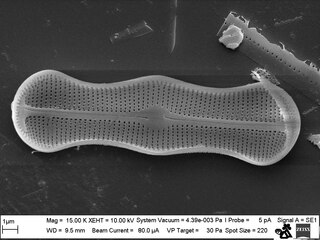
Diatom Achanthes trinodis

Stand-alone cell of Bacillaria paxillifer
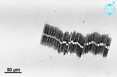
Colonial group of Bacillaria paxillifer
Three diatom species were sent to the International Space Station, including the huge (6 mm length) diatoms of Antarctica and the exclusive colonial diatom, Bacillaria paradoxa. The cells of Bacillaria moved next to each other in partial but opposite synchrony by a microfluidics method.[109]
Heterokont chloroplasts appear to derive from those of red algae, rather than directly from prokaryotes as occurred in plants. This suggests they had a more recent origin than many other algae. However, fossil evidence is scant, and only with the evolution of the diatoms themselves do the heterokonts make a serious impression on the fossil record.
The earliest known fossil diatoms date from the early Jurassic (~185 Ma ago),[110] although the molecular clock[110] and sedimentary[111] evidence suggests an earlier origin. It has been suggested that their origin may be related to the end-Permian mass extinction (~250 Ma), after which many marine niches were opened.[112] The gap between this event and the time that fossil diatoms first appear may indicate a period when diatoms were unsilicified and their evolution was cryptic.[113] Since the advent of silicification, diatoms have made a significant impression on the fossil record, with major fossil deposits found as far back as the early Cretaceous, and with some rocks such as diatomaceous earth, being composed almost entirely of them.
The expansion of grassland biomes and the evolutionary radiation of grasses during the Miocene is believed to have increased the flux of soluble silicon to the oceans, and it has been argued that this promoted the diatoms during the Cenozoic era.[114][115] Recent work suggests that diatom success is decoupled from the evolution of grasses, although both diatom and grassland diversity increased strongly from the middle Miocene.[116]
Diatom diversity over the Cenozoic has been very sensitive to global temperature, particularly to the equator-pole temperature gradient. Warmer oceans, particularly warmer polar regions, have in the past been shown to have had substantially lower diatom diversity. Future warm oceans with enhanced polar warming, as projected in global-warming scenarios,[117] could thus in theory result in a significant loss of diatom diversity, although from current knowledge it is impossible to say if this would occur rapidly or only over many tens of thousands of years.[116]
The fossil record of diatoms has largely been established through the recovery of their siliceous frustules in marine and non-marine sediments. Although diatoms have both a marine and non-marine stratigraphic record, diatom biostratigraphy, which is based on time-constrained evolutionary originations and extinctions of unique taxa, is only well developed and widely applicable in marine systems. The duration of diatom species ranges have been documented through the study of ocean cores and rock sequences exposed on land.[118] Where diatom biozones are well established and calibrated to the geomagnetic polarity time scale (e.g., Southern Ocean, North Pacific, eastern equatorial Pacific), diatom-based age estimates may be resolved to within <100,000 years, although typical age resolution for Cenozoic diatom assemblages is several hundred thousand years.
Diatoms preserved in lake sediments are widely used for paleoenvironmental reconstructions of Quaternary climate, especially for closed-basin lakes which experience fluctuations in water depth and salinity.

When diatoms die their shells (frustules) can settle on the seafloor and become microfossils. Over time, these microfossils become buried as opal deposits in the marine sediment. Paleoclimatology is the study of past climates. Proxy data is used in order to relate elements collected in modern-day sedimentary samples to climatic and oceanic conditions in the past. Paleoclimate proxies refer to preserved or fossilized physical markers which serve as substitutes for direct meteorological or ocean measurements.[119] An example of proxies is the use of diatom isotope records of δ13C, δ18O, δ30Si (δ13Cdiatom, δ18Odiatom, and δ30Sidiatom). In 2015, Swann and Snelling used these isotope records to document historic changes in the photic zone conditions of the north-west Pacific Ocean, including nutrient supply and the efficiency of the soft-tissue biological pump, from the modern day back to marine isotope stage 5e, which coincides with the last interglacial period. Peaks in opal productivity in the marine isotope stage are associated with the breakdown of the regional halocline stratification and increased nutrient supply to the photic zone.[120]


The initial development of the halocline and stratified water column has been attributed to the onset of major Northern Hemisphere glaciation at 2.73 Ma, which increased the flux of freshwater to the region, via increased monsoonal rainfall and/or glacial meltwater, and sea surface temperatures.[121][122][123][124] The decrease of abyssal water upwelling associated with this may have contributed to the establishment of globally cooler conditions and the expansion of glaciers across the Northern Hemisphere from 2.73 Ma.[122] While the halocline appears to have prevailed through the late Pliocene and early Quaternary glacial–interglacial cycles,[125] other studies have shown that the stratification boundary may have broken down in the late Quaternary at glacial terminations and during the early part of interglacials.[126][127][128][129][130][120]
The Cretaceous record of diatoms is limited, but recent studies reveal a progressive diversification of diatom types. The Cretaceous–Paleogene extinction event, which in the oceans dramatically affected organisms with calcareous skeletons, appears to have had relatively little impact on diatom evolution.[131]
Although no mass extinctions of marine diatoms have been observed during the Cenozoic, times of relatively rapid evolutionary turnover in marine diatom species assemblages occurred near the Paleocene–Eocene boundary,[132] and at the Eocene–Oligocene boundary.[133] Further turnover of assemblages took place at various times between the middle Miocene and late Pliocene,[134] in response to progressive cooling of polar regions and the development of more endemic diatom assemblages.
A global trend toward more delicate diatom frustules has been noted from the Oligocene to the Quaternary.[118] This coincides with an increasingly more vigorous circulation of the ocean's surface and deep waters brought about by increasing latitudinal thermal gradients at the onset of major ice sheet expansion on Antarctica and progressive cooling through the Neogene and Quaternary towards a bipolar glaciated world. This caused diatoms to take in less silica for the formation of their frustules. Increased mixing of the oceans renews silica and other nutrients necessary for diatom growth in surface waters, especially in regions of coastal and oceanic upwelling.

In 2002, the first insights into the properties of the Phaeodactylum tricornutum gene repertoire were described using 1,000 expressed sequence tags (ESTs).[135] Subsequently, the number of ESTs was extended to 12,000 and the diatom EST database was constructed for functional analyses.[136] These sequences have been used to make a comparative analysis between P. tricornutum and the putative complete proteomes from the green alga Chlamydomonas reinhardtii, the red alga Cyanidioschyzon merolae, and the diatom Thalassiosira pseudonana.[137] The diatom EST database now consists of over 200,000 ESTs from P. tricornutum (16 libraries) and T. pseudonana (7 libraries) cells grown in a range of different conditions, many of which correspond to different abiotic stresses.[138]

In 2004, the entire genome of the centric diatom, Thalassiosira pseudonana (32.4 Mb) was sequenced,[139] followed in 2008 with the sequencing of the pennate diatom, Phaeodactylum tricornutum (27.4 Mb).[140] Comparisons of the two reveal that the P. tricornutum genome includes fewer genes (10,402 opposed to 11,776) than T. pseudonana; no major synteny (gene order) could be detected between the two genomes. T. pseudonana genes show an average of ~1.52 introns per gene as opposed to 0.79 in P. tricornutum, suggesting recent widespread intron gain in the centric diatom.[140][141] Despite relatively recent evolutionary divergence (90 million years), the extent of molecular divergence between centrics and pennates indicates rapid evolutionary rates within the Bacillariophyceae compared to other eukaryotic groups.[140] Comparative genomics also established that a specific class of transposable elements, the Diatom Copia-like retrotransposons (or CoDis), has been significantly amplified in the P. tricornutum genome with respect to T. pseudonana, constituting 5.8 and 1% of the respective genomes.[142]
Diatom genomics brought much information about the extent and dynamics of the endosymbiotic gene transfer (EGT) process. Comparison of the T. pseudonana proteins with homologs in other organisms suggested that hundreds have their closest homologs in the Plantae lineage. EGT towards diatom genomes can be illustrated by the fact that the T. pseudonana genome encodes six proteins which are most closely related to genes encoded by the Guillardia theta (cryptomonad) nucleomorph genome. Four of these genes are also found in red algal plastid genomes, thus demonstrating successive EGT from red algal plastid to red algal nucleus (nucleomorph) to heterokont host nucleus.[139] More recent phylogenomic analyses of diatom proteomes provided evidence for a prasinophyte-like endosymbiont in the common ancestor of chromalveolates as supported by the fact the 70% of diatom genes of Plantae origin are of green lineage provenance and that such genes are also found in the genome of other stramenopiles. Therefore, it was proposed that chromalveolates are the product of serial secondary endosymbiosis first with a green algae, followed by a second one with a red algae that conserved the genomic footprints of the previous but displaced the green plastid.[143] However, phylogenomic analyses of diatom proteomes and chromalveolate evolutionary history will likely take advantage of complementary genomic data from under-sequenced lineages such as red algae.
In addition to EGT, horizontal gene transfer (HGT) can occur independently of an endosymbiotic event. The publication of the P. tricornutum genome reported that at least 587 P. tricornutum genes appear to be most closely related to bacterial genes, accounting for more than 5% of the P. tricornutum proteome. About half of these are also found in the T. pseudonana genome, attesting their ancient incorporation in the diatom lineage.[140]
To understand the biological mechanisms which underlie the great importance of diatoms in geochemical cycles, scientists have used the Phaeodactylum tricornutum and Thalassiosira spp. species as model organisms since the 90's.[144] Few molecular biology tools are currently available to generate mutants or transgenic lines : plasmids containing transgenes are inserted into the cells using the biolistic method[145] or transkingdom bacterial conjugation[146] (with 10−6 and 10−4 yield respectively[145][146]), and other classical transfection methods such as electroporation or use of PEG have been reported to provide results with lower efficiencies.[146]
Transfected plasmids can be either randomly integrated into the diatom's chromosomes or maintained as stable circular episomes (thanks to the CEN6-ARSH4-HIS3 yeast centromeric sequence[146]). The phleomycin/zeocin resistance gene Sh Ble is commonly used as a selection marker,[144][147] and various transgenes have been successfully introduced and expressed in diatoms with stable transmissions through generations,[146][147] or with the possibility to remove it.[147]
Furthermore, these systems now allow the use of the CRISPR-Cas genome edition tool, leading to a fast production of functional knock-out mutants[147][148] and a more accurate comprehension of the diatoms' cellular processes.

Diatomaceous earth consisting of centric (radially symmetric) and pennate (bilaterally symmetric) diatoms suspended in water.
(click 3 times to fully enlarge)
Decomposition and decay of diatoms leads to organic and inorganic (in the form of silicates) sediment, the inorganic component of which can lead to a method of analyzing past marine environments by corings of ocean floors or bay muds, since the inorganic matter is embedded in deposition of clays and silts and forms a permanent geological record of such marine strata (see siliceous ooze).
Diatoms, and their shells (frustules) as diatomite or diatomaceous earth, are important industrial resources used for fine polishing and liquid filtration. The complex structure of their microscopic shells has been proposed as a material for nanotechnology.[149]
Diatomite is considered to be a natural nano material and has many uses and applications such as: production of various ceramic products, construction ceramics, refractory ceramics, special oxide ceramics, for production of humidity control materials, used as filtration material, material in the cement production industry, initial material for production of prolonged-release drug carriers, absorption material in an industrial scale, production of porous ceramics, glass industry, used as catalyst support, as a filler in plastics and paints, purification of industrial waters, pesticide holder, as well as for improving the physical and chemical characteristics of certain soils, and other uses.[150][151][152]
Diatoms are also used to help determine the origin of materials containing them, including seawater.
The deposition of silica by diatoms may also prove to be of utility to nanotechnology.[153] Diatom cells repeatedly and reliably manufacture valves of various shapes and sizes, potentially allowing diatoms to manufacture micro- or nano-scale structures which may be of use in a range of devices, including: optical systems; semiconductor nanolithography; and even vehicles for drug delivery. With an appropriate artificial selection procedure, diatoms that produce valves of particular shapes and sizes might be evolved for cultivation in chemostat cultures to mass-produce nanoscale components.[154] It has also been proposed that diatoms could be used as a component of solar cells by substituting photosensitive titanium dioxide for the silicon dioxide that diatoms normally use to create their cell walls.[155] Diatom biofuel producing solar panels have also been proposed.[156]

  CNN = cloud condensation nuclei, DMS = dimethylsulphide, DMSP = dimethylsulfoniopropionate, VOCs = volatile organic compounds
 dashed arrow: negative effect, solid arrow: positive effects
The main goal of diatom analysis in forensics is to differentiate a death by submersion from a post-mortem immersion of a body in water. Laboratory tests may reveal the presence of diatoms in the body. Since the silica-based skeletons of diatoms do not readily decay, they can sometimes be detected even in heavily decomposed bodies. As they do not occur naturally in the body, if laboratory tests show diatoms in the corpse that are of the same species found in the water where the body was recovered, then it may be good evidence of drowning as the cause of death. The blend of diatom species found in a corpse may be the same or different from the surrounding water, indicating whether the victim drowned in the same site in which the body was found.[157]

The first illustrations of diatoms are found in an article from 1703 in Transactions of the Royal Society showing unmistakable drawings of Tabellaria.[158] Although the publication was authored by an unnamed English gentleman, there is recent evidence that he was Charles King of Staffordshire.[158][159] It is only 80 years later that we find the first formally identified diatom, the colonial Bacillaria paxillifera, discovered and described in 1783 by Danish naturalist Otto Friedrich Müller.[158] Like many others after him, he wrongly thought that it was an animal due to its ability to move. Even Charles Darwin saw diatom remains in dust whilst in the Cape Verde Islands, although he was not sure what they were. It was only later that they were identified for him as siliceous polygastrics. The infusoria that Darwin later noted in the face paint of Fueguinos, native inhabitants of Tierra del Fuego in the southern end of South America, were later identified in the same way. During his lifetime, the siliceous polygastrics were clarified as belonging to the Diatomaceae, and Darwin struggled to understand the reasons underpinning their beauty. He exchanged opinions with the noted cryptogamist G. H. K. Thwaites on the topic. In the fourth edition of On the Origin of Species he stated that "Few objects are more beautiful than the minute siliceous cases of the diatomaceae: were these created that they might be examined and admired under the high powers of the microscope"? and reasoned that their exquisite morphologies must have functional underpinnings rather than having been created purely for humans to admire.[160]
A diatom (Neo-Latin diatoma) is any member of a large group comprising several genera of algae, specifically microalgae, found in the oceans, waterways and soils of the world. Living diatoms make up a significant portion of the Earth's biomass: they generate about 20 to 50 percent of the oxygen produced on the planet each year, take in over 6.7 billion tonnes of silicon each year from the waters in which they live, and constitute nearly half of the organic material found in the oceans. The shells of dead diatoms can reach as much as a half-mile (800 m) deep on the ocean floor, and the entire Amazon basin is fertilized annually by 27 million tons of diatom shell dust transported by transatlantic winds from the African Sahara, much of it from the Bodélé Depression, which was once made up of a system of fresh-water lakes.
Diatoms are unicellular organisms: they occur either as solitary cells or in colonies, which can take the shape of ribbons, fans, zigzags, or stars. Individual cells range in size from 2 to 200 micrometers. In the presence of adequate nutrients and sunlight, an assemblage of living diatoms doubles approximately every 24 hours by asexual multiple fission; the maximum life span of individual cells is about six days. Diatoms have two distinct shapes: a few (centric diatoms) are radially symmetric, while most (pennate diatoms) are broadly bilaterally symmetric. A unique feature of diatom anatomy is that they are surrounded by a cell wall made of silica (hydrated silicon dioxide), called a frustule. These frustules have structural coloration due to their photonic nanostructure, prompting them to be described as "jewels of the sea" and "living opals". Movement in diatoms primarily occurs passively as a result of both ocean currents and wind-induced water turbulence; however, male gametes of centric diatoms have flagella, permitting active movement to seek female gametes. Similar to plants, diatoms convert light energy to chemical energy by photosynthesis, but their chloroplasts were acquired in different ways.
Unusually for autotrophic organisms, diatoms possess a urea cycle, a feature that they share with animals, although this cycle is used to different metabolic ends in diatoms. The family Rhopalodiaceae also possess a cyanobacterial endosymbiont called a spheroid body. This endosymbiont has lost its photosynthetic properties, but has kept its ability to perform nitrogen fixation, allowing the diatom to fix atmospheric nitrogen. Other diatoms in symbiosis with nitrogen-fixing cyanobacteria are among the genera Hemiaulus, Rhizosolenia and Chaetoceros.
Dinotoms are diatoms that have become endosymbionts inside dinoflagellates. Research on the dinoflagellates Durinskia baltica and Glenodinium foliaceum have shown that the endosymbiont event happened so recently, evolutionarily speaking, that their organelles and genome are still intact with minimum to no gene loss. The main difference between these and free living diatoms is that they have lost their cell wall of silica, making them the only known shell-less diatoms.
The study of diatoms is a branch of phycology. Diatoms are classified as eukaryotes, organisms with a nuclear envelope-bound cell nucleus, that separates them from the prokaryotes archaea and bacteria. Diatoms are a type of plankton called phytoplankton, the most common of the plankton types. Diatoms also grow attached to benthic substrates, floating debris, and on macrophytes. They comprise an integral component of the periphyton community. Another classification divides plankton into eight types based on size: in this scheme, diatoms are classed as microalgae. Several systems for classifying the individual diatom species exist.
Fossil evidence suggests that diatoms originated during or before the early Jurassic period, which was about 150 to 200 million years ago. The oldest fossil evidence for diatoms is a specimen of extant genus Hemiaulus in Late Jurassic aged amber from Thailand.
Diatoms are used to monitor past and present environmental conditions, and are commonly used in studies of water quality. Diatomaceous earth (diatomite) is a collection of diatom shells found in the earth's crust. They are soft, silica-containing sedimentary rocks which are easily crumbled into a fine powder and typically have a particle size of 10 to 200 μm. Diatomaceous earth is used for a variety of purposes including for water filtration, as a mild abrasive, in cat litter, and as a dynamite stabilizer.
Dwindling diatoms and the mixed layer Earth's oceans teem with microscopic plants called phytoplankton. But according to a 2015 NASA study, populations of diatoms, the largest type of phytoplankton algae, have declined more than 1 percent per year from 1998 to 2012. Phytoplankton are an essential base of the marine food web and absorb carbon dioxide dissolved in the ocean that originally came from the atmosphere. The tiny organisms occupy the uppermost layer of ocean water, called the mixed layer, where waves and currents continually churn, drawing up nutrients from a deeper layer of water below. Scientists say the phytoplankton declines observed over the 15-year study period are due to the mixed layer becoming shallower, which results in fewer nutrients reaching the diatoms. The reduction in population may reduce the amount of carbon dioxide drawn out of the atmosphere and transferred to the deep ocean for long-term storage. 3D-animation of the diatom Corethron sp.Diatomeoj, aŭ Diatomaceae laŭ la scienca latina nomo, estas tipoj de flavbrunaj unuĉelaj algoj, kies skeleto pristudata mikroskope de la paleobotaniko estas el siliko. Ili vivas en ĉia tipo de akvo kaj estas bonkvalitaj indikiloj pri saleco, temperaturo kaj aliaj kondiĉoj de sia tiama vivmedio. Tradicie oni diferencigis inter la diatomeoj Centraj, kun radia simetrio, kaj la Pennales, kun duflanka simetrio kaj plilongigita formo. Multaj specioj aperas formantaj ĉenojn aŭ aliajn ordajn kunmetaĵojn.
La diatomeoj estas fotosintezuloj, kiuj formas parton de la fitoplanktono. Ili estas flavbrunaj pro la kuniĝo de fotosintezaj pigmentoj, kiuj enhavas klorofilon c1 kaj c2, kiel aliajn pigmentojn.
Ili povas esti koloniukoj kaj de sala akvo kaj de fluanta akvo; same ili povas ĉeesti malfacilegajn kondiĉojn de temperaturo aŭ saleco, aŭ interĉeestas kun aliaj vivuloj.
Ili posedas kovraĵon el siliko, nomitan frustulo, konsistanta el du pecoj kiuj alĝustiĝas kiel skatoleto kaj kovrilo, sed tiu pli malgranda ol tiu ĉi. La frustulo estas delikate ornamita per variaj kaj simetriaj desegnoj kiuj diferencigas la du tipojn de diatomeoj. La frustuloj de la diatomeoj sedimentiĝas pro gravecforto, kiam ĝi estas englutita aŭ kiam mortas la ĉelo, kreante la konsiston de la sedimentaj rokoj kiel la diatomito aŭ la moronito. La ecoj de tiuj materialoj, formitaj de mikroskopaj eroj, komplikegaj sed tre regulaj laŭ grandeco, faris ilin allogaj por diversa uzado, kiel la produktado de la dinamito, kie la nitroglicerino estas facile sorbita, malpliigante la eblecon de eventuala akcidenta eksplodo. La diatomeoj ne aperis ĝis la fino de la Mezozoiko aŭ komence de la Cenozoiko, sed rapide akiris grandan ekologian gravecon, ĉar iĝis granda parto de la unua produkto de maroj.
Endre Dudich, "Ĉu vi konas la Teron? Ĉapitroj el la geologiaj sciencoj", Scienca Eldona Centro de UEA, Budapest, 1983.
.jpg)
Diatomeoj, aŭ Diatomaceae laŭ la scienca latina nomo, estas tipoj de flavbrunaj unuĉelaj algoj, kies skeleto pristudata mikroskope de la paleobotaniko estas el siliko. Ili vivas en ĉia tipo de akvo kaj estas bonkvalitaj indikiloj pri saleco, temperaturo kaj aliaj kondiĉoj de sia tiama vivmedio. Tradicie oni diferencigis inter la diatomeoj Centraj, kun radia simetrio, kaj la Pennales, kun duflanka simetrio kaj plilongigita formo. Multaj specioj aperas formantaj ĉenojn aŭ aliajn ordajn kunmetaĵojn.
La diatomeoj estas fotosintezuloj, kiuj formas parton de la fitoplanktono. Ili estas flavbrunaj pro la kuniĝo de fotosintezaj pigmentoj, kiuj enhavas klorofilon c1 kaj c2, kiel aliajn pigmentojn.
Ili povas esti koloniukoj kaj de sala akvo kaj de fluanta akvo; same ili povas ĉeesti malfacilegajn kondiĉojn de temperaturo aŭ saleco, aŭ interĉeestas kun aliaj vivuloj.
Ili posedas kovraĵon el siliko, nomitan frustulo, konsistanta el du pecoj kiuj alĝustiĝas kiel skatoleto kaj kovrilo, sed tiu pli malgranda ol tiu ĉi. La frustulo estas delikate ornamita per variaj kaj simetriaj desegnoj kiuj diferencigas la du tipojn de diatomeoj. La frustuloj de la diatomeoj sedimentiĝas pro gravecforto, kiam ĝi estas englutita aŭ kiam mortas la ĉelo, kreante la konsiston de la sedimentaj rokoj kiel la diatomito aŭ la moronito. La ecoj de tiuj materialoj, formitaj de mikroskopaj eroj, komplikegaj sed tre regulaj laŭ grandeco, faris ilin allogaj por diversa uzado, kiel la produktado de la dinamito, kie la nitroglicerino estas facile sorbita, malpliigante la eblecon de eventuala akcidenta eksplodo. La diatomeoj ne aperis ĝis la fino de la Mezozoiko aŭ komence de la Cenozoiko, sed rapide akiris grandan ekologian gravecon, ĉar iĝis granda parto de la unua produkto de maroj.
Las diatomeas (taxón Diatomea, Diatomeae o Bacillariophyceae sensu lato) son un grupo de algas unicelulares que constituyen uno de los tipos más comunes de fitoplancton. Contiene actualmente unas 20 000 especies vivas, las cuales son importantes productores dentro de la cadena alimenticia.[3] [4][5] Muchas diatomeas son unicelulares, aunque algunas de ellas coexisten en forma de filamentos o cadenas celulares (e.g. Fragillaria), abanicos (e.g. Meridion), zigzags (e.g. Tabellaria), estrelladas (e.g. Asterionella). Una característica especial de este tipo de algas es que se hallan rodeadas por una pared celular única hecha de sílice opalino (dióxido de silicio hidratado) llamada frústula. Estas frústulas muestran una amplia variedad en su forma, pero generalmente consisten en dos partes asimétricas con una división entre ellas, característica que da nombre al grupo. La evidencia fósil sugiere que las diatomeas se originaron durante o después del periodo Jurásico temprano, aunque los primeros restos corpóreos son del Paleógeno. Las comunidades de diatomeas son una herramienta usada recurrentemente para la vigilancia de las condiciones medioambientales, de la calidad del agua y en el estudio de los cambios climáticos.
Las diatomeas son microorganismos foto-sintetizadores que forman parte del fitoplancton. Como colonizadores, las diatomeas se distinguen por encontrarse en cualquier cuerpo de agua, es decir ambiente marino, dulceacuícola, e incluso terrestre sobre superficies húmedas. Otras se encuentran en ambientes donde existen condiciones extremas de temperatura o salinidad y de igual forma las encontramos interactuando con otros organismos como es el caso con las cianofíceas filamentosas donde existe un epifitismo por parte de las diatomeas. La mayoría son pelágicas (viven en aguas libres), algunas son bentónicas (sobre el fondo marino), e incluso otras viven bajo condiciones de humedad atmosférica. Son especialmente importantes en los océanos, donde se calcula que proporcionan alrededor un 45 % del total de la producción primaria oceánica.[6] La distribución espacial del fitoplancton marino no esta restringida tanto horizontal como verticalmente. Las diatomeas habitan en todos los cuerpos de agua, desde los polos hasta las latitudes más bajas; las regiones polar y subpolar contienen relativamente pocas especies en contraste con la biota templada.[7] Aunque las regiones tropicales exhiben la mayor cantidad de especies, las mayores poblaciones de diatomeas se hallan entre las regiones polar y templada.
Las diatomeas pertenecen a un gran grupo llamado Heterokontophyta, el cual incluye especies tanto autótrofas (e.g algas pardas) como heterótrofas (e.g. oomicetos). Los cloroplastos amarillo-marrones de las diatomeas son típicos de los heteroncontos, con cuatro membranas, clorofilas a, c1 y c2 y pigmentos tales como β-caroteno, fucoxantina, diatoxantina y diadinoxantina. Las reservas de alimento se almacenan como carbohidratos o aceites, que además de servir de reserva, contribuyen a su flotabilidad.
Sus individuos carecen de flagelo, pero están presentes en gametos y usualmente presentan una estructura heteroconta, excepto en que carecen de vellosidades (mastigonemas) característicos de otros grupos. En general, las diatomeas no poseen movimiento propio, no obstante, algunas de ellas pueden desplazarse mediante la contracción del rafe, debido a su pared celular silícea. Las diatomeas tienden a formar cadenas de células, y estas cadenas ayudan a la flotabilidad de las células para así mantenerse en las aguas superficiales con mayor incidencia de luz por más tiempo. Algunas especies regulan activamente su flotabilidad con los lípidos intracelulares para hacer frente al hundimiento.
A pesar de ser generalmente microscópicas, algunas especies de diatomeas pueden alcanzar los dos milímetros de longitud. Las diatomeas están contenidas dentro de una única pared celular de silicato (frústula) compuesta de dos valvas separadas. La sílice biogénica de la que la pared celular se compone es sintetizada intracelularmente por polimerización de monómeros de ácido silícico. Este material es luego secretado hacia el exterior de la célula en donde participa en la conformación de la pared celular. Las valvas de las diatomeas se superponen una a otra, denominadas así entonces, la epivalva (placa superior) e hipovalva (placa inferior). El frústulo aparece delicadamente ornamentado con relieves que forman dibujos variados y perfectamente simétricos, dando lugar a dos tipos de diatomeas, las que tienen simetría radial y las de simetría lateral. Los frústulos de las diatomeas se sedimentan por gravedad cuando es digerida o muere la célula, dando origen a rocas sedimentarias como las diatomitas y moronitas.
Las diatomeas alternan entre la reproducción asexual por división celular, que es la más frecuente, y la reproducción sexual.[8] En la reproducción por división celular, cada célula hija recibe una de las frústulas de la célula padre y la utiliza como frústula mayor (o epiteca), reconstruyendo una frústula menor (o hipoteca). Como consecuencia de este proceso, la célula que recibió la frústula menor resulta en una diatomea de menor tamaño que la original. Por otra parte, la frústula no puede crecer. De esta forma, en la primera generación el 50% de las diatomeas son de menor tamaño que la original y se van haciendo cada vez más pequeñas en cada generación. Se ha observado sin embargo, que algunas especies pueden dividirse sin causar una reducción en el tamaño de célula.[9] El proceso continúa hasta que las células alcanzan una tercera parte de su tamaño máximo.[8] Después de un determinado número de generaciones, las diatomeas se reproducen de forma sexual, produciendo gametos sin frústulas que se fusionan formando una auxospora. Este mecanismo ayuda a restablecer el tamaño original de las diatomeas porque el cigoto crece mucho antes de producir una nueva frústula.
Las células vegetativas de las diatomeas son diploides (2N) y la meiosis genera gametos machos y hembra que entonces se fusionan para formar el cigoto. El cigoto se libera de su cubierta y crece en la forma de una célula esférica cubierta por una membrana orgánica (auxospora). Cuando la auxospora alcanza su medida máxima (la de diatomea inicial), forma en su interior a una diatomea que da comienzo a una nueva generación. También pueden formarse esporas como respuesta a condiciones medioambientales desfavorables, produciéndose la germinación cuándo las condiciones mejoran.[10]
Las diatomeas son mayoritariamente no móviles, aunque el esperma de algunas especies puede ser flagelado, con movimiento normalmente limitado al deslizamiento.[10] En las diatomeas céntricas, los gametos machos son pequeños y tienen un flagelo, mientras que los gametos hembra son grandes e inmóviles (oogamia). Por el contrario, en las diatomeas pennadas ambos gametos carecen de flagelos (isogamia).[8] Se ha documentado que una especie del grupo de las diatomeas pennadas sin rafe es anisógama y, por tanto, se la considera una etapa transicional entre las diatomeas céntricas y las diatomeas pennadas con rafe.[9]
Las diatomeas, Diatomeae (Dumortier, 1821) o Bacillariophyceae (Haeckel, 1878), son clasificadas según la distribución de sus poros y ornamentación. Si las frústulas poseen una simetría radial o trímera se las denomina diatomeas centradas, mientras que si poseen una simetría bilateral y forma alargada se las denomina pennadas.[4] El primer tipo es parafilético con respecto al segundo. Algunas diatomeas pennadas también presentan una fisura a lo largo del eje longitudinal, denominada rafe, que está implicada en los movimientos realizados por la diatomea. Al grupo Bolidophyceae, recientemente descubierto, se le considera una clase aparte. La clasificación más reciente considera cuatro grupos de diatomeas:[11]
DiatomeaCoscinodiscophyceae (P): Centrales radiales.
Mediophyceae (P) : Centrales polares.
PennalesFragilariophyceae (P): Pennales sin rafe.
Bacillariophyceae: Pennales con rafe.
La tierra diatomea es un material constituido por las frústulas de diatomeas fosilizadas, aplicado como fertilizante e insecticida en tierras para cultivo, al ser un producto natural, es inocuo y no presenta riesgos para la salud o contaminación. La tierra diatomea provee micronutrientes al suelo que son de gran importancia para el crecimiento de las plantas, pudiendo incrementar la fertilidad del suelo, actuando sinérgicamente con calcio y magnesio, además reduce la lixiviación de fósforo, nitrógeno y potasio y favorece su absorción en las plantas. La tierra diatomea también actúa como reconstituyente en tierras contaminadas por metales pesados o hidrocarburos, además neutraliza la toxicidad del aluminio en suelos ácidos y reduce la absorción de hierro y manganeso.[12]
Las propiedades de esos materiales, formados por partículas microscópicas, intrincadas y muy regulares en tamaño, los han hecho atractivos para diversos usos, como la fabricación de la dinamita, donde la nitroglicerina es embebida, reduciendo la probabilidad de una explosión accidental.
El polvo de diatomeas se emplea como insecticida en animales y plantas. Actúa deshidratando a insectos hasta matarlos, además corta y perfora el exoesqueleto, hiriéndolos y eliminándolos de forma progresiva y efectiva. Las frústulas de las diatomeas son de origen natural, por lo cual son inocuas para animales y plantas. A diferencia de insecticidas tóxicos, el polvo de diatomeas no puede ingresar en los tejidos animales debido a su tamaño.[13]
Las diatomeas pertenecen a las microalgas oleaginosas debido a que presentan fracciones lipídicas del 25 % (condiciones normales) al 45 % (condiciones de estrés), cultivables en fotobiorreactores (FBR). La producción de biodiésel a partir de diatomeas se da por medio de transesterificación del aceite preveniente de las microalgas. La producción de biodiésel se basa en la producción y captación de biomasa de diatomeas, la cual es deshidratada y sometida a ultrasonidos para que libere sus componentes, posteriormente los lípidos son separados de carbohidratos y proteínas. El aceite obtenido es sometido a transesterificación alcalina, ácida o enzimática para producir glicerol y biodiésel.
Los aceites provenientes de diatomeas son principalmente triglicéridos, que generan mezclas de ésteres de alquilo al convertirse en biocombustible. El cracking térmico o pirólisis, es un proceso alterno a la transesterificación que transforma triglicéridos en otros compuestos orgánicos simples.[14]
Se ha determinado que las diatomeas tienen la capacidad de producir ácidos grasos poliinsaturados (AGPI) en altas concentraciones, como por ejemplo la producción de diatomeas del género Nitzschia de ácido eicosapentanoico (EPA). Nitzschia posee ventajas como la resistencia a temperaturas de hasta -6 °C y ambientes altamente salobres, además su aceite alcanza el 50 % del peso seco de la biomasa.[15]
Las comunidades de diatomeas pueden utilizarse para la determinación de las condiciones ambientales tanto de presente como del pasado y del cambio climático. También pueden utilizarse para determinar la calidad del agua.
La demostración de la presencia de Diatomeas dentro de las cavidades cardíacas o en la médula ósea de cadáveres es utilizada en medicina forense como fuerte indicio de muerte por sumersión (ahogamiento).[16]
En la limnología, que es el estudio de los ecosistemas acuáticos, el estudio de diatomeas permite hacer un rastreo del trascurrir en el tiempo de dichos entornos; un ejemplo puede ser la reconstrucción paleolimnológica de la Laguna Boquete, al norte de Panamá.[17]
Melosira sp.
Cymbella sp.
Licmophora sp.
Las diatomeas (taxón Diatomea, Diatomeae o Bacillariophyceae sensu lato) son un grupo de algas unicelulares que constituyen uno de los tipos más comunes de fitoplancton. Contiene actualmente unas 20 000 especies vivas, las cuales son importantes productores dentro de la cadena alimenticia. Muchas diatomeas son unicelulares, aunque algunas de ellas coexisten en forma de filamentos o cadenas celulares (e.g. Fragillaria), abanicos (e.g. Meridion), zigzags (e.g. Tabellaria), estrelladas (e.g. Asterionella). Una característica especial de este tipo de algas es que se hallan rodeadas por una pared celular única hecha de sílice opalino (dióxido de silicio hidratado) llamada frústula. Estas frústulas muestran una amplia variedad en su forma, pero generalmente consisten en dos partes asimétricas con una división entre ellas, característica que da nombre al grupo. La evidencia fósil sugiere que las diatomeas se originaron durante o después del periodo Jurásico temprano, aunque los primeros restos corpóreos son del Paleógeno. Las comunidades de diatomeas son una herramienta usada recurrentemente para la vigilancia de las condiciones medioambientales, de la calidad del agua y en el estudio de los cambios climáticos.
Ränivetikad ehk diatomeed (Bacillariophyceae ehk Diatomophyceae) on üherakulised või koloniaalsed mikroskoopilised organismid. Liike on hinnanguliselt 30 000 kuni 100 000.[1] Ränivetikaid esineb kõikides veekogudes ja ka mullas.
Ränivetikate iseloomulikuks tunnuseks on rakke kattev keeruka struktuuriga ränipantser.
Ränipantser koosneb kahest poolmest: välimisest suuremast epitheekast ja sisemisest väiksemast hüpotheekast. Kummalgi pantseri poolmel on omakorda kaks osa: lame kaas ja ümbritsev vöö. Kõik pantseri osad on kindla struktuuriga, neid läbivad poorid ja kanalid. Iseloomulikeks pigmentideks on karoteen, ksantofüll, diatomiin ning klorofüll a ja c. Varuaine on enamasti õlitilgakestena.
Ränivetikad paljunevad pooldudes ja suguliselt. Pooldumisel saab kumbki tütarrakk endale pantseri ühe poolme. Alati moodustub juurde pantseri väiksem pool. Seega need rakud, mis saavad endale sisemise poole, vähenevad pidevalt. Niisugune rakkude vähenemine lakkab, kui tütarraku maht on ainult pool kuni kolmandik omaraku esialgsest mahust (suurusest). Siis tekib rakust auksospoor, millega kaasneb raku esialgse suuruse taastumine. Sageli kaasneb aukspooride tekkega suguline protsess.
Üldiselt käsitletakse ränivetikaid klassi tasemel (Bacillariophyceae). Harvem (ja ka enamikus varasemates allikates), kui on kõrvale jäetud suhteliselt uus stramenopiilide (heterokontide) hõimkond, siis on ränivetikaid (siis ränivetiktaimed) käsitletud hõimkonna tasemel (Bacillariophyta).
Pantseri kuju alusel jaotatakse ränivetikad kahte klassi: radiaalsümmeetrilise ehitusega ketasränivetikad (Centrales) ja kahekülgse ehk bilateraalse sümmeetriaga sulgränivetikad (Pennales).
.jpg)
Ränivetikad ehk diatomeed (Bacillariophyceae ehk Diatomophyceae) on üherakulised või koloniaalsed mikroskoopilised organismid. Liike on hinnanguliselt 30 000 kuni 100 000. Ränivetikaid esineb kõikides veekogudes ja ka mullas.
Diatomeak eskeleto silizeoa duten protofitorik garrantzitsuenak dira. Gehienek fotosintesia burutzen dute eta beraz zona fotikoan bizi dira. Ur gazi zein gezatan bizi dira eta planktoniko eta bentonikoak izan daitezke.
Alga zelulabakar fotosintetikoak dira, eta fitoplanktonaren osagai oso garrantzitsua.
Diatomeoak Baciollariophyta dibisioan sartzen dira eta marroi-urre koloreko alga zelulabakarrak dira. Beraien teka silize opalinoz osaturiko bi piezaz osaturik dago. Teka honi frustula izena ematen zaio eta frustularen bariabilitate eta morfologia erabiltzen da bereizketa egiteko. Frustularen tamaina 1etik 1000 mikra artekoa da.
Dinoflagelatuak eta silikoflagelatuak ez bezala, diatomeoek ez dituzte flageloak garatzen, nahiz eta batzuk ernalkuntza sexualeko aldi motz batean garatzen duten. Hau dela eta diatomeo planktoniko guztiak pasiboki sakabanatzen dira itsasoko uretan eta horretarako flotagarritasuna errazten dituzten bereizgarriak garatu behar dituzte. Adibide gisa frustularen forma ezberdinak edo azalera eta bolumenaren arteko erlazioa. Flotagarritasuna handitzeko koloniak eratzen dituzte eta zelularen grabitate espezifikoa igotzen duten gantz metaketak sortzen dituzte.
Kolonietan bizi direnean euren artean gai muzilaginotsu batez elkartzen dira. Bakartiak direnean gai muzilaginotsu hau substratuari heltzeko erabiltzen dute.
Diatomeoen eskeletoa (frustula) kontutan hartuz, simetriaren araberako bi talde daude:
Frustulan, gainjartzen diren bi kuxku daude, hipokuskua eta epikuskua. Biak batzen diren lekua gerrikoa da. Gerrikoa bi zatitan bana daiteke: epigerrikoa edo epizigulua eta hipogerrikoa edo hipozigulua.
Diatomeo zentral gehienak oso arinak dira, poro edo aureola asko dituztelako. Poro hauen artean ornamentaziorik gabeko guneak egon daitezke, hialinoak. Gune hialino hauek marginalki koka daitezke eraztun marginal hialino gunea eratuz, edo poro eta aureolaren artean. Beste apaingarriak izan dezakete: aranteak eta zetak (iletxoak) adibidez.
Forma penaletan, zentralek dituzten ezaugarriez gain, saihetsak eta errateak ager daitezke. Erratzak ildo longitudinalak dira eta taxonomian oso garrantzitsuak. Askotan pseudoerrateak izan ahal dituzte, hauek gune marjinalean jartzen diren ildoak dira.
175 genero fosil ezberdin ezberdintzen dira. Genero guzti horietatik %70 itsastarrak dira, %17a ur gezatakoak eta %13 bietan bizi daitezke eta leku batetik bestera mugitu. Gaur eguneko itsasoetan diatomeoak lehen ekoizle garrantzitsuenak dira eta diatomeoan ugaritasunak itsasoko gune emankorrenak adierazten digu. Ugaritasun handiko uretan fosilizatzen direnean litifikatu eta diatomitak sortzen dira. Diatomita hauek porositate handia dute eta filtro naturalak egiteko erabiltzen dira.
Izaki fotosintetikoak direnez, gune fotikoan bizi dira, hau da, 100 metroko sakonera baino gutxiagoan eta izan ahal dira plantonikoak edo bentonikoak. Mikrofosil karetsuak ez bezala latitude altuetan aurki daitezke. Tenperatura ez da mugatzailea.
Izaki zelulabakar, eukariotak eat diploideak dira. Bakarka edo kolonietan bizi dira. Gorputzeko atal bigunak bi kuxku silizeoen barruan kokatzen dira. Kuxku hauek frustula izendatzen dira. Bi kuxku hauen kanpoaldean gai muzilaginotsua jartzen du. Beraien zelula kuxkuen barnealdean itsatsirik dago, protoplasma, bakuolak... eta beste organuluez gain nukleoa inguratzen duen zitoplasma osatzen dute.
Klorofila a eta c ditu eta karotenoideak dute, hori delarik kolore marroi-urre hori ematen diona.
Nutrienteak ugariak direnean eta argi baldintzak onuragarriak direnean bloomak gertatzen dira, bloom hauek ernalkuntza asexualarekin ematen dira.
Diatomeoek ernalkuntza asexuala daukate. Asexualki bi zatitan banatzen dira eta mitosi edo fisio hau 8 aldiz eman dezakete egun berean. Ernalkuntza asexual honetan, ama bi zatitan banatzen da eta kuskuaren zati bakoitza izango da semearen epikuskua. Epikuskuarekin garatzen dena hipokuskua da eta hipokusku hau amak zuenaren tamaina berekoa izango da. Hipokuskuarekin geratzen dena, hipokuskua epikuskuan bihurtuko da, ondorioz hipokusku berri txikiago bat sortuko du.
Prozesu honetan, belaunaldiz belaunaldi tamaina txikiagotuz dihoaz. Momentu batean frustulan ez da zitoplasma sartuko, orduan ernalkuntza sexualarekin hasiko dira.
Inguruko baldintzak onuragarriak ez direnean (janari faltagatik, argi faltagatik), diatomeo planktonikoek zelula begetatiboak sortzen dituzte, Estratoblasto izenekoak. Hauek dentsitate handia dute eta horregatik itsaso hondoan metatuko dira hurrengo upwellinga eman arte.
Korrontearekin azaleraino doaz eta estratoblastoetatik irteten dira (baldintza hobeak direnez germinatzen dute) diatomeoak sortuz.
Faktore paleomugatzaileak aztertzen dira non bizi diren jakiteko. Lehen aipatutako faktore mugatzialeak erabiltzen dira paleoingurua ezagutzeko, baina prozesu tafonomikoak ere beharrezkoak dira, lurperatze tasa azkarra ez badugu, diatomeoak asko murrizten direlako.
Diatomeoen artean itsastarrak dira gehien erabiltzen direnak ikerketak egiteko.
Diatomeoak algetatik banatuta daude eta Bacillartophyceae dibisioan sartzen dira. Autore batzuek honen barruan Crysophita klasea sortzen dute eta bertan sartu Diatomeoak. Proposaturiko sailkapena honako hau da:
Bi azpiklase horien barruan 8 ordena ezberdin proposatu dira:
Lehen diatomeoak Behe Kretazeoan agertu ziren eta zentralak ziren. Maastrichtiarrean pennalak agertu ziren. Hasieran forma ugarienak zentralak ziren eta batez ere Miozenotik gaur egunera pennalen proportzioa ugaritzen joan da. Diatomeak talde moduan krisialdi handiena jasan zuten K/T muga eta batez ere Kuaternarioan izotz masen fluktuazioekin aldaketak eman dira.
Ur gezako lehen diatomeoak Behe Eozenoan agertu ziren, Parisko Eozeno arroan. Batzuen ustez, fase erregresiboan, transgresio baten ostean, ur gazia kontinentean geratu ziren harrapatuta eta hauetatik batera ur gazikoak. Gero ur gezara moldatzeko, batzuk eboluzionatu zuten. Garai horretako bulkanismoak garrantzia izan zuen, ingurunera silizea botatzen delako, frustularentzako erabili zutena ur gezako formek.
Izaki fotosintetikoak dira, ondorioz argia eta karbono dioxidoa behar dute, dena den, diatomeo batzuk gai dira argirik gabe materia organikoa ateratzen. Argia eta karbono dioxidoaz gain, nitrogenoa, fosforoa eta silizea ere dira faktore mugatzaileak. Faktore hauek urtaroekin erlazioa dutenez, diatomeoen kontzentrazioa ere aldatzen da urtaroekin, upwelling prozesuekin ere.
Upwelling prozesuak materia jangarria igotzen da azalera, beraz nutrienteen kopurua giotzen da eta diatomeen kontzentrazioa ere handitzen da. Prozesu hauek lakuetan ere ematen dira, adibidez haizea dagoenean, honek olatuak sortzen ditu, nutrienteak azaleratzen dira eta prozesu bera ematen da. Hau urtaroekiko erlatiboa da.
Normalean arroka konpaktuetan aurkitu ohi dira fosilduak, ondorioz ateratzeko Azido klorhidrikoa eta Azido fosforikoa erabili behar da. Latitude altuetako arroketan aztertzen dira, bertan ez baitago mikroforaminiferorik. Zailak izan ohi dira ikertzen eta horregatik bakarrik errazen suertatzen direnak aztertzen dira.
.jpg)
Diatomeak eskeleto silizeoa duten protofitorik garrantzitsuenak dira. Gehienek fotosintesia burutzen dute eta beraz zona fotikoan bizi dira. Ur gazi zein gezatan bizi dira eta planktoniko eta bentonikoak izan daitezke.
Alga zelulabakar fotosintetikoak dira, eta fitoplanktonaren osagai oso garrantzitsua.
Piilevät eli diatomit (Bacillariophyta) on ruskeisiin leviin kuuluva leväryhmä, johon kuuluu yleisimmän käsityksen mukaan noin 20 000 – 100 000 lähinnä yksisoluista lajia. Ryhmä on lähteestä riippuen esitetty sekä kaari- että alakaaritason ja myös luokkatason taksonina. Piileviä esiintyy kaikissa maailman vesissä; etenkin viileissä merissä ne muodostavat merkittävän osan kasviplanktonista. Piilevät ovat saaneet nimensä piidioksidista koostuvasta soluseinästään, joka muodostaa levälle kaksiosaisen kuoren. Piilevät ovat tavallisesti väriltään ruskehtavia ja muodoltaan silmiinpistävän symmetrisiä.
Piilevät lisääntyvät lähinnä suvuttomasti jakautumalla kahtia, jolloin rasian tavoin limittäin sijaitsevat piikuoren puolikkaat, kansi ja pohja, erkanevat toisistaan ja näin syntyneet tytärsolut kasvattavat pienemmän kuoripuoliskon eli pohjan alkuperäisen sisään. Sukupolvien kuluessa uusien piilevien koko pienenee jopa 60 prosenttia muutamassa kuukaudessa, kunnes minimikoon saavutettuaan levät synnyttävät (yleensä suvullisesti) itiöitä, jotka kasvavat jälleen normaalikokoisiksi soluiksi ja sykli alkaa uudelleen.
Vanhimmat tunnetut piilevien fossiilit ovat peräisin varhaiselta jurakaudelta; eniten fossiileja syntyi liitukaudella. Kuolleiden piilevien piikuoret ovat aikojen kuluessa valuneet valtamerten pohjiin ja muodostaneet sinne piimaata.
Piileviä esiintyy sekä planktisina että alustalla kasvavina. Piilevillä ei niiden sukusolumuotoja lukuun ottamatta ole uintisiimoja, joten planktonissa elävät solut pyrkivät vajoamaan valottomaan alusveteen, ellei jatkuva veden sekoittuminen palauta niitä takaisin päällysveteen. Joillain lajeilla piikuoresta esiin pistävät pitkät piikit aiheuttavat virtausvastusta ja hidastavat vajoamista. Alustalla kasvavat lajit kykenevät liikkumaan hitaasti alustaansa pitkin erittämällä kuoressaan olevista huokosista limaa, joka työntää solua eteenpäin.[2]
Piilevät eli diatomit (Bacillariophyta) on ruskeisiin leviin kuuluva leväryhmä, johon kuuluu yleisimmän käsityksen mukaan noin 20 000 – 100 000 lähinnä yksisoluista lajia. Ryhmä on lähteestä riippuen esitetty sekä kaari- että alakaaritason ja myös luokkatason taksonina. Piileviä esiintyy kaikissa maailman vesissä; etenkin viileissä merissä ne muodostavat merkittävän osan kasviplanktonista. Piilevät ovat saaneet nimensä piidioksidista koostuvasta soluseinästään, joka muodostaa levälle kaksiosaisen kuoren. Piilevät ovat tavallisesti väriltään ruskehtavia ja muodoltaan silmiinpistävän symmetrisiä.
Piilevät lisääntyvät lähinnä suvuttomasti jakautumalla kahtia, jolloin rasian tavoin limittäin sijaitsevat piikuoren puolikkaat, kansi ja pohja, erkanevat toisistaan ja näin syntyneet tytärsolut kasvattavat pienemmän kuoripuoliskon eli pohjan alkuperäisen sisään. Sukupolvien kuluessa uusien piilevien koko pienenee jopa 60 prosenttia muutamassa kuukaudessa, kunnes minimikoon saavutettuaan levät synnyttävät (yleensä suvullisesti) itiöitä, jotka kasvavat jälleen normaalikokoisiksi soluiksi ja sykli alkaa uudelleen.
Vanhimmat tunnetut piilevien fossiilit ovat peräisin varhaiselta jurakaudelta; eniten fossiileja syntyi liitukaudella. Kuolleiden piilevien piikuoret ovat aikojen kuluessa valuneet valtamerten pohjiin ja muodostaneet sinne piimaata.
Piileviä esiintyy sekä planktisina että alustalla kasvavina. Piilevillä ei niiden sukusolumuotoja lukuun ottamatta ole uintisiimoja, joten planktonissa elävät solut pyrkivät vajoamaan valottomaan alusveteen, ellei jatkuva veden sekoittuminen palauta niitä takaisin päällysveteen. Joillain lajeilla piikuoresta esiin pistävät pitkät piikit aiheuttavat virtausvastusta ja hidastavat vajoamista. Alustalla kasvavat lajit kykenevät liikkumaan hitaasti alustaansa pitkin erittämällä kuoressaan olevista huokosista limaa, joka työntää solua eteenpäin.
Les Bacillariophyta, ou Diatomées, sont une division de microalgues unicellulaires (de deux micromètres à un millimètre) présentes dans tous les milieux aquatiques et majoritaires dans certains biofilms[2] (avec une préférence pour les eaux froides) et enveloppées par un squelette externe siliceux nommé frustule. Elles peuvent vivre isolées ou en colonie, être libres ou fixées. Les formes pélagiques appartiennent au phytoplancton, les formes benthiques appartiennent au microphytobenthos. Les diatomées sont un constituant majeur du phytoplancton participant à 50 % de la production primaire océanique globale[3].
Ces micro-algues produisent à elles seules un quart de l'oxygène que nous respirons et jouent un rôle primordial dans la vie des écosystèmes marins, puisqu'elles sont à la base des réseaux alimentaires de nombreuses espèces. Selon Mann et Droop (1996)[4] il y existerait probablement plus de 200 000 espèces de diatomées, dont ils affirment que 10 % ont été nommés, soit environ 20 000 espèces. Mais une étude de 2012[5] souligne que bien que les estimations du nombre total d'algues varient considérablement selon les auteurs (entre 30 000 et un million d'espèces), il est plus probable que chez les diatomées, le nombre total d'espèces se situe en fait bien autour de 20 000 dont environ 8 000 restaient alors à découvrir et à décrire. Et début 2018, 14 803 espèces[6] de Bacillariophyta sont répertoriées dans AlgaeBase.
Le mot Bacillariophyta dérive du grec βακτηριον / baktirion, « petit baton ; bâtonnet », et φυτό / phytó, plante, littéralement « plante (en forme de) bâtonnet », bien que beaucoup de microalgues de ce groupe n'ont pas du tout cette forme. Le mot « diatomée » étant drivé du grec διατομ / diatom, « couper en deux ; séparer ; diviser », en référence au frustule de ces organismes, enveloppe siliceuse formée de deux valves emboîtées.
Les diatomées sont des algues jaunes et brunes unicellulaires caractérisées par le fait qu'elles sont les seuls organismes unicellulaires à posséder une structure externe siliceuse enveloppant totalement la cellule. Transparente et rigide, cette enveloppe, appelée frustule, possède une architecture complexe qui définit l'espèce dans la nomenclature. La silice qui compose le frustule dite silice biogénique, est amorphe, c'est-à-dire qu'elle est faiblement cristallisée. La formation de la silice biogénique, ou silicification implique des molécules organiques qui vont guider la formation du frustule et y inclure des composants organiques[7]. Le frustule est formé de deux thèques emboîtées à symétrie remarquable. Les diatomées se divisent en deux groupes en fonction de la symétrie de leur frustule. Les centriques, à symétrie radiale, dont la forme générale est celle d'une boite de camembert et les pennées dont le frustule possède une symétrie bilatérale ou transversale.
Comme toute algue, la cellule d'une diatomée possède des chloroplastes pour sa photosynthèse. Les échanges avec le milieu extérieur se font au travers de nombreux orifices très fins qui traversent la frustule et qui sont disposés en lignes (droites ou courbes) ou en réseau, selon un motif propre à l'espèce. Ces ornementations sont ainsi utilisées pour la classification.
Le frustule est composé de deux parties ou thèques s'emboîtant l'une dans l'autre, à la manière d'une boîte. Chacune de ces deux parties est elle-même composée de deux éléments. Le premier, la valve, plus ou moins bombée (rarement plate), correspond à la face du « couvercle » ou du « fond » de la « boîte » ; à sa périphérie peut se situer une zone oblique ou verticale, le manteau, qui fait la liaison avec le second élément. Ce second élément, le cingulum, est une paroi verticale entourant la valve qui peut être formé d'une simple bande siliceuse ou comporter plusieurs bandes ou segments cingulaires (copulae). L'épithèque est constituée de l'épivalve et de l'épicingulum et correspond à la partie du frustule correspondant au « couvercle » ; l'hypothèque, plus petite, est constituée de l'hypocingulum et de l'hypovalve, et désigne le « fond » (voir la figure, d'après Van den Hoek, 1995). Le volume interne du frustule, occupé par la cellule, peut être partiellement subdivisé par des cloisons, perforées ou incomplètes, portées par les valves ou par les bandes cingulaires.
Chez de nombreuses diatomées pennales, une fente, de longueur variable, parcourant les deux valves ou une seule, souvent en leur milieu, est appelée « raphé » et constitue un canal de communication avec l'extérieur et sert à la locomotion par excrétion du mucilage (voir les schémas du frustule de P. viridis, d'après Van den Hoek et al., 1995). Le raphé est interrompu en son milieu par un épaississement siliceux, le nodule central, et possède un nodule terminal à chaque extrémité. Si le raphé est en position médiane, il est situé dans une zone sans ornementations, l'aire longitudinale. Le nodule central est, lui, localisé dans l'aire centrale. Les diatomées dites monoraphidées sont dépourvues de raphé sur la valve supérieure et présentent une ornementation différente sur les deux valves. Les diatomées centrales[a] et certaines diatomées pennales sont dites araphidées (ou araphides) car elles ne possèdent aucun raphé.
C'est notamment dans les familles des Surirellaceae et des Nitzschiaceae que l'évolution du raphé a abouti à la création d'une structure en forme de tuyau, le canal-raphé. À l'opposé du côté où s'ouvre le raphé, la paroi du tube est percée d'orifices débouchant à l'intérieur du frustule (pores internes) et séparés par des piliers siliceux, les fibules. Souvent, le canal-raphé est situé au faîte d'une carène valvaire plus ou moins développée.
La paroi des valves est constituée d'une seule couche siliceuse ou de deux couches superposées entre lesquelles se trouvent des espaces délimités par des cloisons transversales. Les diatomées pennales tiennent leur nom du fait que les ornements de leurs valves sont souvent disposés à la manière des dents d'un peigne alors qu'ils sont souvent radiaux chez les diatomées centrales. Ces ornementations sont propres à chaque espèce et sont donc gouvernées par les gènes. Les bandes cingulaires sont souvent dépourvues d'ornements. Ces ornementations de la frustule, qui apparaissent comme des stries, des côtes, des perles ou une dentelle, présentent une esthétique d'une extrême finesse dont la beauté attire de nombreux diatomistes.
Ces « ornements » correspondent en fait à des regroupements de fines perforations ou aréoles avec un diamètre de l'ordre du micromètre. Ces perforations ont souvent une extrémité (du côté soit externe soit interne de la paroi valvaire), partiellement obturé par une fine dentelle siliceuse qualifiée de crible. Ces orifices servent aux échanges entre la cellule et le milieu extérieur. Chez certaines diatomées, il existe des pores très fins (porelles) regroupés dans des structures limitées par un anneau marginal épaissi, les ocelles. Les diatomées pennales peuvent posséder des pores de plus gros diamètre à l'extrémité des valves permettant l'écoulement de substances mucilagineuses sécrétées par la cellule.
À la surface des valves se trouvent de petites structures silicifiées tubulaires : les « processus ». Il en existe quatre ou cinq types. C'est à leur niveau que certaines espèces extrudent des filaments de chitine.
Les diatomées sont des organismes unicellulaires la plupart du temps photosynthétiques. Elles sont donc généralement autotrophes et utilisent l'énergie de la lumière grâce à la chlorophylle a et la chlorophylle c contenues dans leurs chloroplastes par photosynthèse. Les chlorophylles sont situés dans les chloroplastes, qui peuvent être présentés comme de petites granules dispersés dans la cellule ou avoir une forme de plaque ou de bande ou d'étoile et sont d'un nombre variable (entre un et plusieurs dizaines). Par la présence de pigments de carotènes et de xanthophylles (dont la fucoxanthine), les chloroplastes sont jaunes, vert olive ou bruns.
Certaines espèces peuvent malgré tout présenter une hétérotrophie plus ou moins importante. Celle-ci peut être facultative et temporaire lorsque la diatomée est en présence de molécules organiques assimilables et que l'éclairement est insuffisant. Quelques rares espèces (notamment en eaux eutrophes et peu éclairées des zones littorales et estuariennes) ne sont cependant pas douées de photosynthèse et ont donc une hétérotrophie obligatoire.
Le squelette externe des diatomées, très élaboré, est couvert d'un réseau de pores pour optimiser la capture de la lumière. Ce procédé a attiré l'attention de nanotechniciens. Greg Rorrer, ingénieur chimiste, cultive des diatomées avec des matériaux semi-conducteurs implantés dans leur squelette, injectées ensuite dans des cellules photovoltaïques, elles créent une unité de production d'énergie 50 % plus efficace.
Comme toute cellule, les diatomées possèdent une membrane organique périphérique à leur cytoplasme, un noyau, et les différents organites nécessaires à son fonctionnement (mitochondries, systèmes membranaires et tubulaires intracytoplasmique…). La cellule des diatomées centrales est en partie occupée par une vacuole dont la densité du volume est proche de celle de l'eau ambiante. La plupart des diatomées pennales possèdent deux vacuoles situées de part et d'autre de la zone cytoplasmique médiane entourant le noyau. Des globules lipidiques sont en outre présents en nombre variable, ceux-ci servent de réserve mais peuvent aussi refléter un mauvais état physiologique de la cellule ; les lipides représentent généralement 4 à 8 % du poids sec mais peuvent parfois en représenter 40 % chez certaines espèces ou dans certaines situations.
Les diatomées sécrètent des substances mucilagineuses à travers des pores spéciaux. Ces excrétions jouent un rôle important dans leur locomotion, leur flottaison (pour les espèces planctoniques), leur reproduction et dans la constitution et la protection des colonies. Certaines peuvent émettre des filaments de chitine. Les diatomées peuvent parfois sécréter des molécules susceptibles d'agir sur d'autres organismes (bactéricides, antibiotiques, substances toxiques pour les œufs d'invertébrés marins, voire des toxines nocives pour l'Homme).
À moins de posséder un raphé sur la valve en contact avec le substrat, les diatomées pennales peuvent se mouvoir de manière autonome. Elles sont notamment attirées par la lumière sauf si cette dernière est trop intense. Les mécanismes permettant cette locomotion ne sont pas totalement élucidés mais il semble néanmoins qu'une substance mucilagineuse adhère au substrat et s'écoule à travers le raphé dans la direction opposée au mouvement. Les diatomées les plus rapides peuvent atteindre des vitesses de 20 μm/s soit 7,2 cm/h. Sans avant ni arrière, les diatomées inversent périodiquement le sens du mouvement généralement.
Les diatomées centrales n'ont pas de raphé et ne peuvent donc se déplacer sur un support.
La multiplication cellulaire végétative est la principale méthode de multiplication des diatomées ; elle ne fait pas intervenir de processus sexué. Lorsque les conditions sont favorables à leur prolifération, les diatomées se multiplient par bipartition (la cellule mère donne deux cellules filles), ce qui peut se faire de manière très rapide.
Cette bipartition est particulière : chaque diatomée fille conserve une des thèques de la diatomée mère comme épithèque propre, et reconstitue la thèque manquante. Les deux thèques n'ayant pas la même taille, les diatomées filles sont de tailles différentes : celle qui est issue de la grande thèque a la même taille que la diatomée mère, tandis que celle issue de la petite thèque est légèrement plus petite. Ce processus se reproduit à chaque cycle.
Il y a d'abord formation de l'hypocingulum de chacune des deux nouvelles thèques. La division du noyau et du cytoplasme est suivie par la formation de deux hypovalves qui sont totalement silicifiées dans les dix à vingt minutes suivant la division. Une fois les deux frustules achevés, les deux cellules filles se séparent, ou restent associées chez les espèces coloniales.
La manière dont la paroi siliceuse est élaborée n'est pas complètement élucidée mais elle peut schématiquement s'expliquer comme suit. La cellule extrait les traces d'acide silicique du milieu. Un système membranaire particulier forme des vésicules sous la membrane cellulaire dans lesquelles est polymérisé l'acide silicique et où la silice s'accumule. Leur nombre va croissant au fur et à mesure de la quantité de silice produite, les vésicules finissent par fusionner et les néoformations du frustule sont alors transférés à l'extérieur. À la fin de la formation du frustule, une partie du cytoplasme reste associé à la partie siliceuse pour former une mince couche organique, tandis que se reforme en dessous une nouvelle membrane cellulaire.
Dans tous les cas, l'œuf résultant de la fusion des gamètes, appelé auxospore, s'entoure d'une épaisse paroi mucilagineuse et grossit considérablement avant de sécréter un frustule et donc devenir une nouvelle diatomée de grande taille.
Une exception a été décrite chez une espèce de diatomée (Coscinodiscus wailesii), la restauration de la taille se fait par croissance cellulaire végétative plutôt que par formation d'un auxospore (Nagai et al., 1995).
Si les conditions environnementales deviennent défavorables (diminution de l'éclairement, de la température, de la teneur en sels nutritifs, etc.), de nombreuses espèces de diatomées centrales (surtout planctoniques) et quelques diatomées pennales, forment des structures de résistance, les hypnospores ou spores de résistance, qui peuvent tenir un état de vie ralenti durant quelques semaines.
Ces structures sont formées par contraction du cytoplasme en une masse dense, de couleur foncée, qui secrète une membrane silicifiée, constituée de deux valves identiques ou différentes. Ces spores peuvent rester dans le frustule parental ou non et peuvent ressembler à la diatomée initiale ou avoir une morphologie différente. Lors de la germination, la spore réinvestit le frustule parental ou en secrète un nouveau.
Des cellules de très petite taille peuvent se former à l'intérieur du frustule de certaines diatomées centrales, au nombre de 8 à 128. La nature de ces microspores est inconnue et fait l'objet de plusieurs théories.
De nombreuses espèces vivent isolées, soit libres soit fixées par du mucilage à un support (inerte ou vivant) mais il existe également des espèces coloniales aussi bien chez les diatomées centrales que pennales. À la suite de la multiplication végétative, les cellules filles restent associées, soit grâce à des substances mucilagineuses ou à des filaments de chitine, soit par l'intermédiaire de dents, d'épines ou de soies, afin de former progressivement une colonie. Selon l'espèce et le mode de liaison, la colonie peut prendre une multitude de formes (ruban, étoile, chaînette, éventail, « arbuste », zigzag ou tube muqueux à l'intérieur duquel les diatomées sont mobiles). Chaque diatomée de la colonie reste autonome et peut survivre si la colonie est fragmentée.
Plusieurs hypothèses sont avancées quant à l'avantage de ces colonies : influence sur la flottabilité des espèces planctoniques et sur l'absorption des sels nutritifs, défense contre le broutage et le zooplancton.
L'aptitude de certaines diatomées à former des constructions multicellulaires a amené certains diatomistes du XIXe siècle (par exemple, Jean Deby en 1888) à proposer que les diatomées étaient fondamentalement des organismes pluricellulaires.
Les diatomées sont répandues dans tous les milieux aquatiques (que ce soit en eau douce, salée ou saumâtre) ou même seulement humides et sont soit planctoniques (vivent en suspension dans le milieu liquide) soit benthiques (vivent sur le fond ou sur des supports variés). La mer contient de nombreuses espèces planctoniques de diatomées, isolées ou en colonie, et surtout centrales. En eau douce, elles sont essentiellement benthiques et sont plutôt des diatomées pennales et coloniales. En général, les espèces benthiques sont d'ailleurs souvent pennales.
La présence des diatomées dans un milieu est liée à plusieurs paramètres physico-chimiques (lumière, sels minéraux, mais aussi le pH, la salinité et les teneurs en oxygène et en matière organique). Elles sont surtout présentes dans les mers froides où des courants ascendants ramènent en surface les nitrates et les phosphates dissous, alors qu'elles se font plus rares dans les mers tropicales, pauvres en éléments minéraux. Elles abondent également dans les zones où ont lieu des remontées d'eau froides (« upwellings »), riches en sels minéraux. Des variations à long terme se superposent parfois aux variations saisonnières (entre deux et plusieurs années selon le lieu), dues à des fluctuations de la température, des courants et des paramètres chimiques de l'eau à cause des évolutions climatiques. Ces variations peuvent avoir des conséquences sur les populations de poissons et donc sur les pêches.
Variations saisonnières : Elles sont normales, car les taux de croissance et de reproduction des espèces varient selon la disponibilité en lumière et nutriments, mais aussi selon la turbulence de l'eau. Par exemple, au printemps, sous l'effet de la température et de la chaleur, certaines espèces peuvent se développer de façon abondante formant des efflorescences dans les lacs. Les petites diatomées sont présentes surtout au printemps et en automne. Les diatomées plus grosses sont ubiquitaires (elles sont présentes dans tous les océans). Elles se déposent sur les fonds et objets immergés formant un amas, plus ou moins gélatineux, de couleur brune. En eaux douces, dormantes ou courant doucement, les diatomées centrales peuvent constituer d'épais entrelacs de longs et fins filaments bruns (à la différence des algues aquatiques, de couleur verte). Il existe des espèces capables de coloniser les milieux les plus inhospitaliers (mares temporaires, mousse humide sur les arbres, glace, effluents pollués). Une température de 0 °C n'est pas mortelle tant que l'eau (salée) ne gèle pas.
Les diatomées ayant besoin de lumière, elles se situent près de la surface de l'eau. À la surface même, l'intensité lumineuse est trop élevée et de nombreuses espèces se limitent à une profondeur où seul un tiers de la lumière est filtrée (entre trois et quarante mètres). Comme le reste du phytoplancton, chaque espèce de diatomée a un besoin d'énergie lumineuse pour compenser l'énergie dépensée pour survivre, lorsqu'une certaine profondeur dite de compensation est franchie, la cellule dépérit. Cette profondeur varie selon la nébulosité et les saisons. Malgré leur densité légèrement supérieure à celle de l'eau, les diatomées ne s'enfoncent pas car elles possèdent des mécanismes adaptatifs morphologiques et physiologiques pour faire varier leur densité (donc leur flottabilité). Les mécanismes morphologiques possibles peuvent être un amincissement de la frustule, l'allongement de sa forme en baguette fine, la présence de mucus, de soies, de filaments et d'épines qui jouent un rôle de parachute, voire l'organisation coloniale en rubans ou en chaînettes. Les mécanismes physiologiques influent sur le métabolisme : échanges sélectifs des ions (les ions chlorure et potassium sont plus légers que les ions sulfate et magnésium) et production de mucus dont la densité est plus faible que celle de la cellule.
La diversité remarquable des diatomées est d'ailleurs utilisée pour indiquer la qualité des cours d'eau : les diatomées constituent un excellent bioindicateur. Elles sont utilisées en routine dans tous les pays d'Europe, mais également aux États-Unis, au Canada, etc. En France, l'indice de polluosensibilité spécifique (IPS) (Coste, 1982) ainsi que l'indice biologique diatomées (IBD) (Afnor, 2000) sont utilisés. La pollution croissante de l'eau ainsi que l'utilisation intempestive d'engrais chimiques et d'herbicides ont transformé et appauvri les populations de diatomées (H. Germain, 1981). Si l'impact sur l'écosystème côtier n'est pas encore totalement saisi, il semblerait que les littoraux en subissent les conséquences par la modification de la composition algale du plancton, provoquant des blooms ou efflorescence algale de phytoplancton toxique. Les dinoflagellés (consommateur de diatomées, comme les ciliés) sont les principaux concernés mais depuis quelque temps, certaines espèces de diatomées sont également mises en cause. La prolifération d'une espèce de diatomées, même si celle-ci n'est pas toxique, peut être responsable de mortalité élevée chez les invertébrés marins (entre autres dans les zones d'élevages conchylicoles) en consommant tout l'oxygène du milieu et surtout, à la fin du bloom, par l'accumulation et la décomposition de leur matière organique qui provoque une désoxygénation des eaux profondes.
Notamment par la masse qu'elles représentent, les diatomées constituent le groupe d'algues le plus important du phytoplancton marin (constitué par ailleurs de dinoflagellés, de coccolithophoridés et d'algues flagellés). Dans les mers et les océans, elles assurent environ 40 % de la production primaire[8], jouant un rôle majeur dans la vie des écosystèmes marins. Elles sont à la base des réseaux alimentaires qui aboutissent aux poissons, aux crustacés et à plusieurs espèces de coquillages économiquement importants. À part quelques exceptions, ce sont typiquement des « algues fourrages », contrairement à d'autres algues comme les dinoflagellés qui sont toxiques pour les vertébrés et les invertébrés.
En effet, comme l'ensemble des éléments phytoplanctoniques, elles sont des producteurs de matière organique alors que les autres êtres vivants, dépourvus de chlorophylle, ne sont que des transformateurs de la matière produite par le phytoplancton. Au printemps, dans les eaux arctiques, la masse des diatomées est de 2 à 20 g/m3 d'eau. Chaque année, 200 à 600 g de carbone, sous forme de dioxyde de carbone, sont incorporés dans la matière vivante par mètre carré d'océan. La production annuelle de matière organique (sèche) dans les océans est estimée à environ 25 milliards de tonnes.
Fabriquant de la matière vivante à partir d'éléments minéraux, les diatomées sont à la base des réseaux trophiques. Elles sont consommées par les animaux herbivores du plancton : petits crustacés (copépodes et euphausiacés), larves de crustacés, alevins de poissons, et quelques poissons ayant un régime herbivore (anchois, sardines…). Ces espèces sont ensuite elles-mêmes consommées par des prédateurs supérieurs. En mer tempérée, on[Qui ?] estime que pour 1 kg de plancton végétal consommé, 2,5 g de poisson carnivore est produit.
En plus de la production de matière organique, l'activité photosynthétique des diatomées et des autres algues phytoplanctoniques a deux effets importants sur le fonctionnement de la biosphère. D'une part, elle réduit le dioxyde de carbone présent dans l'atmosphère en fixant une partie des dix milliards de tonnes que l'ensemble des activités humaines rejette chaque année. Les diatomées fixent entre 25 et 50 % du carbone total sur Terre[9]. En raison du rôle important des diatomées dans le cycle du carbone sur Terre, les génomes de Thalassiosira pseudonana et de Phaeodactylum tricornutum ont été séquencés. D'autre part, étant productrice d'oxygène, l'activité photosynthétique du phytoplancton favorise l'oxygénation des eaux et fait de l'océan une immense source d'oxygène. On estime qu'environ un tiers de l'oxygène produit par les végétaux sur Terre provient du phytoplancton[10].
L'importance des diatomées est grande également pour les espèces animales benthiques. En ces lieux, le plancton végétal constitue, avec les détritus organiques, les bactéries voire le plancton animal, une source de nourriture importante pour les espèces d'invertébrés filtreurs (moules, huîtres, balanes, etc.).
En mer comme en eau douce, les diatomées benthiques, associées souvent à d'autres espèces microscopiques, forment sur les roches et les plantes des revêtements organiques souvent minces et peu apparents dont de nombreuses espèces d'invertébrés brouteurs se nourrissent (oursins, bigorneaux, patelles, etc.).
De récents développements dans la recherche sur l'influence des diatomées sur le réseau trophique montrent qu'elles ne se contentent pas d’être mangées par les crustacés et les autres animaux. Les diatomées ont développé de nombreux moyens de défense contre leurs prédateurs. Ces armes sont soit chimiques (production de composés toxiques à court ou long terme pour les crustacés) soit mécaniques. En effet, leurs thèques en silice les rendraient moins ingérables pour le microplancton. La possibilité que les diatomées émettent des signaux chimiques en cas d’attaque est aussi à l'étude. Il est très probable qu'une cellule en train d’être mangée par un crustacé produise un signal pour que les autres diatomées puissent renforcer leurs défenses. Il existe au moins cinq espèces de diatomées marines toxiques pour l'Homme, celles-ci peuvent être responsables de troubles digestifs ou neurologiques (souvent ingérés en consommant des coquillages intoxiqués). Au moins deux de ces espèces prolifèrent de plus en plus sur les côtes françaises depuis 1999. La recherche sur les interactions entre les diatomées, et plus généralement tout le phytoplancton, avec leurs prédateurs est à ses débuts, mais cela promet de remettre en question la vision classique et parfois simpliste de l’écologie marine.
En plus de soutenir le réseau trophique, les diatomées contribuent à l'exportation de carbone de l'océan de surface jusqu'à la profondeur. Les travaux de Tréguer et al. (2018)[8] ont montré qu'elles représentent aussi environ 40 % du carbone particulaire exporté en profondeur dans le cadre de la pompe biologique.
La dénomination « diatomée » a été retenue après que Augustin Pyrame de Candolle (1778-1841) eut, en 1805, baptisé Diatoma un genre de diatomée d'eau douce. En grec, diatomos signifie « coupé en deux ». De Candolle a pu faire allusion au fait que le frustule peut se dissocier en deux (ou plus exactement, l'aptitude de la diatomée mère à se diviser pour donner deux diatomées filles identiques). Les diatomées forment l'une des classes des algues jaunes et brunes (ou chromophytes), caractérisées par la présence de pigments associés à la chlorophylle qui donnent aux chloroplastes une couleur brune ou jaune.
Les premières classifications étaient fondées sur des observations au microscope photonique et furent depuis corrigées avec l'apparition du microscope électronique. En fait, avec la masse croissante d'informations, les diatomistes privilégient tels ou tels caractères et il existe encore beaucoup d'incertitudes sur leur classification : plusieurs systèmes de classification sont proposés actuellement (le plus récent date des années 1980-1990). Ainsi le nombre d'espèces et de genres est très imprécis, oscillant entre 5 000 et 12 000 pour les espèces, et entre 250 et 300 pour les genres.
Selon AlgaeBase (8 mai 2022)[11] :
Parmi les ordres qui se situent sous ces classes, les diatomées étaient classiquement divisées en deux ordres :
Les diatomées centrales présentent une symétrie radiale et ont un frustule qui a la forme soit d'un disque, plus ou moins épais, soit d'un tube, cylindrique ou non. Les pennales présentent une symétrie bilatérale et un frustule plus ou moins allongé. Les diatomées centrales et les diatomées pennées araphides sont paraphylétiques.
À partir de ces deux symétries de base (radiale ou bilatérale) s'ajoutent de nombreuses variantes parfois si marquées qu'elles masquent plus ou moins la morphologie de base. Ainsi, la forme en disque ou cylindre n'est pas toujours évidente chez les diatomées centrales et les diatomées pennales peuvent avoir une forme en croissant, en baguette, en S, en vrille, être resserrés au milieu ou avoir deux extrémités dissemblables (frustules hétéropolaires). Le raphé peut être plus ou moins excentré, faire le tour de chaque valve ou être très court et limité aux extrémités valvaires. Ces variations morphologiques au sein d'une même espèce peuvent être suffisamment importantes pour que les systématiciens les considèrent comme des variétés. Pour la même raison, de nombreux genres (comme Navicula par exemple) présentent un passage insensible d'une espèce à une autre.
La classification phylogénétique comprend les Bacillariophycidae et les Eunotiophycidae.
Les diatomées sont apparues au cours de l'ère secondaire. Les plus anciens fossiles connus datent du début du jurassique (~185 millions d'années ; Kooistra et Medlin, 1996) bien que des récents faits génétiques (Kooistra et Medlin, 1996) et sédimentaires (Schieber, Krinsley et Riciputi, 2000) suggèrent une origine plus ancienne. Medlin et al. (1997) suggèrent que leur origine pourrait se rapporter à la fin du permien (extinction massive), après quoi de nombreuses niches écologiques marines furent ouvertes. Les principaux dépôts fossiles de diatomées remontent au début du Crétacé, un type de roche (appelé Kieselguhr) est composé presque entièrement de ceux-ci.
Les diatomées centrales sont apparues il y a 150 Ma, les pennales il y a 70 Ma et les pennales avec raphé 20 Ma plus tard. Elles sont devenues depuis les principales constituantes du plancton marin et d'eau douce.
Les frustules de diatomées étant imputrescibles, leur prolifération intense et leur accumulation pendant des millions d'années ont formé des gisements parfois considérables de tourbe siliceuse ou de roches appelées diatomite (constituées à plus de 80 % par les frustules).
La diatomite (connue aussi sous le nom de tripoli, de farine fossile ou de Kieselguhr) est de couleur claire, tendre, légère et à porosité élevée. La diatomite possède un intérêt économique : elle est utilisée, une fois nettoyée et réduite en poudre, comme support dans certaines techniques de filtration et de clarification (raffinage du sucre, filtration du vin…) ou les micropores des frustules servent de tamis ultrafin (0,7 à 2 µm). Elle est aussi utilisée comme adjuvant dans de très nombreux produits : peintures, bitumes, détergents, décolorants, désodorisants, engrais, etc. Lors du grattage d'une allumette, c'est grâce à l'ultrastructure microperforée des frustules de diatomées que les gaz issus de la combustion du soufre s'échappent sans que le bout de l'allumette n'explose. Nobel utilisa la diatomite pour stabiliser la nitroglycérine afin de fabriquer de la dynamite. De par leur faible densité, ces roches servirent à bâtir le dôme de la cathédrale Sainte-Sophie à Constantinople en 532 (32 mètres de haut).
Ses poussières sont cancérigènes.
Les gisements les plus importants se situent aux États-Unis : ceux de l'ouest, d'origine marine, s'étendent de San Francisco jusqu'au sud de la Californie, ceux de l'est du Maryland à la Virginie. En France, des gisements d'origine d'eau douce sont exploités en Auvergne et dans le Gard. Actuellement, des boues à diatomées, futures diatomites, se déposent dans les mers notamment en mer froide mais aussi, en plus faible quantité, dans les grands lacs comme le lac de Lugano, le lac Pavin ou le lac Léman.
Dans des conditions géologiques différentes, les diatomées ont également participé à la formation du pétrole, issu de la substance accumulée durant des millions d'années par des animaux qui vivaient dans la masse d'eau ainsi que celle des végétaux dont les diatomées constituaient une grande partie. Des gisements pétrolifères en Californie et en Roumanie ont ainsi une roche composée quasi-uniquement de frustules.
Diatomites et pétrole permettent de saisir combien les diatomées, malgré leur taille microscopique, ont joué un rôle non négligeable dans la mise en place des roches sédimentaires.
Les premières études sur les diatomées remontent au XVIIIe siècle : en 1703, le chercheur hollandais Antoni van Leeuwenhoek (1632-1723), en utilisant l'un des premiers microscopes sur une goutte d'eau, découvre une structure que l'on a identifiée a posteriori comme étant une diatomée mais qu'il prit pour un cristal. Pendant tout le XVIIIe siècle, les observations sur les diatomées se multiplièrent, laissant perplexes les observateurs quant à la nature végétale ou animale de ces organismes aquatiques. À la fin du siècle, une à deux dizaines de diatomées étaient connues.
En 1817, Christian Ludwig Nitzsch (1782-1837) publie le résultat de ses recherches approfondies sur les diatomées[12]. Il observe notamment la multiplication des diatomées, mais les considère plutôt comme des animaux du fait de leur capacité à se mouvoir. En 1832, Christian Gottfried Ehrenberg (1795-1876) publie un ouvrage illustré de dessins très précis décrivant 144 espèces de diatomées[13]. Durant la seconde moitié du XIXe siècle, de nombreuses nouvelles espèces sont découvertes et décrites, notamment grâce aux expéditions océanographiques dans les mers froides.
Au début du XXe siècle, mille espèces sont connues rien que pour les eaux marines françaises et la classification des diatomées prend forme[14]. Cette classification s'affine ensuite pendant la seconde moitié du XXe siècle grâce à divers auteurs qui proposeront des classifications plus ou moins différentes les unes des autres. En même temps, l'écologie de ces cellules se développe. Au début des années 1960 l'utilisation du microscope électronique à balayage va permettre d'observer de très près la surface des cellules, relançant l'étude de la structure ultrafine des frustules et fournissant de nouvelles bases pour une classification. Parallèlement, des études sur les molécules qu'elles produisent et sur leurs gènes sont développées soit dans l'optique de manipulations génétiques futures soit pour appuyer la classification sur des critères non plus morphologiques mais génétiques.
La nécessité d'optimiser les moyens d'observation des frustules des diatomées pour les diatomistes du XIXe siècle a été la principale cause des améliorations qu'opticiens et physiciens ont apportées à la construction du microscope, et surtout à la conception des objectifs. De ce point de vue, les diatomées ont joué un rôle primordial dans l'avancement de la microbiologie, permettant de disposer de matériels performants lorsque l'étude des microbes fut abordée.
Il existe en France un groupement d'intérêt scientifique « Diatomées des eaux continentales », et une Association des Diatomistes de langue française (ADLaF) incluant un conseil scientifique. Au niveau international, l'International Diatom Society (IDS) peut fédérer des chercheurs et travaux du monde entier.
On dénombre aujourd'hui cent mille espèces de Bacillariophytes, la majorité sont photosynthétiques. Par ailleurs, elles sont toujours caractéristiques de l'étendue d'eau dans laquelle elles ont été prélevées : deux échantillons de deux lacs différents seront forcément dissemblables l'un de l'autre.
Au microscope, selon sa position, une diatomée est vue soit de dessus (vue valvaire) soit de côté (vue connective).
En mytiliculture et en ostréiculture traditionnelles, les diatomées participent à la croissance des moules et des huîtres qui se nourrissent de celles que la mer contient. Des systèmes d'élevage intégré sont à l'étude pour en améliorer le rendement.
Certaines espèces de diatomées produisent des molécules intéressantes, telles que des antibiotiques et des substances antitumorales (Haslea ostrearia par exemple). Elles produisent des acides gras nécessaires aux animaux et aux humains qui ne peuvent pas les produire eux-mêmes. Par ailleurs, la teneur en lipides de certaines espèces en fait une source potentielle de biodiesel.
La structure poreuse particulière de l'exosquelette des diatomées, régulière et hyperfine, ainsi que sa biocompatibilité, permet d'envisager leur exploitation dans le domaine des nanotechnologies, pour la mise au point de système d'administration de molécules thérapeutiques notamment. En effet, les substances pharmaceutiques peuvent être chargées dans les pores du frustule par simple adsorption. De plus, la surface de l'exosquelette peut être fonctionnalisée par ingénierie génétique ou chimique, pour permettre l'optimisation de la diffusion des composés actifs ou encore pour cibler des cellules ou organes spécifiques (avec l'ajout d'anticorps par exemple). Les diatomées pourraient ainsi être une alternative naturelle aux nanomatériaux en silice poreuse synthétiques, économique et respectueuse de l'environnement[15].
Les diatomées répondant à la plupart des paramètres physico-chimiques et selon des paramètres de chaque espèce, elles sont de très bons indicateurs de la qualité des eaux. En France, un indice biologique diatomées (IBD), prenant en compte 209 espèces benthiques a été élaboré en 1994 par les Agences de l'eau et le Cemagref (IRSTEA depuis 2012).
Dans le domaine de la médecine légale, les diatomées sont utilisées quand une victime est retrouvée dans un cours d'eau ou un lac pour déterminer s'il y a eu noyade ou immersion du corps après le décès en fonction de leur concentration dans les tissus. De même, leur examen et la détermination de leurs espèces, permet de localiser avec précision le lieu du décès.
En médecine vétérinaire, la terre de diatomées a été utilisée dans des traitements naturels contre le pou rouge des volailles (Dermanyssus gallinae). Les cristaux microscopiques adhèrent aux soies et à la cuticule des poux, provoquant des fuites liquidiennes et une ankylose des articulations. Par léchage des pattes, l'animal ingère ces cristaux, ce qui provoque le même effet abrasif dans son tube digestif, le tout conduisant à la mort de l'insecte. Cette propriété insecticide de la terre de diatomées est aussi utilisée en droguerie domestique, un poudrage de ce produit formant un barrage aux insectes rampants.
La calcination de diatomées produit une silice d'environ 92 % de pureté, vendue sous le nom de « Clarcel », qui est utilisée comme agent de filtration dans de nombreux procédés chimiques ou pharmaceutiques.
Depuis plusieurs années, la diatomée calcinée à 850 °C est utilisée comme absorbant des hydrocarbures, sur les routes, dans les usines chimiques, les ateliers mécaniques. De plus en plus, les formes calcinées à 850 °C tendent à être utilisées dans les élevages d'animaux comme absorbant d'humidité. Comme additif dans l'alimentation des animaux, pour remplacer les produits chimiques dans les centres de collecte des céréales pour le contrôle des parasites, dans la lutte contre les parasites des cultures et de la vigne, contre les punaises de lit (diatomée amorphe non calcinée). La terre de diatomée non calcinée est autorisée (Suisse) dans l'alimentation bio (FiBL 2013) des animaux de rente (sous le nom de Diacellite Nutri).
Les diatomées fossiles ont été utilisées pour stabiliser la nitroglycérine et constituer la dynamite.
Le terme générique tellurine désignait autrefois l'ensemble des diatomées fossiles dans l'industrie des abrasifs.
Selon AlgaeBase (15 août 2017)[1] :
Selon ITIS (15 août 2017)[16] :
Les Bacillariophyta, ou Diatomées, sont une division de microalgues unicellulaires (de deux micromètres à un millimètre) présentes dans tous les milieux aquatiques et majoritaires dans certains biofilms (avec une préférence pour les eaux froides) et enveloppées par un squelette externe siliceux nommé frustule. Elles peuvent vivre isolées ou en colonie, être libres ou fixées. Les formes pélagiques appartiennent au phytoplancton, les formes benthiques appartiennent au microphytobenthos. Les diatomées sont un constituant majeur du phytoplancton participant à 50 % de la production primaire océanique globale.
Ces micro-algues produisent à elles seules un quart de l'oxygène que nous respirons et jouent un rôle primordial dans la vie des écosystèmes marins, puisqu'elles sont à la base des réseaux alimentaires de nombreuses espèces. Selon Mann et Droop (1996) il y existerait probablement plus de 200 000 espèces de diatomées, dont ils affirment que 10 % ont été nommés, soit environ 20 000 espèces. Mais une étude de 2012 souligne que bien que les estimations du nombre total d'algues varient considérablement selon les auteurs (entre 30 000 et un million d'espèces), il est plus probable que chez les diatomées, le nombre total d'espèces se situe en fait bien autour de 20 000 dont environ 8 000 restaient alors à découvrir et à décrire. Et début 2018, 14 803 espèces de Bacillariophyta sont répertoriées dans AlgaeBase.
Alga glas micreascópach aoncheallach atá flúirseach i ngnáthóga mara is fionnuisce. Bíonn blaosc sheachtrach (fruistín) air, le silice inti, a bhíonn ornáideach go minic agus débhlaoscach. Atáirgeann sé go coitianta trí dheighilt ina dhá leath (eamhnú dénártha). Tagann an dath glas ó líocha clóraifille ann.
As diatomeas[1] ou bacilarofíceas[2] (do grego dia/tomos, 'dividido' ou 'cortado en dous') son un dos principais grupos de algas, e un dos tipos máis comúns de organismos do fitoplancto. A maioría das diatomeas son unicelulares, pero poden existir tamén en forma de colonias con forma de filamento ou fita (por exemplo, Fragilaria), abanos (por exemplo, Meridion), zigzag (por exemplo, Tabellaria), ou formas estreladas (por exemplo, Asterionella). As diatomeas son autótrofas, é dicir, produtores nas cadeas tróficas dos ecosistemas. Unha característica das células das diatomeas é que están dentro dunha parede celular feita de sílice (dióxido de silicio hidratado) chamada frústulo ou frústula. Estes frústulos teñen unha gran diversidade de formas, pero xeralmente constan de dúas metades asimétricas cunha división entre elas, característica da que procede o nome do grupo. A evidencia fósil suxire que se orixinaron durante, ou antes, do período Xurásico inferior. As comunidades de diatomeas son moi utilizadas para a monitorización das condicións ambientais pasadas e presentes, e úsanse frecuentemente en estudos da calidade da auga. Algunhas diatomeas poden moverse por medio de flaxelos, pero a maioría non.
Hai máis de 200 xéneros de diatomeas vivas, e estímase que hai unhas 100.000 especies.[3][4][5][6] As diatomeas son un grupo amplamente distribuído, que se pode encontrar nos océanos, augas doces, solos e superficies húmidas. A maioría son seres peláxicos do océano aberto, pero algunhas son bentónicas e viven formando películas superficiais na interface auga-sedimento, ou mesmo en condicións atmosféricas húmidas. Son especialmente importantes nos océanos, onde contribúen a unha boa parte da produción primaria oceánica. A distribución espacial das especies do fitoplancto mariño está restrinxida tanto horizontal coma verticalmente. As diatomeas están presentes en todos os océanos desde os polos aos trópicos; as rexións polares e subpolares conteñen relativamente poucas especies comparadas coa biota temperada. Aínda que as rexións tropicais teñen gran número de especies, as poboacións das zonas polares a temperadas son de maior tamaño [7][8] Normalmente son microscópicas, pero algunhas especies de diatomeas poden atinguir os 2 milímetros de lonxitude.
As diatomeas pertencen a un gran grupo chamado heterocontas, no que se inclúen tanto autótrofos (por exemplo, algas douradas, quelpo ou kelp) e heterótrofos (por exemplo, mofos mucosos). Os seus cloroplastos castaño amarelados son típicos das heterocontas, e presentan catro membranas e conteñen pigmentos como o carotenoide fucoxantina. As células normalmente carecen de flaxelos, pero estes aparecen nos gametos e teñen a típica estrutura heteroconta, excepto que non teñen os "pelos" (mastigonemas) característicos doutros grupos. A maioría das diatomeas non son móbiles, aínda que algunhas se moven por flaxelos. Como a súa relativamente pesada parede celular fai que tendan a afundirse na auga, as formas planctónicas oceánicas xeralmente dependen de que as turbulencias creadas polo vento nas capas superiores das augas mesturen estas e as manteñan así suspendidas na zona fótica superficial. Algunhas especies regulan activamente a súa flotabilidade coa acumulación de lípidos intracelulares de baixa densidade que contrarrestan a trendencia ao afundimento. Unha característica das diatomeas é que teñen ciclo da urea, no cal se parecen aos animais.[9]
As células das diatomeas están contidas nunha parede celular exclusiva delas feita de sílice formada por dúas valvas ou metades. A silice disolta nos océanos é un nutriente que as diatomeas deben absorber para formar as súas paredes. A sílice bioxénica que compón a parede celular sintetízase intracelularmente por polimerización de monómeros silícicos. Este material é despois expulsado ao exterior da célula e engadido á parede celular. As paredes celulares das diatomeas tamén se chaman frústulos, frústulas ou testas, e as súas dúas valvas encaixan unha sobre a outra como as dúas metades dunha placa de Petri. Na maioría das especies, cando unha diatomea se divide para formar dúas células fillas, cada célula filla conserva unha das metades e produce outra metade nova máis pequena para que encaixe dentro da outra. Como resultado disto, en cada ciclo de división o tamaño medio das células das diatomeas dunha poboación é cada vez menor. Unha vez que esas células atinguen un certo tamaño mínimo, en vez de seguir dividíndose vexetativamente, forman unha auxospora. Esta aumenta o seu tamaño orixinando unha célula máis grande, que volve outra vez a realizar divisións vexetativas minguando de tamaño. A produción de auxosporas está case sempre unida á meiose e a reprodución sexual.
A morte e descomposición das diatomeas orixina materia orgánica e inorgánica (en forma de sedimentos silíceos) que sedimentan, e o compoñente inorgánico pode servir para analizar as condicións dos medios mariños do pasado tomando mostras de perforacións dos fondos oceánicos ou de limos de baías, xa que a materia inorgánica é duradeira e queda integrada nos sedimentos de arxilas e limo, formando un rexistro xeolóxico permanente de estratos.
O estudo das diatomeas é unha póla da ficoloxía, e os ficoloxistas especializados en diatomeas chámanse diatomistas.
A clasificación das heterocontas aínda non está firmemen´te establecida, e poden ser tratadas como división (ou filo), reino, ou unha categoría intermedia. De acordo con isto, os grupos como as diatomeas poden clasificarse como clase (normalmente chamada Diatomophyceae) ou división (xeralmente chamada Bacillariophyta), cos correspondentes cambios no rango dos seus subgrupos.
As diatomeas divídense tradicionalmente en dúas ordes:
Unha clasificación máis recente [4] divídeas en tres clases:
Probablemente haberá ulteriores revisións da clasificación no futuro consonte se vaian comprendendo mellor as relacións entre os distintos grupos.[10]
As diatomeas xeralmente varían en tamaño desde 2 a 200 μm,[3] e a súa parede celular silícea é importante na clasificación.[8] Esta parede silícea pode presentar un variado patrón de poros, costelas, pequenas espiñas, cristas marxinais e elevacións; todas as cales son utilizadas para delimitar os xéneros e as especies. A parede celular consta de dúas metades ou valvas, cada unha das cales está formada por unha placa plana e unha beira marxinal de conexión ou banda a modo de faixa. Unha das metades, a hipoteca, é lixeiramente máis pequena ca a outra ou epiteca. A morfoloxía das diatomeas varía. Aínda que a forma da célula máis tipica é circular, algunhas células poden ser triangulares, cadradas, ou elípticas.
As células están illadas ou xuntas formando colonias de varias clases, que poden estar unidas por estruturas silíceas; almofadiñas mucilaxinosas, ou pedúnculos; tubos mucilaxinosos; masas amorfas de mucílago e filamentos de polisacárido (quitina), que son secretados a través de procesos que sobresaen. Os principais pigmentos das diatomeas son as clorofilas a e c, beta-caroteno, fucoxantina, diatoxantina e diadinoxantina.[3] As diatomeas son principalmente fotosintéticas. Hai unhas poucas que son heterótrofas obrigadas, e outras que poden vivir heterotroficamente en ausencia de luz, con tal de que teñan dispoñible unha fonte apropiada de carbono orgánico. Os produtos de almacenamento son crisolaminarina e lípidos.[8]
Para unha maior comprensión da taxonomía das diatomeas pode consultarse Round e Crawford (1990) [4] e Hoek et al. (1995) [11].
As diatomeas planctónicas de auga doce e mariña mostran tipicamente un tipo de vida no que hai unha explosión da poboación seguida dun declive. Cando as condicións na capa superior das augas (con nutrientes e luz) son favorables (por exemplo, ao comezo da primavera) as poboacións crecen [12] dominando rapidamente as comunidades do fitoplancto (floración). Por esta razón clasifícanse como oportunistas estrategas do r (é dicir, organismos cunha ecoloxía definida por unha alta taxa de crecemento, r).
Cando as condicións se fan desfavorables, xeralmente pola falta de nutrientes, as diatomeas adoitan afundirse máis nas augas e abandonan as capas superiores (declive poboacional). Este afundimento está inducido pola perda do control de flotabilidade, a síntese de mucilaxe que adhire as diatomeas unhas a outras, ou pola produción de pesadas esporas de repouso. Ao saíren das capas superficiais escapan das condicións desfavorables para o crecemento, como a presenza de animais que as comen e altas temperaturas (que incrementarían o seu metabolismo). As células que chegan a augas máis profundas ou aos fondos de augas menos profundas poden descansar alí ata que as condicións se tornen favorables outra vez. En alta mar, moitas células que se afunden pérdense nas profundidades, pero parte da poboación persiste preto da termoclina.
Finalmente, as diatomeas nestas poboacións en repouso volven ás capas superiores onde as correntes verticais as levan. Na maioría das circunstancias, estas correntes tamén repoñen os nutrientes na capa superior, o que posibilitará un novo episodio de explosión poboacional. No mar aberto (lonxe das áreas onde hai continuas correntes ascendentes [13]), este ciclo de floración e declive e regreso ás condicións axeitadas para unha nova floración ocorre normalmente nun ciclo anual, e as diatomeas só son dominantes durante o comezo do verán. Nalgúns lugares, pode haber unha floración tamén en outono, causada pola rotura da estratificación das augas do verán e o arrastre de nutrientes nun momento en que os niveis de iluminación son aínda suficientes para o crecemento. Como a mestura vertical das augas se incrementa, e os niveis de luz van decrecendo a medida que se aproxima o inverno, estas floracións son menores e de menor duración ca as da primavera.
En mar aberto, a situación que normalmente causa o fin das floracións primaverais de diatomeas é a falta de silicio. A diferenza doutros nutrientes, este é o único requirimento importante para o crecemento das diatomeas que non é rexenerado eficientemente no ecosistema planctónico, como si o son o nitróxeno ou o fósforo. Isto pode observarse en mapas de concentracións de nutrientes na superficie, xa que os nutrientes declinan seguindo gradientes e o silicio é o primeiro en esgotarse, seguido normalmente polo nitróxeno e o fósforo.
Debido a este ciclo de floración e declive, crese que as diatomeas xogan un papel moi importante na exportación do carbono das augas superficiais [13][14]. Ademais xogan un importante papel na regulación do ciclo bioxeoquímico do silicio nos océanos actuais.[15][16]
Moitos investigadores cren que o uso do silicio polas diatomeas é a chave do seu éxito ecolóxico. No estudo clásico de Egge & Aksnes (1992) [17] encontrouse que a dominancia das diatomeas no fitoplancto estaba directamente relacionada coa dispoñibilidade de ácido silícico, e cando as concentracións eran superiores a 2 milimoles/ m3, encontraron que as diatomeas representaban máis do 70% da comunidade do fitoplancto. Raven (1983) [18] indicou que en comparación coas paredes celulares de composición orgánica, os frústulos de sílice requiren menos enerxía para sintetizarse (entre 0 5 e o 10 % do gasto para formar unha parede celular orgánica similar), o que potencialmente é un significativo aforro de enerxía. Outros investigadores [19] suxeriron que a sílice bioxénica das paredes celulares das diatomeas actúa como un efectivo axente amortecedor do pH, facilitando a conversión do bicarbonato a CO2 disolvido (que é máis rapidamente asimilado). As diatomeas teñen taxas de crecemento maiores ca outras algas dun tamaño similar.[12]
As diatomeas viven practicamente en todos os ambientes que conteñan auga. Isto inclúe non só os océanos, lagos e ríos, senón tamén o solo húmido.
As diatomeas non son móbiles; porén, os gametos dalgunhas especies poden ser flaxelados, pero normalmente a mobilidade adoita estar limitada ao escorregamento.[8] A reprodución entre estes organismos é principalmente asexual por fisión binaria, e cada célula filla recibe unha das metades do frústulo da célula parental. Despois as células fillas forman a outra metade de tamaño menor (hipoteca).
Esta forma de división provoca unha diminución do tamaño das células en sucesivas divisións, ata que chegan a un terzo do seu tamaño máximo.[3] Porén, observouse que certos taxons teñen a capacidade de dividirse sen reducir o seu tamaño.[20] Para recuperar o seu tamaño normal as diatomeas realizan a reprodución sexual e a formación de auxosporas.[3] As células vexetativas das diatomeas son diploides (2N) e cando ten lugar a meiose, orixínanse gametos masculinos e femininos, haploides, que se fusionan formando cigotos. O cigoto despréndese do seu frústulo ou teca silícea e crece formando unha gran célula esférica cuberta por unha membrana orgánica, a auxospora. Dentro da auxospora fórmase a nova célula de diatomea co seu tamaño máximo, que iniciará novas xeracións vexetativas. Poden tamén formarse esporas de repouso como resposta ás condicións desfavorables do medio, que xerminan cando as condicións melloran.[8]
Nas diatomeas céntricas, os pequenos gametos masculinos teñen un flaxelo e os femininos son grandes e non móbiles (oogamia). Nas diatomeas pinnadas ambos os gametos carecen de flaxelos (isogamia).[3] Certas especies de aráfidos foron documentadas como anisógamos e son por iso considerados como representantes dun estado transicional entre as diatomeas céntricas e pinnadas.[20]
Os cloroplastos das Heterokonta parecen derivar dos das algas vermellas, en lugar de directamente de procariotas como ocorreu nas plantas. Isto suxire que teñen unha orixe máis recente que moitas outras algas. Porén, a evidencia fósil é escasa, e só cando apareceron as diatomeas as heterocontas empezaron a deixar un rexistro fósil importante.
O fósil máis antigo coñecido de diatomeas data do Xurásico inicial (~185 millóns de anos),[21] aínda que as evidencias dos reloxos moleculares [21] e as sedimentarias [22] suxiren que tiveron unha orixe anterior. Sinalouse que a súa orixe pode estar relacionada coa extinción en masa do final do Pérmico (~250 millóns de anos), despois da cal moitos nichos ecolóxicos quedaron libres.[23] O intervalo entre este acontecemento e o momento en que apareceron diatomeas fósiles pode indicar un período no que as diatomeas aínda non estaban silicificadas e a súa evolución foi críptica.[24] Unha vez que empezaron a formar paredes silicificadas, as diatomeas empezaron a deixar unha importante impresión no rexistro fósil, e os maiores depósitos de diatomeas fósiles atópanse no Cretáceo inferior, e algunhas rochas (terra de diatomeas, diatomita, kieselguhr) están compostas case enteiramente delas.
Aínda que as diatomeas puideron existir desde o Triásico, o momento da aparición do ciclo do silicio é máis recente. Antes do Fanerozoico (antes de hai 544 millóns de anos), crese que eran procesos bacterianos ou inorgánicos os que regulaban lixeiramente o ciclo do silicio no océano.[25][26][27] Posteriormente, o ciclo foi dominado (e máis fortemente regulado) polos radiolarios e esponxas silíceas, os primeiros como organismos do zooplancto, e os segundos como filtradores sedentarios principalmente das plataformas continentais.[28] Nos últimos 100 millóns de anos, pénsase que se incrementou o control sobre o ciclo do silicio, e que isto deu lugar a un aumento da importancia ecolóxica das diatomeas.
Porén, o momento preciso da toma de control do ciclo polas diatomeas non está claro, e diferentes autores teñen interpretacións contrapostas do rexistro fósil. Algunhas evidencias, como o desprazamento das esponxas silíceas das plataformas continentais,[29] suxiren que isto ocorreu no Cretáceo (de hai 146 millóns de anos a 65 millóns de anos), pero as evidencias procedentes dos radiolarios suxiren que non tivo lugar ata o Cenozoico (desde hai 65 millóns de anos ata o presente).[30] A expansión do bioma de praderías e a radiación evolutiva das herbas durante o Mioceno crese que incrementou o fluxo de silicio soluble nos océanos, e que isto favoreceu ás diatomeas durante a era cenozoica.[31][32] Porén, os traballos sobre a variación da biodiversidade das diatomeas durante o Cenozoico indica, polo contrario, que o éxito das diatomeas non está asociado coa evolución das plantas herbáceas, e que as diatomeas eran máis diversas antes da diversificación das herbas.[33] De todos os xeitos, sen ter en conta os detalles do momento do predominio das diatomeas no ciclo do silicio, está claro que esta revolución ecolóxica produciu un maior control sobre o ciclo bioxeoquímico do silicio.
O rexistro fósil das diatomeas foi establecido polo exame dos frústulos silíceos recollidos en sedimentos mariños e terrestres. De todos modos, a bioestratigrafía de diatomeas, que está baseada na aparición e extinciónn restrinxidas no tempo de determinados taxons, só está ben desenvolvida e é aplicable amplamente nos sistemas mariños. A duración das especies de diatomeas foi ben documentada por medio do estudo de mostras oceánicas e secuencias de rochas que quedaron expostas en terra.[34] Onde as biozonas de diatomeas están ben establecidas e calibradas coa escala de tempo da polaridade xeomagnética (por exemplo, no Océano Antártico, Pacífico Norte, e Pacífico oriental ecuatorial), as estimacións da idade baseadas nas diatomeas poden ser resoltas cunha precisión de <100.000 anos, aínda que a resolución típica para o Cenozoico é de varios centos de miles de anos.
O rexistro de diatomeas do Cretáceo está limitado, pero estudos recentes revelan unha progresiva diversificación dos tipos de diatomeas. O evento de extinción Cretáceo–Paleóxeno, o cal afectou nos océanos drasticamente a organismos con esqueletos calcarios, parece que tivo un impacto relativamente pequeno sobre a evolución das diatomeas.[35]
Aínda que non se observaron extincións en masa de diatomeas mariñas no Cenozoico, si houbo épocas de cambios evolutivos relativamente rápidos nos conxuntos de diatomeas mariñas preto do límite entre Paleoceno e Eoceno [36] e entre o Eoceno e Oligoceno.[37] Outros cambios nos conxuntos de diatomeas tiveron lugar en varios momentos entre o Mioceno medio e o final do Plioceno,[38] en resposta a un progresivo arrefriamento das rexións polares e o desenvolvemento de conxuntos máis endémicos de especies de diatomeas. Detectouse unha tendencia global cara á formación de frústulos máis delicados desde o Oligoceno ao Cuaternario.[34] Isto coincide cunha cada vez máis vigorosa circulación nas superficies oceánicas e nas augas profundas producida por aumentos nos gradientes térmicos latitudinais ao comezo da expansión do casquete de xeo maior da Antártida e o progresivo arrefriamento do Neoxeno e Cuaternario que levaría a un mundo con glaciares nos dous polos. Isto levou ás diatomeas a captar a sílice máis competitivamente (por exemplo, usar menos sílice na formación dos frústulos). Un incremento das correntes de mestura nos océanos renova a sílice e outros nutrientes necesarios para o crecemento das diatomeas nas augas supeficiais, especialmente en rexións de afloramentos costeiros ou oceánicos.
As diatomeas vivas poden atoparse adheridas en grandes cantidades a algas filamentosas, ou formando masas xelatinosas sobre plantas mergulladas. A alga Cladophora esta cuberta frecuentmente da diatomea elíptica Cocconeis; a alga Vaucheria adoita estar cuberta con pequenas diatomeas. As diatomeas aparecen sobre pedras e paus mergullados formando cubertas castañas, e poden tamer fluír nas correntes dos ríos.
A superficie do barro dunha poza, canal de rego, ou lagoa sempre ten diatomeas. Pode facerse que emerxan enchendo unha xerra con auga e barro dunha poza, envolvéndoa en papel opaco e deixando que a luz directa do sol dea na superficie da auga. Nun período de 24 horas as diatomeas saen á superficie formando unha escuma e poden ser illadas.
Como as diatomeas forman unha parte importante do alimento dos moluscos, tunicados, e peixes, o tracto alimentario destes animais a miúdo contén tipos de diatomeas que non son doadas de obter doutro modo. As diatomeas mariñas poden recollerse directamente de mostras de auga, pero as formas bentónicas poden obterse rascando percebes, cunchas de ostras, e outras cunchas.[39]
Os ESTs (Expressed Sequence Tag, Marcador de secuencia expresada), que se utilizan para identificar xenes transcritos e descubrir xenes, foron utilizados para estudar o xenoma das diatomeas. Os primeiros puntos de vista sobre as propiedades do repertorio de xenes de P. tricornutum describíronse utilizando 1.000 ESTs.[40] Despois, o número de ESTs estendeuse a 12.000 e construíuse a base de datos de ESTs de diatomeas para facer análises funcionais.[41] Estas secuencias foron usadas para facer análises comparativas entre P. tricornutum e os supostamente completos proteomas da alga verde Chlamydomonas reinhardtii, a alga vermella Cyanidioschyzon merolae, e Thalassiosira pseudonana.[42] A base de datos de ESTs de datomeas agora contén 200.000 ESTs de células de P. tricornutum (16 librarías) e T. pseudonana (7 librarías) que creceron en diferentes condicións, moitas das cales corresponden a diferentes estreses abióticos (dispoñible en http://www.biologie.ens.fr/diatomics/EST3/).[43]
Secuenciáronse por completo os xenomas das diatomeas céntricas, Thalassiosira pseudonana (32,4 Mb),[44] e da pennada Phaeodactylum tricornutum (27,4 Mb),[45]. A comparación destes dous xenomas encontrou que o xenoma de P. tricornutum contiña poucos xenes comparado co de T. pseudonana (10.402 contra 11.776) e non se puido detectar unha gran sintenia (coincidencia na orde dos xenes) entre os dous xenomas. Os xenes de T. pseudonana presentan unha media de ~1,52 intróns por xene comparado con 0,79 en P. tricornutum, o que suxire que houbo unha ampla ganancia de intróns recentemente nas diatomeas céntricas.[45][46] Malia a relativamente recente diverxencia evolutiva (90 millóns de anos), a amplitude da diverxencia molecular entre as diatomeas céntricas e pennadas indica unha taxa evolutiva máis rápida nas Bacillariophyceae en comparación con outros grupos eucarióticos.[45] A xenómica comparativa tamén estableceu que unha determinada clase de elementos transpoñibles, chamados retrotransposóns similares aos Diatom Copia (ou CoDis), foron ampliados significativamente no xenoma de P. tricornutum con respecto ao de T. pseudonana, xa que constitúen o 5,8 e o 1% dos respectivos xenomas.[47]
A xenómica das diatomeas proporcionou moita información sobre a extensión e a dinámica do proceso de transferencia de xenes endosimbiótica. A comparación das proteínas de T. pseudonana coas homólogas doutros organismos suxire que centos delas teñen homólogas moi próximas na liñaxe das Plantae. A transferencia endosimbiótica de xenes cara aos xenomas das diatomeas pode ilustrarse polo feito de que o xenoma de T. pseudonana codifica seis proteínas que están estreitamente relacionadas con xenes do xenoma de Guillardia theta, unha (criptomónada) nucleomorfa. Catro destes xenes tamén se encontran nos xenomas de plastos de algas vermellas, o que demostra unha sucesiva transferencia simbiótica de xenes desde os plastos das algas vermellas ao núcleo das algas vermellas e ao núcleo das heterocontas hospedadoras.[44] Análises filoxenómicas máis recentes dos proteomas das diatomeas forneceron moitas evidencias da existencia dun endosimbionte similar ás prasinófitas no antepasado común dos cromalveolados, o que se apoia no feito de que o 70% dos xenes de diatomeas que tiveron a súa orixe nas Plantae proceden da liñaxe verde e de que tales xenes se encontran tamén no xenoma doutras stramenopiles. por tanto, propúxose que os cromalveolados son o produto dunha endosimbiose secundaria en serie, primeiro con algas verdes e despois con algas vermellas que conservaron as pegadas xenómicas dos previos (pero desprazados) plastos verdes.[48] Porén, as análises filoxenómicas dos proteomas das diatomeas e a historia evolutiva dos cromalveolados seguramente poderá tirar proveito de datos de xenómica complementaria de liñaxes pouco secuenciados como as algas vermellas.
Ademais da transferencia de xenes endosimbiótica, a transferencia horizontal de xenes pode ocorrer independentemente dos episodios endosimbióticos. A publicación do xenoma de P. tricornutum informou que polo menos 587 xenes de P. tricornutum parecían estar máis relacionados con xenes bacterianos, que supoñían máis do 5% do proteoma de P. tricornutum. Aproximadamente a metade destes tamén se encontran no xenoma de T. pseudonana, o que testemuña a súa antiga incorporación á liñaxe das diatomeas.[45]
A deposición de sílice polas diatomeas pode tamén ser de utilidade para a nanotecnoloxía.[49] As células das diatomeas poden fabricar repetidamente e de forma fiable valvas de varias formas e tamaños, o que potencialmente pode ser aproveitado para facer que as diatomeas fabriquen estruturas a micro ou nanoescala, que poden utilizarse en diversos dispositivos e procesos, como os seguintes: sistemas ópticos; nanolitografía de semicondutores; e mesmo para utilizar as valvas como vehículo para a administración de medicamentos. Utilizando un apropiado procedemento de selección artificial, as diatomeas que producen valvas con formas e tamaños determinados poderían obterse no laboratorio, e despois utilizarse en cultivos en quimiostato para a produción en masa de compoñentes a nanoescalas.[50] Tamén se propuxo que as diatomeas poderían usarse como compoñentes das células solares, substituíndo o dióxido de titanio fotosensible polo dióxido de silicio, que usan para a formación da parede celular.[51]
As diatomeas ou bacilarofíceas (do grego dia/tomos, 'dividido' ou 'cortado en dous') son un dos principais grupos de algas, e un dos tipos máis comúns de organismos do fitoplancto. A maioría das diatomeas son unicelulares, pero poden existir tamén en forma de colonias con forma de filamento ou fita (por exemplo, Fragilaria), abanos (por exemplo, Meridion), zigzag (por exemplo, Tabellaria), ou formas estreladas (por exemplo, Asterionella). As diatomeas son autótrofas, é dicir, produtores nas cadeas tróficas dos ecosistemas. Unha característica das células das diatomeas é que están dentro dunha parede celular feita de sílice (dióxido de silicio hidratado) chamada frústulo ou frústula. Estes frústulos teñen unha gran diversidade de formas, pero xeralmente constan de dúas metades asimétricas cunha división entre elas, característica da que procede o nome do grupo. A evidencia fósil suxire que se orixinaron durante, ou antes, do período Xurásico inferior. As comunidades de diatomeas son moi utilizadas para a monitorización das condicións ambientais pasadas e presentes, e úsanse frecuentemente en estudos da calidade da auga. Algunhas diatomeas poden moverse por medio de flaxelos, pero a maioría non.
Alge kremenjašice ili Dijatomeje (lat. ''Bacillariophyceae, Diatomeae) pripadaju skupini protista. Njihova stanična stijenka građena je od kremena ili silicijeva dioksida koju nazivamo frustulum. Na stanici razlikujemo gornju i donju polovicu, a one su sa strane vezane pleurom.
Razmnožavaju se vegetativno i spolno. Kod vegetativnog načina se valve razmaknu, a protoplazma se podijeli te nastupi mitoza jezgre. Obe nove stanice dobiju po jednu valvu od roditeljske stanice koja postaje gornja ili veća valva, dok manju valvu stvaraju same. Na ovaj način postepeno dolazi do smanjivanja veličine stanice. Kada stanica dode do veličine kod koje se ne može dalje dijeliti, nastupa posebni način dijeljenja tzv. auksosporulacija i spolni proces. Kod auksosporulacije dolazi do stvaranje zigote - velike auksospore koja zatim luči veću ljušturu. Zapaženo je spolno razmnožavanje muškim spolnim stanicama koje imaju po jedan bič. U nepovoljnim uvjetima može doći do stvaranja trajnih stadija (spora) tvrdom silificiranom ljušturicom koja ima karakterističe nastavke kod različitih vrsta. Trajne spore padnu na morsko dno gdje mogu preživjeti nepovoljnu godišnju sezonu i aktivirati se kada nastupe povoljni uvjeti.
Hrane se autotrofno. Sadrže pigmente klorofil a i c, karotenoid i ksantofil (fukoksantin). Produkt fotosinteze je krizolaminarin, polimer glukoze.
Na Zemlji ima oko 100.000 tisuća vrsta algi kremenjašica razvrstanih u 250 rodova. Staništa su im razna: na kopnu, u stajaćim i tekućim vodama, u slobodnoj vodi (plankton) ili na dnu (bentos). Mogu i obrastati potopljene predmete. Općenito obitavaju u hladnijim morima, a u temperiranim morima su bolje zastupljene u hladnijem dijelu godine. S obzirom na strukturu ljušturice dijelimo ih na Centricae koje imaju radijalnu strukturu u odnosu na centralnu točku i Pennatae koje su izdužene. Prve su pretežno planktonske dok druge prevladavaju u bentosu.
Nakon ugibanja stanice, njezina ljuštura pada na dno, a tijekom milijuna godina na dnu se stvara dijatomejska zemlja. Alge kremenjašice upotrebljavaju se za proizvodnju izolacijskih materijala, izradu paste za zube, te brušenje kovina.
|wikivrstehr = Algama kremenjašicama |wikizvor = |wikizvor_autor = |wječnik = |wikiknjige = |wikicitat = }}
Alge kremenjašice ili Dijatomeje (lat. ''Bacillariophyceae, Diatomeae) pripadaju skupini protista. Njihova stanična stijenka građena je od kremena ili silicijeva dioksida koju nazivamo frustulum. Na stanici razlikujemo gornju i donju polovicu, a one su sa strane vezane pleurom.
Razmnožavaju se vegetativno i spolno. Kod vegetativnog načina se valve razmaknu, a protoplazma se podijeli te nastupi mitoza jezgre. Obe nove stanice dobiju po jednu valvu od roditeljske stanice koja postaje gornja ili veća valva, dok manju valvu stvaraju same. Na ovaj način postepeno dolazi do smanjivanja veličine stanice. Kada stanica dode do veličine kod koje se ne može dalje dijeliti, nastupa posebni način dijeljenja tzv. auksosporulacija i spolni proces. Kod auksosporulacije dolazi do stvaranje zigote - velike auksospore koja zatim luči veću ljušturu. Zapaženo je spolno razmnožavanje muškim spolnim stanicama koje imaju po jedan bič. U nepovoljnim uvjetima može doći do stvaranja trajnih stadija (spora) tvrdom silificiranom ljušturicom koja ima karakterističe nastavke kod različitih vrsta. Trajne spore padnu na morsko dno gdje mogu preživjeti nepovoljnu godišnju sezonu i aktivirati se kada nastupe povoljni uvjeti.
Hrane se autotrofno. Sadrže pigmente klorofil a i c, karotenoid i ksantofil (fukoksantin). Produkt fotosinteze je krizolaminarin, polimer glukoze.
Na Zemlji ima oko 100.000 tisuća vrsta algi kremenjašica razvrstanih u 250 rodova. Staništa su im razna: na kopnu, u stajaćim i tekućim vodama, u slobodnoj vodi (plankton) ili na dnu (bentos). Mogu i obrastati potopljene predmete. Općenito obitavaju u hladnijim morima, a u temperiranim morima su bolje zastupljene u hladnijem dijelu godine. S obzirom na strukturu ljušturice dijelimo ih na Centricae koje imaju radijalnu strukturu u odnosu na centralnu točku i Pennatae koje su izdužene. Prve su pretežno planktonske dok druge prevladavaju u bentosu.
Nakon ugibanja stanice, njezina ljuštura pada na dno, a tijekom milijuna godina na dnu se stvara dijatomejska zemlja. Alge kremenjašice upotrebljavaju se za proizvodnju izolacijskih materijala, izradu paste za zube, te brušenje kovina.
Diatom (dari bahasa Yunani dia yang berarti ' through ' dan tomos yang berarti ' cutting ') adalah suatu kelompok besar dari alga plankton yang termasuk paling sering ditemui. Kebanyakan diatom adalah bersel tunggal, walaupun beberapa membentuk rantai atau koloni. Sel diatom dilapisi dinding sel unik yang terbuat dari silika. Diatom memiliki klorofil dan mampu berfotosintesis.
Secara biologi Sebuah diatom. Wisuda bernomor adalah 11 mikrometer terpisah
Ada lebih dari 200 genera diatom yang hidup, dan diperkirakan bahwa ada sekitar 100.000 spesies yang tersisa [2] [3] [4] [5] Diatom adalah kelompok luas. Dan dapat ditemukan di lautan, di air tawar, dalam tanah dan pada permukaan basah. Kebanyakan hidup di perairan terbuka pelagically, meskipun beberapa hidup sebagai film permukaan pada antarmuka air-sedimen (bentik), atau bahkan di bawah kondisi atmosfer lembap. Mereka terutama penting di samudera, di mana mereka diperkirakan berkontribusi hingga 45% dari keseluruhan produksi primer kelautan [Referensi?] Distribusi spasial spesies fitoplankton laut dibatasi secara horisontal dan vertikal [6] [7]. Biasanya mikroskopis., beberapa spesies diatom dapat mencapai sampai 2 milimeter panjangnya. Beberapa spesies air tawar diatom.
Diatom milik kelompok besar yang disebut heterokonts, termasuk autotrof (misalnya emas ganggang, rumput laut) dan heterotrof (misalnya air cetakan). Kekuningan-coklat mereka kloroplas adalah khas dari heterokonts, dengan empat membran dan berisi pigmen seperti fucoxanthin karotenoid. Individu biasanya kekurangan flagela, tetapi mereka hadir dalam gamet dan memiliki struktur heterokont biasa, kecuali mereka tidak memiliki rambut (mastigonemes) karakteristik dalam kelompok lain. Kebanyakan diatom adalah non-motil, meskipun beberapa berpindah melalui dera. Sebagai dinding sel mereka relatif padat menyebabkan mereka untuk siap tenggelam, bentuk planktonik di perairan terbuka biasanya bergantung pada pencampuran turbulen lapisan atas oleh angin untuk menjaga mereka dihentikan di perairan permukaan yang diterangi matahari. Beberapa spesies aktif mengatur daya apung mereka dengan lipid intraselular untuk tenggelam kontra. Sebuah fitur diatom adalah siklus urea, yang menghubungkan mereka evolusi dengan hewan. [8]
Sel diatom yang terkandung dalam dinding sel yang unik silikat (asam silikat) yang terdiri dari dua katup yang terpisah (atau kerang). Silika biogenik bahwa dinding sel terdiri dari yang disintesis secara intraseluler oleh polimerisasi monomer asam silikat. Bahan ini kemudian diekstrusi dengan eksterior sel dan ditambahkan ke dinding. Dinding sel diatom juga disebut frustules atau tes, dan dua katup biasanya tumpang tindih satu atas yang lain seperti dua bagian dari sebuah cawan petri. Pada sebagian besar spesies, ketika diatom membelah menghasilkan dua sel anak, setiap sel menyimpan salah satu dari dua bagian dan tumbuh setengah lebih kecil di dalamnya. Akibatnya, setelah setiap siklus pembelahan ukuran rata-rata sel diatom dalam populasi semakin kecil. Setelah sel-sel tersebut mencapai ukuran minimum tertentu, bukan hanya membagi vegetatif, mereka membalikkan penurunan ini dengan membentuk sebuah auxospore. Ini memperluas dalam ukuran untuk menimbulkan sebuah sel jauh lebih besar, yang kemudian kembali ke ukuran-divisi berkurang. Auxospore produksi hampir selalu dikaitkan dengan meiosis dan reproduksi seksual.
Dekomposisi dan pembusukan diatom mengarah ke organik dan anorganik (dalam bentuk silikat) sedimen, komponen anorganik yang dapat menyebabkan metode analisis lingkungan laut masa lalu oleh corings laut atau teluk lumpur lantai, karena materi anorganik tertanam di endapan tanah liat dan silts dan bentuk catatan geologi permanen strata laut seperti.
Klasifikasi Seleksi dari 1904 der Ernst Haeckel Kunstformen Natur (Bentuk Seni Alam), menunjukkan pennate (kiri) dan sentris (kanan) frustules. Bumi diatom sebagai dilihat di bawah pencahayaan bidang terang pada mikroskop cahaya. Tanah diatom adalah lembut, mengandung silika, batuan sedimen terdiri dari dinding sel diatom dan mudah hancur menjadi bubuk halus. Sampel ini terdiri dari campuran sentris (radial simetris) dan (bilateral simetris) pennate diatom. Ini gambar partikel tanah diatom dalam air berada pada skala 6,236 piksel / pM, gambar mencakup seluruh wilayah sekitar 1,13 0,69 mm dengan.
Klasifikasi heterokonts masih gelisah, dan mereka dapat diperlakukan sebagai divisi (atau filum), kerajaan, atau sesuatu di-antara. Dengan demikian, kelompok-kelompok seperti diatom dapat peringkat mana saja dari kelas (biasanya disebut Diatomophyceae) ke divisi (biasanya disebut Bacillariophyta), dengan perubahan yang sesuai dalam jajaran subkelompok mereka.
Diatom secara tradisional dibagi menjadi dua perintah:
centric diatom (Centrales), yang radial simetris pennate diatom (Pennales), yang simetris bilateral. Yang pertama paraphyletic yang kedua.
Sebuah klasifikasi yang lebih baru [3] membagi diatom menjadi tiga kelas:
centric diatom (Coscinodiscophyceae) pennate diatom tanpa rafe (Fragilariophyceae) dengan rafe (Bacillariophyceae)
Hal ini kemungkinan akan ada revisi lebih lanjut sebagai pemahaman hubungan mereka meningkat [9].
Diatom umumnya berkisar dalam ukuran dari ca. 2-200μm, [2] dan terdiri dari dinding sel terutama terdiri dari silika [7] Hal ini mengandung silika dinding dapat sangat bermotif dengan berbagai pori-pori, tulang rusuk, punggung menit, pegunungan marjinal dan elevasi;. Semua yang dapat digunakan untuk menggambarkan genera dan spesies. Sel itu sendiri terdiri dari dua bagian, masing-masing berisi sebuah piring dasarnya datar, atau katup dan band marjinal menghubungkan, atau korset. Satu setengah, hypotheca, sedikit lebih kecil dari setengah lainnya, epitheca. Morfologi diatom bervariasi. Meskipun bentuk sel biasanya melingkar, beberapa sel bisa segitiga, persegi, atau elips.
Sel soliter atau disatukan dalam koloni dari berbagai jenis, yang mungkin dihubungkan oleh struktur silika, bantalan lendir, atau batang; tabung lendir, massa amorf dari lendir dan benang dari polisakarida (kitin), yang disekresikan melalui proses melangkah. Pigmen utama dari diatom adalah klorofil a dan c, beta-karoten, fucoxanthin, diatoxanthin dan diadinoxanthin [2]. Diatom terutama fotosintesis. Beberapa, bagaimanapun, adalah heterotrof obligat, sementara yang lain dapat hidup heterotrophically dalam ketiadaan cahaya, menyediakan sumber karbon organik yang sesuai tersedia. Produk penyimpanan chrysolaminarin dan lipid. [7]
Putaran & Crawford (1990) [3] dan Hoek dkk. (1995) [10] menyediakan cakupan yang lebih komprehensif taksonomi diatom.
Studi tentang diatom merupakan cabang dari phycology, dan phycologists mengkhususkan diri dalam diatom disebut diatomists.
Diatom (dari bahasa Yunani dia yang berarti ' through ' dan tomos yang berarti ' cutting ') adalah suatu kelompok besar dari alga plankton yang termasuk paling sering ditemui. Kebanyakan diatom adalah bersel tunggal, walaupun beberapa membentuk rantai atau koloni. Sel diatom dilapisi dinding sel unik yang terbuat dari silika. Diatom memiliki klorofil dan mampu berfotosintesis.
Secara biologi Sebuah diatom. Wisuda bernomor adalah 11 mikrometer terpisah
Ada lebih dari 200 genera diatom yang hidup, dan diperkirakan bahwa ada sekitar 100.000 spesies yang tersisa [2] [3] [4] [5] Diatom adalah kelompok luas. Dan dapat ditemukan di lautan, di air tawar, dalam tanah dan pada permukaan basah. Kebanyakan hidup di perairan terbuka pelagically, meskipun beberapa hidup sebagai film permukaan pada antarmuka air-sedimen (bentik), atau bahkan di bawah kondisi atmosfer lembap. Mereka terutama penting di samudera, di mana mereka diperkirakan berkontribusi hingga 45% dari keseluruhan produksi primer kelautan [Referensi?] Distribusi spasial spesies fitoplankton laut dibatasi secara horisontal dan vertikal [6] [7]. Biasanya mikroskopis., beberapa spesies diatom dapat mencapai sampai 2 milimeter panjangnya. Beberapa spesies air tawar diatom.
Diatom milik kelompok besar yang disebut heterokonts, termasuk autotrof (misalnya emas ganggang, rumput laut) dan heterotrof (misalnya air cetakan). Kekuningan-coklat mereka kloroplas adalah khas dari heterokonts, dengan empat membran dan berisi pigmen seperti fucoxanthin karotenoid. Individu biasanya kekurangan flagela, tetapi mereka hadir dalam gamet dan memiliki struktur heterokont biasa, kecuali mereka tidak memiliki rambut (mastigonemes) karakteristik dalam kelompok lain. Kebanyakan diatom adalah non-motil, meskipun beberapa berpindah melalui dera. Sebagai dinding sel mereka relatif padat menyebabkan mereka untuk siap tenggelam, bentuk planktonik di perairan terbuka biasanya bergantung pada pencampuran turbulen lapisan atas oleh angin untuk menjaga mereka dihentikan di perairan permukaan yang diterangi matahari. Beberapa spesies aktif mengatur daya apung mereka dengan lipid intraselular untuk tenggelam kontra. Sebuah fitur diatom adalah siklus urea, yang menghubungkan mereka evolusi dengan hewan. [8]
Sel diatom yang terkandung dalam dinding sel yang unik silikat (asam silikat) yang terdiri dari dua katup yang terpisah (atau kerang). Silika biogenik bahwa dinding sel terdiri dari yang disintesis secara intraseluler oleh polimerisasi monomer asam silikat. Bahan ini kemudian diekstrusi dengan eksterior sel dan ditambahkan ke dinding. Dinding sel diatom juga disebut frustules atau tes, dan dua katup biasanya tumpang tindih satu atas yang lain seperti dua bagian dari sebuah cawan petri. Pada sebagian besar spesies, ketika diatom membelah menghasilkan dua sel anak, setiap sel menyimpan salah satu dari dua bagian dan tumbuh setengah lebih kecil di dalamnya. Akibatnya, setelah setiap siklus pembelahan ukuran rata-rata sel diatom dalam populasi semakin kecil. Setelah sel-sel tersebut mencapai ukuran minimum tertentu, bukan hanya membagi vegetatif, mereka membalikkan penurunan ini dengan membentuk sebuah auxospore. Ini memperluas dalam ukuran untuk menimbulkan sebuah sel jauh lebih besar, yang kemudian kembali ke ukuran-divisi berkurang. Auxospore produksi hampir selalu dikaitkan dengan meiosis dan reproduksi seksual.
Dekomposisi dan pembusukan diatom mengarah ke organik dan anorganik (dalam bentuk silikat) sedimen, komponen anorganik yang dapat menyebabkan metode analisis lingkungan laut masa lalu oleh corings laut atau teluk lumpur lantai, karena materi anorganik tertanam di endapan tanah liat dan silts dan bentuk catatan geologi permanen strata laut seperti.
Klasifikasi Seleksi dari 1904 der Ernst Haeckel Kunstformen Natur (Bentuk Seni Alam), menunjukkan pennate (kiri) dan sentris (kanan) frustules. Bumi diatom sebagai dilihat di bawah pencahayaan bidang terang pada mikroskop cahaya. Tanah diatom adalah lembut, mengandung silika, batuan sedimen terdiri dari dinding sel diatom dan mudah hancur menjadi bubuk halus. Sampel ini terdiri dari campuran sentris (radial simetris) dan (bilateral simetris) pennate diatom. Ini gambar partikel tanah diatom dalam air berada pada skala 6,236 piksel / pM, gambar mencakup seluruh wilayah sekitar 1,13 0,69 mm dengan.
Klasifikasi heterokonts masih gelisah, dan mereka dapat diperlakukan sebagai divisi (atau filum), kerajaan, atau sesuatu di-antara. Dengan demikian, kelompok-kelompok seperti diatom dapat peringkat mana saja dari kelas (biasanya disebut Diatomophyceae) ke divisi (biasanya disebut Bacillariophyta), dengan perubahan yang sesuai dalam jajaran subkelompok mereka.
Diatom secara tradisional dibagi menjadi dua perintah:
centric diatom (Centrales), yang radial simetris pennate diatom (Pennales), yang simetris bilateral. Yang pertama paraphyletic yang kedua.Sebuah klasifikasi yang lebih baru [3] membagi diatom menjadi tiga kelas:
centric diatom (Coscinodiscophyceae) pennate diatom tanpa rafe (Fragilariophyceae) dengan rafe (Bacillariophyceae)Hal ini kemungkinan akan ada revisi lebih lanjut sebagai pemahaman hubungan mereka meningkat [9].
Diatom umumnya berkisar dalam ukuran dari ca. 2-200μm, [2] dan terdiri dari dinding sel terutama terdiri dari silika [7] Hal ini mengandung silika dinding dapat sangat bermotif dengan berbagai pori-pori, tulang rusuk, punggung menit, pegunungan marjinal dan elevasi;. Semua yang dapat digunakan untuk menggambarkan genera dan spesies. Sel itu sendiri terdiri dari dua bagian, masing-masing berisi sebuah piring dasarnya datar, atau katup dan band marjinal menghubungkan, atau korset. Satu setengah, hypotheca, sedikit lebih kecil dari setengah lainnya, epitheca. Morfologi diatom bervariasi. Meskipun bentuk sel biasanya melingkar, beberapa sel bisa segitiga, persegi, atau elips.
Sel soliter atau disatukan dalam koloni dari berbagai jenis, yang mungkin dihubungkan oleh struktur silika, bantalan lendir, atau batang; tabung lendir, massa amorf dari lendir dan benang dari polisakarida (kitin), yang disekresikan melalui proses melangkah. Pigmen utama dari diatom adalah klorofil a dan c, beta-karoten, fucoxanthin, diatoxanthin dan diadinoxanthin [2]. Diatom terutama fotosintesis. Beberapa, bagaimanapun, adalah heterotrof obligat, sementara yang lain dapat hidup heterotrophically dalam ketiadaan cahaya, menyediakan sumber karbon organik yang sesuai tersedia. Produk penyimpanan chrysolaminarin dan lipid. [7]
Putaran & Crawford (1990) [3] dan Hoek dkk. (1995) [10] menyediakan cakupan yang lebih komprehensif taksonomi diatom.
Studi tentang diatom merupakan cabang dari phycology, dan phycologists mengkhususkan diri dalam diatom disebut diatomists.
Kísilþörungar (fræðiheiti: Bacillariophyceae) eru stór flokkur heilkjarna þörunga og ein algengasta gerð plöntusvifa. Þeir eru flestir einfruma en mynda stundum sambú með öðrum kísilþörungum. Skel þeirra er úr kísli og finnast þeir nánast í öllu vatnsrænu umhverfi, þ.á m. í ferskvatni, sjó, jarðvegi og í raun nánast alls staðar þar sem raka er að finna.
Þeir eru óhreyfanlegir eða hafa eingöngu takmarkaða hreyfigetu eftir undirlagi með því að seyta slímugu efni eftir saumnum. Þar sem kísilþörungar eru frumbjarga lífverur eru þeir bundnir við ljóstillífunarbeltið (vatnsdýpt niður á 200 m eftir skýrleika). Til eru bæði botnþörungar og svifþörungar. Gullþörungar eru þörungategund sem mynda þolhjúp úr frymisneti, varðveita olíu í stað sterkju, hafa tvískipta frumuveggi og mynda kísil á einhverju stigi lífs síns. Kísilþörungar eru venjulega í kringum 20-200 míkrómetrar í þvermál eða á lengd þrátt fyrir að stundum geti þeir orðið allt að 2 millimetra langir. Hólf þeirra geta verið stök eða mörg saman (fest saman með mörgum slímugum þráðlum eða böndum sem mynda langar keðjur). Kísilþörungar geta verið svo margir og svo vel varðveittir að þeir myndi setlög sem eingöngu eru gerð úr skeljum þeirra (kísilgúr) og eru þessi setlög nothæf og arðbær þar sem þau eru notuð í síur, málningu, tannkrem og margt fleira.
Fyrst áreiðanlegu vísbendingar um kísilþörunga í jarðsögunni eru frá árjúra og elsta og best varðveitta flóran er frá árkrít. Elstu ferskvatnsþörungarnir koma ekki fram fyrr en á eósen, en á míósen hafa bæði sjávar- og ferskvatnsþörungar orðnir útbreiddir og svipar mörgum flokkum til núlifandi tegunda.
Kísilþörungar eru, ásamt skoruþörungum og kalksvifþörungum í meirihluta af plöntusvifinu. Þeir tilheyra flokknum Bacillariophyceae. Kísilþörungar eru einfruma þörungar sem hafa einskonar glerkenndan frumuvegg úr kísilvökva (kísildíoxíð, SiO2) raðað í lífræn fylki. Veggurinn samanstendur úr tveimur pörtum: annar parturinn er minni og skarast þeir líkt og lokuð petriskál. Veggurinn verndar þörunginn fyrir sterkum kjálkum rándýra. Lifandi kísilþörungur þolir gífurlegan þrýsting, eða á við 1,4 milljón kg/m². Þetta þol kísilþörunga má rekja til fjölda af hola og raufa í vegg þeirra, ef veggirnir væru sléttir myndi þurfa 60% minna afl til að kremja þá. Þessi göt hafa einnig það hlutverk að auðvelda upptöku nauðsynlegra efna úr umhverfinu og eigi síður losun úrgangsefna.
Þörungarnir skipta sér með frumuskiptingu, þá losnar skelin í sundur og hver helmingur myndar „lok“ á nýja frumu. Að þessu gefnu gefur að skilja að eftir margar frumuskiptingar hefur frumum með minna ummál en móðurfruman fjölgað. Þetta er leyst með kynæxlun: risagró myndar nýja stóra skel í kjölfar samruna kynfrumna.
Þar sem þeir eru svipulausir hafa þeir mjög litla hreyfigetu og synda ekki sjálfir. Eðlisþyngd þeirra er meiri en eðlisþyngd sjávar en hafa margar tegundir svokallaða „bursta“ sem dregur úr sökkhraða þeirra, en flothæfni og lóðrétt blöndun sjávar í yfirborðslaginu hefur líka áhrif. Þessi blanda verður til þess að þörungarnir ná gjarnan að haldast í þeim hluta sjávar sem birtan nær til. Engu að síður eru þeir bæði áberandi í svifinu og meðal botnlægra tegunda. Þekkt er að kísilþörungar myndi dvalargró á strandsvæðum. Þó þessi skilyrði henti þeim ekki til vaxtar þá hjálpar það þeim að lifa af. Þeir eru ríkjandi á kaldari hafsvæðum norðan og sunnan til á hnettinum, eða þar sem endurnýjun næringarefna er stöðug. Þó eru kísilþörungar einnig í öðrum hlutum hafsins og einnig í ferskvatni. Eru þeir sú tegund svifþörunga sem fyrstir eru til að hasla sér völl á vorin.
Kísilþörungar hafa áhrif á koltvísýringsmyndun hnattrænt með ljóstillífun sinni og þá sérstaklega þegar næg næringarefni eru í boði og þeir fjölga sér hratt; eru í blóma. Kísilþörungar eru mikilvæg fæða fyrir ýmsar aðrar frumverur og hryggleysingja, en í blóma sleppa þó margir þeirra við þau örlög. Þegar þeir óétnu, eða eftirlifendur, deyja þá sökkva þeir á hafsbotninn. Þeir sem enda á hafsbotninum eru frekar ólíklegir til niðurbrots af bakteríum og öðrum sundrurum og þar af leiðandi helst kolefni þeirra á botninum frekar en að leysast upp sem koltvísýringur. Niðurstaðan er sú að koltvísýringnum sem kísilþörungarnir innbyrða með ljóstillífuninni er dælt niður á hafsbotninn.
Með það í huga að lækka styrk koldíoxíðs í andrúmsloftinu vilja sumir vísindamenn ýta undir slíkan blóma kísilþörunga með því t.d. að auðga hafið með nauðsynlegum næringarefnum einsog járni. Aðrir efast þó og benda á að erfitt sé að lesa í heildaráhrif slíkra inngripa í vistfræðilegu samfélagi þar sem prófunin sé smá og hafi skilað misjöfnum niðurstöðum. Þótt frumurnar séu oft stakar eiga þær einnig til að tengjast saman og geta myndað langar keðjur. Þetta er eitt af því sem einkennir tegundir og notað er við greiningu, en einnig eru lögun skálarinnar og mynstrið sem holur og rákir mynda greiningaratriði. Oft er talað um tvo hópa kísilþörunga; staflaga og hringlaga, og er þá verið að vísa í mynstrið ofan á skelinni. Raðist það út frá línu er talað um staflaga kísilþörung en hringlaga ef það raðast út frá punkti.
Kísilþörungar eru mjög fjölbreyttur hópur frumvera. Þar sem þeir eru stór hluti plöntusvifa gæti eitt fötufylli af yfirborði sjávar innihaldið milljónir kísilþörunga. Fornar leifar af kísilveggjum þörunganna er eitt helsta innihaldsefni í jarðefninu kísilgúr. Er þetta jarðefni mikið notað til síunar á vökva en einnig sem fylliefni í margvíslegum iðnaði. Þar sem skeljarnar eru mjög harðar nýtist skeljaduftið einnig sem slípiefni t.d. í tannkremi og bílabóni. Þess má geta að stærstur hluti bjórs í Evrópu er síaður í gegnum kísilgúr, en einnig er sykurvökvi, matarolía, flugvélabensín og blóð í blóðbönkum síað með hjálp hans. Hann er notaður sem fylliefni meðal annars í málningu, pappír og í plastiðnaði. Kísilgúr er líka nýttur til lyfjagerðar og í snyrtivörur, en lyfjatöflur eru til að mynda gerðar úr sampressuðum kísilgúr sem lyfinu er svo blandað í.
Kísilþörungar (fræðiheiti: Bacillariophyceae) eru stór flokkur heilkjarna þörunga og ein algengasta gerð plöntusvifa. Þeir eru flestir einfruma en mynda stundum sambú með öðrum kísilþörungum. Skel þeirra er úr kísli og finnast þeir nánast í öllu vatnsrænu umhverfi, þ.á m. í ferskvatni, sjó, jarðvegi og í raun nánast alls staðar þar sem raka er að finna.
Þeir eru óhreyfanlegir eða hafa eingöngu takmarkaða hreyfigetu eftir undirlagi með því að seyta slímugu efni eftir saumnum. Þar sem kísilþörungar eru frumbjarga lífverur eru þeir bundnir við ljóstillífunarbeltið (vatnsdýpt niður á 200 m eftir skýrleika). Til eru bæði botnþörungar og svifþörungar. Gullþörungar eru þörungategund sem mynda þolhjúp úr frymisneti, varðveita olíu í stað sterkju, hafa tvískipta frumuveggi og mynda kísil á einhverju stigi lífs síns. Kísilþörungar eru venjulega í kringum 20-200 míkrómetrar í þvermál eða á lengd þrátt fyrir að stundum geti þeir orðið allt að 2 millimetra langir. Hólf þeirra geta verið stök eða mörg saman (fest saman með mörgum slímugum þráðlum eða böndum sem mynda langar keðjur). Kísilþörungar geta verið svo margir og svo vel varðveittir að þeir myndi setlög sem eingöngu eru gerð úr skeljum þeirra (kísilgúr) og eru þessi setlög nothæf og arðbær þar sem þau eru notuð í síur, málningu, tannkrem og margt fleira.
Fyrst áreiðanlegu vísbendingar um kísilþörunga í jarðsögunni eru frá árjúra og elsta og best varðveitta flóran er frá árkrít. Elstu ferskvatnsþörungarnir koma ekki fram fyrr en á eósen, en á míósen hafa bæði sjávar- og ferskvatnsþörungar orðnir útbreiddir og svipar mörgum flokkum til núlifandi tegunda.
Le diatomee sono alghe unicellulari non flagellate, comparse nel Cretaceo, circa 145 milioni di anni fa. Rappresentano una delle più importanti classi di microalghe in ambiente marino e di acqua dolce.
Costituiscono un'importante fonte di ossigeno per il pianeta Terra, producendo il 25% circa dell'ossigeno immesso in atmosfera[2].
Le diatomee sono provviste di una caratteristica parete, detta frustolo, costituito da silice e componenti organiche. Il frustulo è formato da due teche, di cui quella superiore (rossa) è più grande e ricopre quella inferiore (blu) come il coperchio di una scatola. Ognuna delle due teche è formata da una parte appiattita più o meno bombata o depressa, detta valva, e da una parte laterale, detta ameba.
Sulla faccia valvare sono presenti perforazioni (areole, pori) ed elevazioni o processi (rimoportulae, fultoportulae) la cui struttura e posizione sono importanti caratteri sistematici. In alcuni gruppi di diatomee pennate è presente il cosiddetto rafe, una fessura longitudinale che attraversa la faccia valvare in posizione centrale o eccentrica.
I cloroplasti possono essere singoli o numerosissimi e sono avvolti da quattro membrane in quanto sono derivati da un evento di endosimbiosi secondaria tra un eucariote eterotrofo e un'alga rossa ancestrale. I tilacoidi sono impilati in gruppi di tre. I pigmenti fotosintetici sono costituiti da clorofilla e clorofilla-c (c1 e c2). Sono presenti anche i pigmenti accessori tra cui il β-carotene e alcune xantofille tra cui la più importante è la fucoxantina che conferisce un colore brunastro.
Le diatomee hanno spesso un grande vacuolo che occupa dal 35 fino al 60% del volume cellulare con la funzione di riserva di nutrienti, controllo del galleggiamento e dell'aumento di volume.
Le sostanze di riserva sono la crisolaminarina (β-1,3 glucano), che si accumula nel citoplasma in speciali vacuoli, e oli.
Le diatomee sono tradizionalmente suddivise in due ordini: le Biddulphiales (diatomee centriche) e le Bacillariales (diatomee pennate), caratterizzate dall'avere le ornamentazioni della valva arrangiate rispettivamente secondo una simmetria assiale o bilaterale. Le diatomee pennate sono suddivise in arafidee (prive di rafe), birafidee (rafe su entrambe le valve) e monorafidee (rafe su una sola valva).
Le diatomee sono prive di flagelli; l'unico stadio flagellato del loro ciclo vitale è rappresentato dal gamete maschile delle centriche. Tuttavia alcune di esse possono muoversi su superfici solide, mediante secrezione (dal rafe o dalle rimoportulae) di filamenti mucopolisaccaridici, che si attaccano al substrato provocando uno scivolamento su di esso, come se la diatomea fosse provvista di un cingolo.
Le diatomee si riproducono abitualmente per via vegetativa. Dopo la divisione mitotica ciascuna delle cellule figlie eredita una delle emiteche materne. Quindi la nuova emiteca viene formata internamente alla teca materna (che fungerà sempre da epiteca). Di conseguenza, una delle due cellule avrà taglia inferiore e con il tempo si avrà una diminuzione della taglia media della popolazione. La taglia ottimale viene ripristinata mediante un processo detto auxosporulazione che in genere passa attraverso la riproduzione sessuata. Le diatomee possiedono un ciclo vitale diplonte: la fase vegetativa è 2n e la meiosi avviene alla gametogenesi. La riproduzione sessuata in genere viene indotta quando le cellule hanno raggiunto una taglia minima e avviene con modalità differenti nelle diatomee centriche (oogamia) e nelle pennate (isogamia). Dopo la fecondazione lo zigote aumenta di volume e raggiunge una taglia ottimale: si forma così l'auxospora, una cellula di grandi dimensioni con una parete più o meno silicizzata, all'interno della quale si formerà la cellula iniziale, che formerà le nuove teche.
Molte diatomee sia planctoniche sia bentoniche possono formare colonie, in cui le cellule si uniscono tramite strutture diverse: denti marginali, denti terminali, spine e sete, filamenti chitinosi, tubi silicei, cuscinetti mucosi. Le colonie rappresentano una strategia di difesa contro il pascolo, influenzano il galleggiamento nelle forme planctoniche e nelle diatomee pennate facilitano la riproduzione sessuata a causa della vicinanza tra le cellule.
Sono distribuite principalmente in ambiente acquatico: in mare, in ambienti salmastri e di acqua dolce (laghi, fiumi, fontane) ma possono trovarsi anche sui terreni umidi e in ambienti estremi (lagune ipersaline, ghiaccio). Le diatomee sono una componente importante sia del fitoplancton che del microfitobenthos. Delle 100.000 specie di diatomee attualmente riconosciute, solo 10.000 sono planctoniche e la diversità maggiore è espressa nel microfitobenthos.
Le diatomee planctoniche hanno sviluppato vari adattamenti per poter vivere in colonna d'acqua, considerato che per la presenza della parete silicea (simile al vetro) tendono ad affondare più di altri organismi planctonici: molte hanno forma allungata, presenza di sete, prolungamenti etc. che aumentano il rapporto superficie/volume favorendo il galleggiamento; inoltre le forme marine posseggono un grosso vacuolo in cui sono accumulati ioni leggeri a concentrazioni maggiori che nell'acqua circostante e le forme dulcicole formano colonie all'interno di involucri gelatinosi.
Le diatomee svolgono un importante ruolo ecologico per il loro contributo alla produzione primaria e per il ruolo nella catena trofica (rappresentano infatti una fonte di cibo importante per gli animali marini quanto lo sono le piante per gli ambienti terrestri).
La ricerca di diatomee nelle vie aeree e nei tessuti corporei è un esame abituale per suffragare l'ipotesi di morte per annegamento in acqua. Dato un corpo rinvenuto in acqua, la presenza di diatomee, non solo a livello bronchiale ed alveolare ma anche a livello parenchimale extrapolmonare (fegato, cervello, rene, midollo osseo etc.) sta a significare che il soggetto è annegato. In caso contrario il corpo si è trovato in acqua a morte già sopraggiunta. Le diatomee sono utili anche per stabilire il PMI (Post-Mortem Interval) e cioè il tempo trascorso dalla morte, grazie al tempo di colonizzazione diverso caratteristico delle varie specie di alga. Per questo motivo è molto importante tipizzare la popolazione algale dello specchio d'acqua dove il corpo è stato ritrovato e quella, come nel caso di un fiume, dove si presume che il corpo sia transitato prima del rinvenimento.
I grandi depositi di diatomee fossili formano spessi sedimenti noti come diatomite (o "farina fossile"), che viene utilizzata per le sue proprietà abrasive (ad esempio nei dentifrici) o filtranti (nelle piscine). Insieme alla nitroglicerina è il principale ingrediente della dinamite, ove funge da stabilizzante. Viene anche usata per la lavorazione del vetro e, in passato, per l'affilatura dei coltelli e per la costruzione di vasi.
Le diatomee sono alghe unicellulari non flagellate, comparse nel Cretaceo, circa 145 milioni di anni fa. Rappresentano una delle più importanti classi di microalghe in ambiente marino e di acqua dolce.
Costituiscono un'importante fonte di ossigeno per il pianeta Terra, producendo il 25% circa dell'ossigeno immesso in atmosfera.
Diatoma (ex lingua graeca διάτέμνειν = "findere") est alga, creberrimum species phytoplancti. Plurimae diatomae unicellulares, sed etiam vivere possunt colonias formans, quae habent formam filamentorum vel taeniae (e.g. Fragillaria), flabelli (Meridion), formam litterae Z (Tabellaria), vel formam coloniae stellatae (Asterionella). Cellulae diatomarum habent testam silicatam (dioxidum hydratum siliconis) quae frustula dicitur. Hae frustulae multas formas diversas habent, formosas et decoras, consistent duabus asymmetris partibus cum fissuram inter invicem, ideo nomen gregis.
Fossilia inveniuntur ab aevo Iurassico. Diatomae communitates indicare possunt conditiones circumiectorum, plerumque usui sunt ad qualitatem aquae pervestigendam. Diatomae habitant oceanos, aquam dulcem, tellurem et alia loca humida. Valde minutae sunt, nonnullae species quidem crescunt ad 2 mm longae. Diatomae heteroconta sunt, partim autotrophice (e.g. fucus) partim heterotrophice vivunt, atque chloroplasta subfusca habent. Gametes adhuc habent flagella et structuram heterocontam (mastigonemes), quam adultes amittere solent, ideo plurimae diatomae non moveri possunt.
Diatomae vivantes saepe inveniuntur adhaerens filamentis algarum, vel fuscas massas gelatinosas formans in variis plantis vel rebus aliis submersis. Colligi possunt diatomae in receptaculo aquae plenum cum luto, involutum papyro nigro, superficies aquae sole exposita. Intra diem diatomae in superficiem emergent.
Diatomae etiam producere iubere structurae desideratae ad Nanotechnologiam conantur.
pagina anglica
Diatoma (ex lingua graeca διάτέμνειν = "findere") est alga, creberrimum species phytoplancti. Plurimae diatomae unicellulares, sed etiam vivere possunt colonias formans, quae habent formam filamentorum vel taeniae (e.g. Fragillaria), flabelli (Meridion), formam litterae Z (Tabellaria), vel formam coloniae stellatae (Asterionella). Cellulae diatomarum habent testam silicatam (dioxidum hydratum siliconis) quae frustula dicitur. Hae frustulae multas formas diversas habent, formosas et decoras, consistent duabus asymmetris partibus cum fissuram inter invicem, ideo nomen gregis.
Fossilia inveniuntur ab aevo Iurassico. Diatomae communitates indicare possunt conditiones circumiectorum, plerumque usui sunt ad qualitatem aquae pervestigendam. Diatomae habitant oceanos, aquam dulcem, tellurem et alia loca humida. Valde minutae sunt, nonnullae species quidem crescunt ad 2 mm longae. Diatomae heteroconta sunt, partim autotrophice (e.g. fucus) partim heterotrophice vivunt, atque chloroplasta subfusca habent. Gametes adhuc habent flagella et structuram heterocontam (mastigonemes), quam adultes amittere solent, ideo plurimae diatomae non moveri possunt.
Diatomae vivantes saepe inveniuntur adhaerens filamentis algarum, vel fuscas massas gelatinosas formans in variis plantis vel rebus aliis submersis. Colligi possunt diatomae in receptaculo aquae plenum cum luto, involutum papyro nigro, superficies aquae sole exposita. Intra diem diatomae in superficiem emergent.
Diatomae etiam producere iubere structurae desideratae ad Nanotechnologiam conantur.
Titnagdumbliai arba titnaginiai dumbliai (lot. Bacillariophyta) – eukariotų (Eukaryota) domeno pirmuonių (Protista) karalystės skyriaus dumbliai.
Titnagdumbliai yra vienaląsčiai arba kolonijiniai. Jie turi kietą apvalkalą, kurio sudėtyje yra titnago.
Paplitę gėluosiuose bei sūriuosiuose vandenyse. Nusėdę į vandens dugną jie gamina uolieną, kuri naudojama statybos pramonėje.
Lietuvoje paplitę:
Klasė. Centradumblainiai (Centrophyceae)
Klasė. Plunksnadumblainiai (Pennatophyceae)
Kramaļģes jeb diatomas (Bacillariophyceae) ir dabā plaši izplatīta aļģu grupa. Tās ir sastopamas gan saldūdeņos, gan jūrās, gan augsnē. Kramaļģes ir mikroskopiskas, ar nepabruņotu aci saskatāmas tikai ja savairojas masveidā. Kramaļģes no citām aļģēm atšķiras ar izturīgu silīcija dioksīda apvalku - krama bruņām. Aļģēm ejot bojā, krama vāciņi uzkrājas ūdenstilpēs un veido biezus slāņus, ko sauc par diatomeju zemi.
Kramaļģu izmērs ir ļoti atšķirīgs, lielākā zināmā suga ir Ethmodiscus rex, kas sastopama siltos okeāna ūdeņos, sasniedz pat 2 milimetru garumu, savukārt dažas Nitzschia ģints sugas kramaļģes ir tikai 2 mikrometrus garas[1]. Vairums kramaļģu ir vienšūnas, retāk koloniju aļģes. Šūnu veido dzīvs protoplasts (šūnas iekšējā vide) un ārējās krama bruņas, ko veido divi vāciņi, aptverot viens otru tāpat kārbiņai. Ārējais vāciņš (epitēka) ir platāks, bet iekšējais vāciņš (hipotēka)- šaurāks [2]. Kramaļģu vāciņi var būt diskveida, cilindrveida, laivveidīgi u.c. Starp vāciņiem iespējama sprauga (rafe), kurā notiek citoplazmas strāvošana, kas kramaļģēm ļauj pārvietoties. Vāciņu virsmu klāj poras, kas šūnai nodrošina vielu apmaiņu ar apkārtējo vidi. Bruņu iekšpusē vai ārpusē veidojas uzbiezinājumi - tā sauktās ribas. Gan ribu, gan poru izvietojumam ir liela nozīme kramaļģu noteikšanā.
Kramaļģes, kā jau visas vienšūnas aļģes, vairojas daloties. Katra jaunā šūna saņem vienu krama vāciņu no mātes šūnas, bet otru pieaudzē klāt pati. Zīmīgi, ka vienmēr pieaug apakšējais vāciņš jeb hipotēka. Tādejādi kramaļģu izmērs pakāpeniski samazinās, līdz šūna sasniedz minimālo izmēru. Tad seko dzimumvairošanās process, kā rezultātā veidojas auksozigota. Auksozigota ir īpaša, kramaļģēm raksturīga dzimumvairošanās stadija, ar kuras palīdzību aļģe atgūst sākotnējos izmērus [2].
Kramaļģes ir autotrofi zemākie augi, kas barības vielas ražo fotosintēzes ceļā. No fotosintētiskajiem pigmentiem kramaļģu šūnās sastopami hlorofils a un b, β-karotīns, fukoksantīns, diatoksantīns un diadinoksantīns [3]. Rezerves barības vielas kramaļģes uzkrāj brūnganu eļļas pilienu veidā, kam ir zivju smaka.
Balstoties uz krama vāciņu simetriju, kramaļģes iedala 2 klasēs: centriskajās un plūksnainajās kramaļģēs.
Centriskajām kramaļģēm vāciņi ir apaļi līdz eliptiski, tām nav rafes, veģetatīvajā stāvoklī tās ir nekustīgas. Kā dzīves vidi centriskās kramaļģes izvēlas jūras, ir daudz fosilo sugu no terciāra. Klasi iedala trīs rindās:
Aļģes ar īsām, cilindriskām šūnām, bieži ar dzeloņveida izaugumiem. Saldūdeņos bieži sastopama Melosira varians, Aulacoseira granulata, Cycotella ģints kramaļģes.
Sastopamas galvenokārt jūrās. Bruņas parasti pagarinātas, galos gari izaugumi. Baltijas jūrā sastopama Rhizosolenia alata, Latvijas saldūdeņos - Rh. longiseta[4].
Šūnas kārbveida vai cilindriskas. Jūras planktonā bieži sastopamas Chaetoceras ģits aļģes, no kurām Rīgas Jūras līcim raksturīgas Ch. wighamii un Ch. danicus.
Plūksnainajām kramaļģēm raksturīga bilaterālā simetrija, šūnas ir lineāras līdz lancetiskas, ar plūksnainu zīmējumu, ko veido ribiņas un punktu rindas. Pennales, kurām ir rafe spēj aktīvi kustēties. Šīs klases kramaļģes sastopamas galvenokārt saldūdeņos. Klasi iedala divās rindās:
Šīm kramaļģēm nav īstās rafes, var būt tikai pseidorafe. Sugas ļoti daudzveidīgas. Tabellaria ģints kramaļģes ar gļotvielu palīdzību sasaistās kopā un var veidot garas zig-zag veida ķēdītes. latvijā ļoti parastas sugas ir T. fenestrata un T. flocculosa. Meridion circulare šūnas ir ķīļveida un veido vēdekļveida kolonijas, bieži sastopamas arī Latvijā. Asterionella sugas veido zvaigžņveida kolonijas, to šūnas ir šauras, ar paresninātiem galiem. Latvijas saldūdeņos sastopama Asterionella formosa.
Rafes kramaļģēm raksturīga rafe, kas var būt tikai vienam vāciņam vai abiem. Šai rindai pieder arī Eunotia ģints kramaļģes, kurām ir nepilnīgi attīstīta rafe. Latvijas purvu ūdeņos bieži sastop Eunotia minor. Labi attīstīta rafe ir navīkulveidīgajām kramaļģēm. Pie tām pieder ģintis Navicula, Pinnularia, Gyrosigma un Cymbella. Citas rafes kramaļģēm piederošās ģintis ir: Epithemia, Nitzschia, Surirella, Achnanthes, Cocconeis.
Vairums kramaļģu mīt ūdeņos. Tās var būt gan bentiskas (apdzīvo ūdentilpes grunti), gan planktoniskas (brīvi peld ūdens masā), gan epifītiskas (veido apaugumus uz zemūdens augiem, priekšmetiem, akmeņiem u.c. substrātiem). Bentiskajām un epifītiskajām kramaļģēm raksturīga aktīva pārvietošanās spēja, tās slīd pa substrāta virsmu, meklējot optimālākos apstākļus dzīvības procesu nodrošināšanai. Kramaļģes var augt arī uz mitrām klintīm un augsnes. Tās var veidot apaugumus uz ūdens dzīvniekiem, piem., Cocconeis seticola veido masveida apaugumus uz antarktiskajām valzivīm [4].
Kramaļģes, to lielā skaita un plašās izplatības dēļ ir nozīmīga aļģu grupa. Tām ir liela loma kā primārās produkcijas ražotājām ūdenstilpēs. Daudzas kramaļģu sugas specializējas noteiktiem dzīves apstākļiem (pH, temperatūra, ūdens ķīmiskais sastāvs). Zinot šīs indikatorsugas, ir iespējams noteikt ūdens kvalitāti.
Kramaļģes jeb diatomas (Bacillariophyceae) ir dabā plaši izplatīta aļģu grupa. Tās ir sastopamas gan saldūdeņos, gan jūrās, gan augsnē. Kramaļģes ir mikroskopiskas, ar nepabruņotu aci saskatāmas tikai ja savairojas masveidā. Kramaļģes no citām aļģēm atšķiras ar izturīgu silīcija dioksīda apvalku - krama bruņām. Aļģēm ejot bojā, krama vāciņi uzkrājas ūdenstilpēs un veido biezus slāņus, ko sauc par diatomeju zemi.
Diatomeeën of kiezelwieren (Bacillariophyta)' vormen, binnen de supergroep Chromalveolata, een stam van eencellige algen met een extern skelet van kiezel (siliciumdioxide, SiO2). Diatomeeën zijn eukaryote algen.
De stam telt ongeveer 10.000 soorten diatomeeën, die variëren in grootte van 10 tot 100 micrometer. Kiezelwieren zijn (net als planten) voor hun organische voedingsstoffen zelfvoorzienend, en staan aan het begin van de voedselketen. Ze leveren ongeveer de helft van de primaire voedselproductie in de oceanen.
Kiezelwieren hebben een exoskelet (extern kiezelskelet) dat uit twee helften bestaat, die als doos en deksel in elkaar passen. Een van de helften heeft een spleet (rafe), waardoor het kiezelwier voedingsstoffen uit zijn milieu opneemt en afvalstoffen van zijn stofwisseling afscheidt.
De voortplanting geschiedt vooral langs vegetatieve weg, door celdeling. De skelethelft afkomstig van de moedercel is daarbij altijd de buitenste schil. Dit heeft tot gevolg dat de dochtercel die uit de grootste skelethelft is ontstaan uiteindelijk even groot zal worden als de moedercel, maar dat de dochtercel die uit de kleinere schil ontstaat iets kleiner zal blijven. Op den duur zouden sommige cellen daardoor kleiner en kleiner worden, maar dat wordt voorkomen doordat af en toe een fase van geslachtelijke voortplanting wordt ingelast, waarbij de daaruit voortkomende cellen weer de maximale grootte kunnen bereiken. Zodra er voldoende nutriënten in het water aanwezig zijn, kunnen diatomeeën zich snel vermenigvuldigen. Er wordt dan van diatomeeënbloei gesproken
Diatomeeën hebben een enigszins bruingroene kleur, omdat de groene kleur van het chlorofyl door andere kleurstoffen wordt gemaskeerd. Kiezelwieren vormen het voedsel van een groot deel van het (heterotrofe) zoöplankton.
Onder gunstige omstandigheden kunnen de exoskeletten van afgestorven kiezelwieren zich ophopen tot zogenaamd 'diatomeeënslik', ook diatomeeënmat genoemd.[1] Dit slik kan zowel in zee (marien) als in zoet water (lacustrien) worden afgezet. Diatomeeënslik is ook uit afzettingslagen in de diepzee bekend. Boven de grondwaterspiegel kan uit dit slik, door ontwatering en compactie, diatomeeënaarde ontstaan.
Omdat diatomeeën goede milieu-indicatoren zijn en snel reageren op milieuveranderingen, worden fossiele diatomeeën veel gebruikt bij paleo-ecologisch onderzoek. Er bestaat in Nederland een lange paleo-ecologische onderzoekstraditie. Milieureconstructies die werden gebruikt voor het tekenen van de geologische kaart van Nederland, zijn gebaseerd op onderzoek aan afzettingen van diatomeeën uit het Holoceen en het Laat Pleistoceen.
Fossiele overblijfselen tonen aan dat diatomeeën sinds het Jura-tijdperk overvloedig op aarde aanwezig zijn. Mogelijkerwijs is de uitstervingscatastrofe, aan het eind van het voorafgaande Perm-tijdvak, een belangrijke factor geweest bij de opkomst van deze groep autotrofe, eukaryote eencelligen.
Sinds enige tijd staan diatomeeën in de belangstelling bij chemici. Diatomeeën bouwen hun siliciumhoudende skelet mede met behulp van enzymen en andere eiwitten, op een manier die veel nauwkeuriger gereguleerd is dan de industriële synthese van siliciumhoudende organische materialen (siliconen). Bovendien vinden de natuurlijke, biochemische reacties, dankzij de natuurlijke enzymwerking in de kiezelwieren, plaats onder "normale" temperatuur, druk en pH, terwijl de industriële synthese plaatsvindt bij hoge temperaturen, hoge druk, en zeer hoge of juist lage pH-waarden. Scheikundigen hopen de biochemische processen die diatomeeën gebruiken te reproduceren in het laboratorium.[2]
In de forensische geneeskunde wordt de aanwezigheid van diatomeeën in de luchtwegen gebruikt als indicatie voor dood door verdrinking.
Een diatomeeënmat
Diatomeeën of kiezelwieren (Bacillariophyta)' vormen, binnen de supergroep Chromalveolata, een stam van eencellige algen met een extern skelet van kiezel (siliciumdioxide, SiO2). Diatomeeën zijn eukaryote algen.
De stam telt ongeveer 10.000 soorten diatomeeën, die variëren in grootte van 10 tot 100 micrometer. Kiezelwieren zijn (net als planten) voor hun organische voedingsstoffen zelfvoorzienend, en staan aan het begin van de voedselketen. Ze leveren ongeveer de helft van de primaire voedselproductie in de oceanen.

Diatomèren Amphoraovalis
Ein diatomé (fleirtal diatomear) eller ein kiselalge er ein mikroskopisk eukaryot alge. Dei fleste av dei er eincella, men somme kan danna kjeder eller enkle koloniar. Diatoméceller er kjenneteikna av å ha ein unik cellevegg av kisel. Slike celler kan vera særs mangfaldige, men består som regel av to asymmetriske sider med ein sprekk mellom. Fossil tyder på at dei dukka opp seinast i juratida.
Ein diatomé (fleirtal diatomear) eller ein kiselalge er ein mikroskopisk eukaryot alge. Dei fleste av dei er eincella, men somme kan danna kjeder eller enkle koloniar. Diatoméceller er kjenneteikna av å ha ein unik cellevegg av kisel. Slike celler kan vera særs mangfaldige, men består som regel av to asymmetriske sider med ein sprekk mellom. Fossil tyder på at dei dukka opp seinast i juratida.
Kiselalger eller diatomeer (Bacillariophyceae) er en klasse av mikroskopiske alger som lever i hav og innsjøer eller i fuktige miljøer på land. Kiselalgene er en av hovedkomponentene i planteplanktonet i norske havområder og i andre hav. Noen arter lever fastsittende på bunnen eller epifyttisk på vannplanter, trestammer og moser. På verdensbasis er det cirka 220 slekter, og omkring 100 000[1] arter er beskrevet (antallet arter er omdiskutert; 10 000–200 000 er nevnt[2]). Cirka 700 av disse lever i norske farvann.[1]
Kiselalgene opptrer i få former, både strekanter, bakformete og lureformete. Det mest karakteristiske ved kiselalgene er hovedveggen, som består av en indre pektinmembran og et ytre kiselskall. Skallet er i to deler, en mindre underdel og en større overdel som slutter om den undre som lokket på en boks. Skallet er gjennomsatt av porer, og gjennom disse kan noen arter sede ut plasmatråder og blir dermed i stand til å bevege seg. Det er vanlig at kiselalgene er kjeda sammen i kolonier.
Kiselalgene formerer seg ukjønnet ved langsgående deling. Delinga foregår innafor skallet i morcella, og det nye individet får det minste skallet som ny overdel. På denne måten fører celledelinga til stadig mindre individer. Når cellestørrelsen kommer ned på 30-40% av maksimumsstørrelse, overtar en kjønna formering som gjenoppretter full størrelse.
Kiselalgene er kjent som fossiler tilbake til mesozoikum. Skallet av marine arter finnes noen steder som mektige avleiringer av diatomitt eller kiselgur.
Kiselalger utgjør sammen med blågrønnbakterier et viktig næringsgrunnlag for mange arter, blant annet flamingoer.
Kiselalgene danner to ordener,
Blant de viktigste kiselalgene i norske hav er slektene
Langs kysten og i fjordene er Skeletonema costatum en viktig art under våroppblomstringa. Seinere på sommeren kommer arter av slekten Rhizosolenia.
Kiselalger eller diatomeer (Bacillariophyceae) er en klasse av mikroskopiske alger som lever i hav og innsjøer eller i fuktige miljøer på land. Kiselalgene er en av hovedkomponentene i planteplanktonet i norske havområder og i andre hav. Noen arter lever fastsittende på bunnen eller epifyttisk på vannplanter, trestammer og moser. På verdensbasis er det cirka 220 slekter, og omkring 100 000 arter er beskrevet (antallet arter er omdiskutert; 10 000–200 000 er nevnt). Cirka 700 av disse lever i norske farvann.
Okrzemki (Bacillariophyceae, Bacillariophyta, Diatomophyceae) – klasa lub gromada jednokomórkowych glonów.
Ściana komórkowa (zwana skorupką) tych organizmów wysycona jest krzemionką stanowiącą ok. połowy suchej masy komórki. Ściana komórkowa składa się z dwóch połówek zachodzących na siebie brzegami i tworzących swojego rodzaju puszkę (theca). Górna część (wieczko, epitheca) zachodzi na dolną (denko, hypotheca). Jest mocno urzeźbiona. Chloroplasty zawierają chlorofile a i c oraz karoten i fukoksantynę.
Żyją w środowisku wodnym (także na śniegu). Ich liczne gatunki występują na całej kuli ziemskiej. Niektóre z nich swobodnie unoszą się w wodzie, inne są przyklejone do np. kamieni śluzowatą substancją. Żyją pojedynczo lub tworzą kolonie.
Zwykle rozmnażają się przez podział komórki. Części skorupki zachowane przez komórki potomne stają się wieczkami, tzn. dobudowywana jest część mniejsza. Prowadzi to do zmniejszania się kolejnych pokoleń. Po osiągnięciu rozmiaru granicznego, następuje pokolenie nieopancerzone (auksospora). Pokolenie to zdolne jest do wzrostu i syngamii. Są diplontami (z wyjątkiem gamet). Czasem tworzą spory przetrwalnikowe.
Procesy płciowe są indukowane obecnością jonów amonowych U niektórych gatunków ich nie zaobserwowano[2].
Są to autotrofy. Produktem fotosyntezy są tłuszcze oraz polisacharydy (chryzolaminaryna). Wytwarzają nawet 25% materii organicznej we Wszechoceanie oraz 25% tlenu na Ziemi. Używane są do oceny jakości wód, mają szerokie zastosowanie przemysłowe, np. przy filtrowaniu win i piwa, dodawane są do farb, mają zastosowanie w medycynie kryminalnej. Z osadów zawierających ich szczątki powstaje ziemia okrzemkowa, powszechnie używana jako składnik dynamitu. Często tworzą zakwity (zwłaszcza podczas cyrkulacji wiosennej i jesiennej w stratyfikowanych jeziorach, gdy z osadów uwalniane są duże ilości rozpuszczonej krzemionki lub późnym latem). Potrafią czynić spustoszenie wśród młodego narybku lokując się na skrzelach, a wydzielające się w czasie fotosyntezy pęcherzyki tlenu wynoszą maleńkie ryby na powierzchnię wody, powodując przez to ich śmierć[3]. Ze względu na dobrze zachowujące się w osadach skorupki, mogą służyć jako bioindykatory służące do odtwarzania dawnych warunków środowiska w badaniach paleoekologicznych[4]. Wskaźniki okrzemkowe oparte na występowaniu i obfitości danych gatunków okrzemek są jednym z parametrów służących do wyznaczania klas jakości wód[5].
Systematyka okrzemek, podobnie jak i innych glonów budzi wśród badaczy kontrowersje. Tradycyjnie grupa ta łączona jest z innymi żółto-brązowymi glonami (choć we wczesnych wersjach systemu Englera klasyfikowana była razem ze sprzężnicami), przy czym dokładne pokrewieństwo jest szacowane w różny sposób. W dwudziestowiecznych systemach bywały zaliczane do typu chryzofitów lub Chromophyta[6]. W systemie Cavaliera-Smitha typ Bacillariophyta należy do podkrólestwa Heterokonta w królestwie Chromista, w innych systemach natomiast zaliczane są do królestwa Protista.
Tradycyjnie dzieli się je na dwie grupy:
Według jednego z dwudziestowiecznych systemów grupy Centriceae i Pennateae miały status rzędu (z końcówką -ales)[6]
W nowszych systemach dzielone są na następujące grupy:
Okrzemki (Bacillariophyceae, Bacillariophyta, Diatomophyceae) – klasa lub gromada jednokomórkowych glonów.
Bacillariophyta, ou diatomáceas (também Diatomea, Diatomeae ou Bacillariophyceae sensu lato), é a designação dada em biologia e taxonomia a um numeroso e biodiverso agrupamento taxonómico de algas unicelulares[12] que na sua presente circunscrição taxonómica contém mais de 20 000 espécies extantes. O grupo caracteriza-se pela presença de um tipo específico de parede celular, designado por frústula, composta por sílica opalina. Constitui um dos tipos mais comuns de fitoplâncton, assumindo na maioria dos ambientes marinhos e lacustres função dominante entre os produtores (organismos autotróficos) que formam a base da cadeia alimentar,[13][14][15] gerando 20 - 50 % do oxigénio molecular libertado na atmosfera terrestre em cada ano.[16]
O termo «diatomácea» deriva do grego clássico diá-tom-os 'cortado a meio', de diá, 'através' ou 'dividido'; e da raiz de tém-n-ō, 'cortar'),[17] uma referência à presença em muitas das espécies de uma rafe que, quando observada ao microscópio óptico, aparenta ser uma comissura que divide a célula em duas metades. Este grupo taxonómico, com filogenia complexa e grande biodiversidade, constitui um dos maiores agrupamentos de algas,[12] especificamente microalgas, presente nos oceanos, lagos, rios e solos de todo o mundo. Por serem tradicionalmente incluídos entre as algas, o estudo destes organismos é considerado parte da ficologia.
Em cada momento as diatomáceas vivas constituem uma porção significativa da biomassa da Terra: geram entre 20 a 50 por cento do oxigénio molecular produzido no planeta em cada ano,[16][18] removendo mais de 6,7 mil milhões de toneladas de silício em cada ano das águas onde vivem,[19] sendo responsáveis pela libertação de cerca de 70% da totalidade do presente nível oxigénio atmosférico da Terra e contribuindo com cerca de metade do material orgânico presente nos oceanos. A sua abundância é tal que as frústulas (conchas) de diatomáceas mortas, apesar de microscópicas, formam depósitos no fundo dos oceanos que nalgumas regiões têm mais de 800 m de espessura.[20] A partir de depósitos fósseis, a bacia do Amazonas é fertilizada anualmente com cerca de 27 milhões de toneladas de poeiras de conchas de diatomáceas transportadas através do Atlântico pelos ventos a partir do Sahara, boa parte com origem na Depressão de Bodélé, uma região que em tempos foi um vasto sistema de lagos de água doce ricos em diatomáceas.[20][21]
A maioria das espécies ocorre na camada eufótica dos mares de águas frias, mas são muito numerosas as espécies de água doce e comuns as espécies que crescem à superfície do solo. Apesar da diversidade dos habitats onde ocorrem diatomáceas, a vasta maioria das espécies flutua nos mares e lagos, representando uma parcela importante do fitoplâncton. Contudo, algumas espécies produzem uma mucilagem adesiva e vivem presas à superfície de organismos marinhos, como macroalgas, moluscos, crustáceos, tartarugas e baleias (como perifíton) e em habitats como a superfície de troncos de árvores, superfície de rochas expostas e mesmo sobre as folhas de plantas (uma forma de epifitismo).[14]
As diatomáceas são organismos unicelulares, ainda que em muitas das espécies coexistam indivíduos isolados com formas coloniais do tipo filamentoso ou em cadeias celulares (p. ex. Fragillaria), em forma de leque (p. ex. Meridion), estruturas em ziguezague (p. ex. Tabellaria) ou formas estreladas (p. ex. Asterionella). Apesar de unicelulares, as diatomáceas são organismos cujas células variam em tamanho entre 2 e 200 μm (micrómetros).[22]
Na presença de nutrientes e luz solar adequados, uma população de diatomáceas dobra aproximadamente a cada 24 horas por reprodução assexuada através de fissão múltipla, tendo cada célula individual uma vida máxima de aproximadamente 6 dias.[23]
Uma característica única da anatomia das diatomáceas, específica e distintiva deste tipo de algas, é apresentarem as suas células rodeadas por uma parede celular única, constituída por dióxido de silício (SiO2), ou sílica opalina (dióxido de silício hidratado), designada por frústula.[24] As frústulas são verdadeiras carapaças microscópicas, exibindo uma ampla variedade nas suas formas e tamanhos, mas sendo geralmente compostas por duas partes assimétricas separadas por uma linha divisória bem marcada, característica que dá o nome ao grupo. As diatomáceas agrupam-se, quanto à sua morfologia, em duas formas distintas: as diatomáceas cêntricas (Centrales), com simetria radial; e diatomáceas penadas (Pennales), com simetria bilateral.
Na maioria das espécies, a frústula é formada por duas partes encaixadas (algo como uma caixa redonda com tampa), com saliências, depressões e poros que permitem contacto da membrana plasmática com o meio exterior. Essa estrutura, associada à coloração estrutural devida à sua nanoestrutura fotónica, confere a muitas espécies de diatomáceas um aspecto iridescente e brilhante, levando-os a serem descritas como «jóias do mar» ou «opalas vivas». Essa iridescência das carapaças siliciosas, associada aos pigmentos fotossintéticos presentes, dá às algas várias colorações, indo do dourado ao castanho-esverdeado.
O movimento nas diatomáceas ocorre principalmente de forma passiva como resultado das correntes de água induzidas pelas diferenças de temperatura e densidade e pelas correntes de turbulência induzidas pelo vento. No entanto, os gâmetas masculinos de diatomáceas cêntricas (Centrales) apresentam flagelos, permitindo movimentos activos na busca de gâmetas femininos.
Organismos autotróficos, semelhantes nesse aspecto às plantas, as diatomáceas convertem energia luminosa em energia química por fotossíntese, embora essa capacidade autotrófica que esses dois grupos de organismos compartilham tenha evoluído independentemente em ambas as linhagens.[25]
Num processo metabólico invulgar entre os organismos autotróficos, as diatomáceas possuem um ciclo da ureia, uma característica que compartilham com os animais, embora esse ciclo seja usado para fins metabólicos diferentes nas diatomáceas. A família Rhopalodiaceae também apresenta uma cianobactéria endossimbionte, denominada corpo esferóide. Esse endossimbionte perdeu as suas propriedades fotossintéticas, mas manteve a capacidade de realizar a fixação de azoto, permitindo que o diatomácea utilize o azoto atmosférico.[25]
As principais substâncias de reserva das diatomáceas são óleos (acumulados em oleoplastos, que em muitas espécies contribuem para facilitar a flutuação.
Em certas regiões do fundos marinho ou lacustres as carapaças de diatomáceas acumularam-se ao longo de centenas de milhar ou mesmo milhões de anos, formando camadas rochosas compactas conhecidas como diatomitos (ou terras de diatomáceas quando sejam rochas pouco consolidadas). São rochas sedimentares moles, constituídas quase exclusivamente por sílica, que são facilmente desintegradas num pó fino que em geral apresenta um tamanho de partícula de 10 a 200 μm. Por serem constituídas por carapaças vitrificadas muito pequenas, com granulosidade finíssima, as terras de diatomáceas são usadas para uma variedade de finalidades, incluindo a confecção de filtros e isolantes, na filtração de água, como abrasivo suave e equipamentos polidores, em camas de gatos e como estabilizador de dinamite.
As terras de diatomáceas são utilizadas desde a Antiguidade Clássica europeia como material de construção, geralmente misturadas com cal. Alguns exemplos de grandes obras construídas com terras de diatomáceas, e que ainda subsistem, são os aquedutos de Roma, os portos de Alexandria e o Canal de Suez.
O registo fóssil conhecido sugere que as diatomáceas se originaram durante ou depois do período Jurássico Inferior, há cerca de 150-200 milhões de anos, apesar dos primeiros restos corpóreos conhecidos estarem datados do Paleogeno. As comunidades de diatomáceas são usadas para monitorizar condições ambientais passadas e presentes, constituindo uma ferramenta frequentemente utilizada para a vigilância das condições ambientais, nomeadamente da qualidade da água e do seu estado trófico, e no estudo das alterações climáticas.
O grupo apresenta enorme biodiversidade, com centenas de milhar de espécies e formas extantes, para além daquelas que apenas são conhecidas do registo fóssil. As suas paredes celulares de sílica, as frústulas, apresentam grande diversidade de formas, muitas delas extraordinariamente intrincadas e ornamentadas, que tornam as diatomáceas um dos grupos de seres vivos microscópicos que mais atenção tem despertado. Esta biodiversidade traduz-se em mais de 250 géneros, não contando os extintos, conhecendo-se mais de 20 00 espécies de diatomáceas, com alguns autores a estimarem que o número total seja superior a 100 000 espécies.[26]
As diatomáceas pertencem a um grande grupo de protistas designado por Stramenopiles (ou Heterokontophyta no anterior sistema de classificação), que inclui conjuntamente espécies autotróficas (como as Phaeophyceae, as algas-castanhas) e heterotróficas (como os fungos aquáticos do grupo Oomycetes, os oomicetes). As diatomáceas são na sua vasta maioria autotróficas, mas algumas espécies são, por adaptação secundária, heterotróficas ou mixotróficas.
As diatomáceas apresentam dimensões que variam entre os 2 e os 200 micrómetros de comprimento.[22] Os seus pirenóides castanho-amarelados, o local da fotossíntese, são típicos dos organismos heterocontes, possuindo quatro membranas plasmáticas e contendo pigmentos como carotenóides e fucoxantinas. Os indivíduos geralmente carecem de flagelos, mas estes estão presentes nos gâmetas masculinos das diatomáceas cêntricas e têm a estrutura heteroconte típica, embora não apresentem mastigonemas.
Os diatomáceas são frequentemente referidos como «jóias do mar» ou «opalas vivas» devido às suas propriedades ópticas.[27] A função biológica daquela coloração estrutural não é clara, mas especula-se que possa estar relacionada com a comunicação, a camuflagem, as trocas térmicas e eventualmente a protecção contra a radiação UV.[28]
Os diatomáceas constroem paredes celulares intricadas, porém porosas, chamadas frústula, compostas principalmente por sílica.[29]:25–30 A parede silicatada[30] pode ser rica em padrões ornamentais, com uma grande variedade de poros, estrias, espinhos minúsculas, nervuras marginais e intumescências, aspectos que podem ser usados para classificar géneros e espécies.
A célula em si consiste em duas metades, cada uma contendo uma placa essencialmente plana, ou teca, e a respectiva conexão marginal, ou faixa de cintura. Uma metade, a hipoteca, é um pouco menor que a outra metade, a epiteca. Embora o formato da célula seja tipicamente circular, algumas células podem ser triangulares, quadradas ou elípticas. A sua característica distintiva é a presença da frústula mineral dura composta de opala (ácido silícico polimerizado e hidratado).
Os cloroplastos de coloração amarelo-acastanhado das diatomáceas são típicos dos heteroncontes, apresentando clorofilas a, c1 e c2 e pigmentos acessórios como o β-caroteno, a fucoxantina e outras xantofilas, entre as quais a neofucoxantina, a diatoxantina e a diadinoxantina.[31] Os cloroplastos apresentam a estrutura típica dos plastos dos heterocontófitos, sendo organelos com quatro membranas plasmáticas, o que aponta para uma origem por endocitobiose secundária.[32][33]
As reservas de alimento são armazenadas como carboidratos ou lípidos, que para além de servirem de reserva, contribuem para flutuabilidade. Os materiais de reserva preferenciais são a crisolaminarina e gotas de lípidos, em oleoplastos que para além da sua função metabólica auxiliam no controlo da flutuação ao permitirem variar a densidade da célula.[34]
Os indivíduos adultos deste grupo não apresentam flagelos, embora estes estejam presentes nos gâmetas e geralmente com a típica estrutura heteroconte, diferenciando-se contudo por carecerem dos mastigonemas características dos restantes grupos de Heterokonta. Em geral, as diatomáceas não apresentam capacidade de movimento próprio, embora algumas espécies possam controlar o seu deslocamento pela contracção repetida da rafe, única região flexível dada a rigidez da parede celular siliciosa que constitui a frústula.
As diatomáceas tendem a formar cadeias de células, as quais ajudam a manter a flutuação das células, essencial para que possam permanecer na zona eufótica, coincidente com as águas superficiais, dada a dependência destas organismos da fotossíntese. Algumas espécies regulam activamente sua flutuabilidade com lípidos intracelulares, reduzindo a sua densidade e assim evitando a subsidência.
Várias espécies são endossimbiontes de extrema importância em cnidários e em muitas das espécies que constituem os recifes de coral, formando parte importante das zooxantelas. Estes endossimbiontes fotossintéticos, que em muitos casos são responsáveis pela coloração dos organismos hospedeiros, fornecem capacidade fixadora de energia solar e são indispensáveis à sobrevivência das espécies heterotróficas a que estão associados.
Algumas espécies apresentam bioluminescência, enquanto outras produzem toxinas que podem penetrar na cadeia trófica através de peixes e de organismos filtradores.
Podem reproduzir-se sexuada ou assexuadamente. Os espécimes adultos carecem, usualmente, de flagelos, ainda que estes estejam presentes nos gâmetas, apresentando a estrutura típica dos heterocontófitos, diferenciando-se contudo pela ausência de mastigonemas, as fibrilhas que estão presentes na maioria dos grupos de heterocontes.
Apesar de ser geralmente microscópicas, algumas espécies de diatomáceas podem alcançar os 200 μm de comprimento. As células das diatomáceas estão contidas dentro de uma única parede celular de silicato (frústula) composta por duas tecas (ou valvas) separadas. A parede celular das diatomáceas, que forma a frústula, é composta, em peso, por cerca de 95% de sílica biogénica e 5% de compostos orgânicos.
A sílica biogénica da parede celular é sintetizada intracelularmente por polimerização de monómeros de ácido silícico. Este material é secretado para o exterior da célula produzindo, em conjugação com os cerca de 5% em peso de material proteico e glúcidos, a estrutura da parede celular.
A maior parte das diatomáceas apresenta uma frústula composta por duas valvas (ou tecas), que se sobrepõem e se encaixam como as partes de uma placa de Petri. A teca maior (ou superior) é designada por epiteca, a menor (ou inferior) é designada por hipoteca. A região de encontro entre as duas valvas é designada por pleura ou fissura pleural.
A frústula pode possuir ornamentações diversas, entre as quais os poros, que estão arranjados em padrões diferentes entre as espécies, não contendo sílica nessas regiões. A frústula aparece delicadamente ornamentada com relevos que formam desenhos variados e perfeitamente simétricos, dando lugar a dois tipos de diatomáceas, as que apresentam simetria radial e a que apresentam de simetria lateral. De acordo com a simetria corporal, podem ser definidos dois grupos: as diatomáceas penadas (simetria bilateral ou Pennales) e as diatomáceas cêntricas (simetria radial ou Centrales).
As frústulas das diatomáceas, por terem densidade superior, sedimentam em meio aquático (marinho e outros) por gravidade quando o organismo morre e a célula é digerida, dando origem, quando em grandes quantidades, a rochas sedimentares como as diatomites e moronites.
Devido a ausência de flagelos, as diatomáceas apresentam uma motilidade que é produzida pela excreção de mucilagem, produzida no citoplasma e libertada através das ornamentações corporais.[31][35]
Entre as Pennales, devido à existência de uma rafe (fenda longitudinal, de onde também é libertada mucilagem), a movimentação ocorre por adesão do substrato, causada pela mucilagem. Entre as Centrales, o deslizamento é proporcionado por canais de sílica, designados por pórtulas, que secretam mucilagem através de uma abertura que se estende do citoplasma até a face externa da teca.[31][35]
As diatomáceas alternam entre a reprodução assexual por divisão celular, a mais frequente, e a reprodução sexual.[22] No primeiro caso o mecanismo de divisão celular é a mitose, enquanto no segundo, como as células vegetativas são diploides, a produção de gâmetas ocorre por meiose.
Na reprodução assexual, isto é na multiplicação celular vegetativa mitótica, de longe a forma de multiplicação mais frequente neste grupo de organismos, as duas tecas da frústula separam-se, a célula divide-se, e cada uma das tecas que a constituíam assume a função de epiteca (teca ou valva maior), reconstituindo uma hipoteca (teca ou valva menor). Em consequência deste mecanismo, na reprodução por divisão celular, cada célula filha recebe uma das tecas da frústula da célula mãe, a qual passa a constituir a teca maior (ou epiteca) do novo organismo, produzindo uma nova teca menor (ou hipoteca).
Como consequência deste processo de multiplicação, a célula filha que recebeu a teca menor resulta necessariamente num espécime de menor tamanho que o original. Por outro lado, a frústula não pode crescer. Desta forma ocorre uma diminuição progressiva do tamanho das células-filhas que recebem a hipoteca, já que logo na primeira geração 50% dos espécimes são de menor tamanho que a diatomácea original e em cada novo ciclo de divisão 50% os espécimes vão sempre diminuindo de tamanho. Apesar de ser esta a tendência geral, contudo já se observou que em algumas espécies a divisão não causa uma redução sensível do tamanho de qualquer das células filhas.[36]
O processo de reprodução assexual continua até que as células alcançam cerca da terça parte do seu tamanho máximo.[22] Quando um tamanho mínimo é atingido, a reprodução sexuada surge para restabelecer o tamanho normal da espécie, produzindo gâmetas sem frústulas que se fundem formando um zigoto que é designado por auxósporo.[31] Em alguns casos, a reprodução sexuada é desencadeada por mudanças nas condições físicas do ambiente ou na disponibilidade de nutrientes.[35] Em qualquer dos casos, este mecanismo ajuda a restabelecer o tamanho original das diatomáceas porque o zigoto cresce rapidamente antes de produzir uma nova frústula.
As células vegetativas das diatomáceas são diploides (número cromossómico = 2n) e a meiose gera gâmetas masculinos e femininos que depois se fundem para formar o zigoto. O zigoto liberta-se da sua camada protectora e cresce em forma de uma célula esférica recoberta por uma membrana orgânica (originando uma forma designada por auxósporo). Quando o auxósporo alcança a sua dimensão máxima (a da diatomácea inicial), forma no seu interior uma diatomácea, completa com a respectiva frústula, que dá início a uma nova geração. Nos casos em que a formação destes esporos ocorre como resposta a condições ambientais desfavoráveis, a germinação dá-se quando as condições melhoram.[29]
As diatomáceas são maioritariamente não móveis, ainda que os zoósporos (o esperma) de algumas espécies possa ser flagelado, com movimento normalmente limitado ao deslizamento.[29] Nas diatomáceas cêntricas, os gâmetas masculinos são mais pequenos que os femininos e apresentam um flagelo, enquanto os gâmetas femininos são grandes e imóveis (oogamia). Pelo contrário, nas diatomáceas penadas ambos os gâmetas carecem de flagelos (isogamia).[22] Foi documentado que uma espécie do grupo das diatomáceas penadas sem rafe é anisógama e, por isso, foi considerada uma etapa de transição entre as diatomáceas cêntricas e as diatomáceas penadas com rafe.[36]
A enorme biodiversidade do grupo e o cosmopolitismo da sua distribuição, aliada à enorme biomassa que agregadamente representa e à sua capacidade fotossintética, fazem das diatomáceas um dos agrupamentos biológicos mais importantes a nível planetário, constituindo parte substancial da base das cadeias tróficas das regiões oceânicas.
As diatomáceas constituem um agrupamento biológico com distribuição cosmopolita, com presença comum nos mais diversos habitats aquáticos ou com elevada disponibilidade de humidade de todas as regiões do planeta. Embora a vasta maioria das espécies ocorra na camada eufótica dos mares de águas frias, o grupo está presente em todos os oceanos, nas massas de água doce, na superfície do solo e numa grande variedade de superfícies húmidas (como a face de rochedos, os troncos de árvores e mesmo a superfície das folhas das plantas) em locais com humidade atmosférica elevada.[35]
Apesar dessa diversidade de habitats, a maioria das espécies são organismos marinhas pelágicos de vida livre presentes na camada eufótica das águas oceânicas. Contudo as diatomáceas são também de ocorrência frequente entre as espécies fitobênticas, ocupando a superfície da interface entre o substrato sedimentar e a água nos fundos marinhos e lacustres pouco profundos e bem iluminados (já que estes organismos necessitam da luz solar para viver).[35] Algumas espécies produzem uma mucilagem adesiva e vivem presas à superfície de organismos marinhos, como macroalgas, moluscos, crustáceos, tartarugas e baleias.
As diatomáceas são especialmente importantes nos oceanos, onde se estima que contribuam com mais de 25% da produção primária planetária,[35] ao assegurarem 45% da produção primária oceânica.[37] O conjunto das diatomáceas é responsável pela libertação de cerca de 70% do oxigénio atmosférico da Terra.
As diatomáceas habitam em todos os tipos de corpos de agua, desde as regiões polares até às latitudes mais baixas, embora as regiões polares e subpolares contenham relativamente poucas espécies em contraste com a diversidade do biota das regiões temperadas e tropicais.[38] Contudo, apesar das regiões tropicais exibirem a maior quantidade de espécies, as maiores populações de diatomáceas ocorrem nas águas marinhas das regiões polar e temperada, onde a disponibilidade de nutrientes é maior.[39]
Algumas espécies formam parte do conjunto de endossimbiontes fotossitéticos, conhecidos por zooxantelas, presentes em organismos marinhos, assumindo extrema importância nos recifes de coral.
Em conclusão, apesar da grande diversidade de habitats que ocupam, a maioria das espécies ocorre na camada eufótica dos mares de águas frias ricas em nutrientes, onde representam uma parcela importante do fitoplâncton. Apesar disso, é grande a diversidade de espécies, mesmo em habitats confinados, como a superfície de troncos de árvores, as faces de rochas expostas e mesmo sobre as folhas de plantas.[14]
As diatomáceas são microorganismos fotossintetizadores que formam parte do fitoplâncton. Como colonizadores, as diatomáceas distinguem-se por estarem presentes em qualquer corpo de água, seja marinho, dulceaquícola ou mesmo sobre superfícies húmidas em ambientes terrestres.
Algumas espécies podem ser consideradas entre os extremófilos, ocorrendo em ambientes onde existem condições extremas de temperatura ou de salinidade. Também ocorrem em formas que depende da interacção com outros organismos, como é o caso do epifitismo das diatomáceas sobre as cianofíceas. Em muitos casos formam uma das componentes mais importantes do perifíton (ou periphyton).
A distribuição espacial do fitoplâncton marinho está limitada, tanto horizontal como verticalmente, essencialmente pela disponibilidade de radiação solar e de nutrientes. Em consequência, as populações de diatomáceas, sendo estas produtores primários, estão limitadas às águas eufóticas onde existam nutrientes disponíveis, sendo nestas frequentemente factor limitante a disponibilidade de micronutrientes como o ferro.
As diatomáceas surgiram provavelmente no início do Triássico como resposta à disponibilização de diversos nichos ecológicos após a grande Extinção Permo-Triássica. Os primeiros fósseis de diatomáceas, que correspondem às primeiras formas silicificadas, surgem em depósitos do Jurássico e com grande diversificação ao longo do registo fóssil posterior. No Cretácico a flora de diatomáceas era muito abundante e há exemplos de formações rochosas, com destaque para as constituídas por diatomitos, compostas quase inteiramente por frústulas de diatomáceas.
Nos campos de petróleo de Santa Maria da Califórnia existe um depósito de diatomito com cerca de 900 m de espessura e próximo a Lompoc, Califórnia, mais de 270 000 t de diatomito são retiradas anualmente para o uso industrial.[35]
As frústulas de sílica das diatomáceas que se ao ao longo de milhões de anos nos sedimentos oceânicos, formando uma substância fina e porosa conhecida por diatomito. Os «diatomitos» e «terras diatomáceas» são materiais constituídos pelas frústulas fossilizadas de diatomáceas, tendo importantes e diversificados usos económicos. Produtos deles derivados são usados em sectores económicos muito diversos, indo desde a utilização como elemento melhorador da fertilidade dos solos até à produção de tijolos e materiais de isolamento térmico, à produção de abrasivos na fabricação de produtos para polimento, a produção de filtros, e o fabrico de cosméticos, de pesticidas e de explosivos.
Para fins agrícolas, produtos derivados dos depósitos de diatomáceas são aplicados como fertilizantes em terras para cultivo, e por serem produtos naturais, são considerados inócuos e sem apresentarem riscos para a saúde ou de contaminação. A terra de diatomáceas fornece micronutrientes ao solo que são de grande importância para o crescimento das plantas, podendo incrementar a fertilidade do solo, actuando sinergicamente com adubos ricos em cálcio e magnésio, para além de reduzir substancialmente a lixiviação de fósforo, azoto e potássio, favorecendo a absorção destes nutrientes pelas plantas. As terra diatomáceas também actuam como reconstituinte em solos contaminadas por metais pesados ou hidrocarbonetos, para além de contribuírem para neutralizar a fitotoxicidade do alumínio em solos ácidos e reduzir a absorção de ferro e manganésio.[40]
Um polvo de diatomáceas é empregado como insecticida em animais e plantas. Actua desidratando os insectos até lhes provocar a morte, para além disso corta e perfura o exoesqueleto, ferindo-os e eliminando-os de forma progressiva e efectiva. As frústulas das diatomáceas são de origem natural, pelo que são inócuas para animais e plantas. Ao contrário de insecticidas tóxicos, o pó de diatomáceas não pode penetrar nos tecidos animais devido ao seu tamanho.[41]
As propriedades desses materiais, formados por partículas microscópicas, intrincadas e muito regulares em tamanho, tornam-nos atractivos para diversos usos, entre os quais o fabrico de dinamite, no qual a nitroglicerina é embebida em diatomite finamente pulverizada, reduzindo a probabilidade de uma explosão acidental por percussão.
Os diatomáceas pertencem ao grupo das microalgas oleaginosas, dado que apresentam fracções lipídicas de até 25% em peso em condições normais, ou até 45% e condições de pressão ambiental (stress ambiental)). Como são organismos cultiváveis em fotobiorreactor (FBR), o seu uso como fonte de lípidos para uso na produção de óleos diversos é cada vez mais atractiva.
A produção de biodiesel a partir de diatomáceas ocorre através da trans-esterificação dos óleos contidos nas microalgas. A produção de biodiesel baseia-se na produção e captura de biomassa das diatomáceas, que é desidratada e submetida a ultra-sons para libertar os seus componentes. Posteriormente, os lipídos são separados dos carbo-hidratos e das proteínas. O óleo obtido é submetido a trans-esterificação alcalina, ácida ou enzimática para produzir glicerol e biodiesel.
Os óleos provenientes de diatomáceas são principalmente triglicerídeos, que geram misturas de ésteres de alquilo ao serem convertidos em biocombustível. O cracking térmico ou pirólise, é um um processo alternativo à trans-esterificação que transforma triglicerídeos em outros compostos orgânicos simples.[42]
Determinou-se que as diatomáceas apresentam a capacidade de produzir ácidos gordos polinsaturados (AGPI) em altas concentrações. Um dos grupos mais promissores é o género Nitzschia, capaz de produzir elevadas concentrações de ácido eicosapentanóico (EPA). As diatomáceas do géneros Nitzschia apresentam adicionalmente vantagens com seja a resistência a temperaturas de até -6 °C e a ambientes de águas salobras, com produções de óleos que chegam aos 50 % do peso seco da sua biomassa.[43]
As comunidades de diatomáceas podem ser utilizadas para a determinação das condições ambientais, tanto presentes como do passado, tendo uso nos estudos das mudanças climáticas. Também são utilizadas como indicador biológico para determinar a qualidade da água.
A demonstração da presença de diatomáceas dentro das cavidades cardíacas ou na medula óssea de cadáveres é utilizada em medicina forense como forte indício de morte por submersão (afogamento).[44]
Em limnologia e em outros estudos dos ecossistemas aquáticos, o estudo das diatomáceas permite fazer um rastreio da evolução das condições da massa de água e dos seus limites passados. Um exemplo desses estudos é a reconstrução paleolimnológica da Laguna Boquete, no norte do Panamá.[45]
Os diatomáceas são classificados como eucariotas, organismos com núcleo celular envolto por uma membrana nuclear, característica que os separa dos procariontes (archaea e bactérias).[46] A classificação destes organismos mantém-se pouco estável, já que muitos dos agrupamentos tradicionais se revelaram parafiléticos. Uma classificação muito comum, mas que não satisfaz os requisitos da moderna cladística, divide o plâncton em oito tipos com base no tamanho: nesse esquema, as diatomáceas são classificadas como microalgas.
As diatomáceas, Diatomeae (Dumortier, 1821) ou Bacillariophyceae (Haeckel, 1878), são classificadas segundo a distribuição dos poros e ornamentação. Se as frústulas apresentam simetria radial ou trímera são designadas por diatomáceas centradas, enquanto que as que apresentam simetria bilateral e forma alongada são denominadas diatomáceas penadas.[14] O primeiro tipo é parafilético em relação ao segundo. Algumas diatomáceas penadas apresentam uma fissura ao longo do eixo longitudinal, denominada rafe, que está implicada no processo de locomoção. O grupo das Bolidophyceae, recentemente descoberto, é considerado por alguns taxonomistas como uma classe separada. A classificação mais recente considera quatro grupos de diatomáceas:[47]
DiatomeaCoscinodiscophyceae (P): Centrales (radiais)
Mediophyceae (P) : Centrales (polares)
Fragilariophyceae (P): Pennales sem rafe.
Bacillariophyceae: Pennales com rafe.
Baseando-se em características estruturais e ultra-estruturais, tanto quanto na fisiologia desses organismos (i.e., mecanismos de reprodução assexuada e sexuada), Round et al. (1990) desenvolveram um sistema de classificação (o qual se encontra descrito logo abaixo). Entretanto, desde então, alguns géneros foram criados e/ou ressuscitados. Alguns desses géneros encontram-se posicionados na classificação abaixo, o que pode diferir do sistema, ipsis literis, de Round e colaboradores.[14]
Melosira sp.
Cymbella sp.
Licmophora sp.
Bacillariophyta, ou diatomáceas (também Diatomea, Diatomeae ou Bacillariophyceae sensu lato), é a designação dada em biologia e taxonomia a um numeroso e biodiverso agrupamento taxonómico de algas unicelulares que na sua presente circunscrição taxonómica contém mais de 20 000 espécies extantes. O grupo caracteriza-se pela presença de um tipo específico de parede celular, designado por frústula, composta por sílica opalina. Constitui um dos tipos mais comuns de fitoplâncton, assumindo na maioria dos ambientes marinhos e lacustres função dominante entre os produtores (organismos autotróficos) que formam a base da cadeia alimentar, gerando 20 - 50 % do oxigénio molecular libertado na atmosfera terrestre em cada ano.
Diatomeele sunt unele din cele mai comune tipuri de fitoplancton. Majoritatea sunt unicelulare, deși pot trăi în colonii sub formă de filament sau funde. Ele sunt producătorii lanțului trofic. O trăsătură aparte a celulei diatomeei constă în aceasta fiind învelită într-un perete celular unic, format din dioxid de siliciu hidratat. Acesta se compune din două părți asimetrice, cu o fisură între ele - de unde și numele algei. Dovezile fosile sugerează că originează din timpul, sau înaintea, perioadei jurasice. Diatomeele sunt folosite pentru monitorizarea condițiilor mediului și în studiul calității apei.
Rozsievky (lat. Bacillariophyceae) sú veľká trieda rôznobičíkatých rias. Sú to jednobunkové mikroskopické riasy s kremičitanovými schránkami - frustulami žijúce v sladkých aj morských vodách, či v pôde. Vyskytujú sa jednotlivo, alebo vytvárajú kolónie rôznych tvarov (vláknité, pásikovité, retiazkovité, vejárovité, kríčkovité, trsovité a iné). V nich bunky k sebe priliehajú, môžu byť spojené pomocou slizovitých vankúšikov či dlhých, vidlicovito delených stopiek. Nietkoré môžu byť uložené v slize alebo tuhých slizových rúrkach.[1]
Schránka (frustula) je inkrustovaná oxidom kremičitým a je rozdelená na dve tvarovo rovnaké časti - téky (misky): vrchnú epitéku a spodnú menšiu hypotéku. Každá polovica má plochú voľnú časť - valvu a prekrývajúci sa bočný pás - pleuru. Ten, na rozdiel od valvy, zvyčajne nebýva štruktúrovaný. Pri mikroskopovaní rozlišujeme pohľad na misku zhora, z valválnej strany (pozoruje sa epivalva na epitéke alebo hypovalva na hypotéke) a pohľad bočný, z pleurálnej strany (pozoruje sa epipleura na epitéke a hypopleura na hypotéke).
Ich odumieraním sa tvoria usadené horniny diatomity. Niektoré druhy sa pohybujú vďaka prúdeniu cytoplazmy v špeciálnej ryhe raphe. Podľa tvaru schránky sa delia na centrické a penátne. Rozsievky sú rozšírené na celom svete, patria medzi významných producentov biomasy. Doteraz bolo opísaných skoro tristo rodov a 10 až 12 tisíc druhov. Množstvo autorov sa však domnieva, že drvivá väčšina druhov rozsievok nebola dodnes opísaná, a že ich počet bude vyše milión, možno až 10 miliónov [2], a sú pravdepodobne najrozšírenejšími vodnými eukaryotmi na svete.
Podľa tvaru misky z valválneho poľadu a jej štruktúry delíme rozsievky do dvoch skupín:
- centrické s lúčovitou symetriou a
- penátne rozsievky s laterálnou symetriou.
Pre centrické rozsievky je charakteristiká lúčovitá symetria podľa stredového bodu (pre lepšiu predstavu môže poslúžiť model Petriho misiek). Elektrónovým mikroskopom sa dá pozorovať bohatá ornamentálnosť misiek (pri pozorovaní svetelným mikroskopom nie sú jednotlivé štruktúry dobre rozpoznateľné).
Penátne rozsievky majú schránky predĺžené v jednom smere. Väčšina z nich býva symetrická podľa troch rovín súmernosti. V strede misky je pozdĺžna štrbina alebo rúrka - rafé. Jej poloha, tvar a vnútorná stavba sú dôležitými rozlišovacími znakmi.[1]
Bunkovú stenu tvorí prevažne amorfný polimerizovaný oxid kremičitý bez krištalickej štruktúry, ďalej bielkoviny, polysacharidy a tukové látky. Rozsievky sú autotrofné, čiže schopné fotosyntézy. Fotosyntetické farbivá sa nachádzajú v drobných diskovitých chloroplastoch. Každý z nich má je okrem svojej dvojitej membrány obalený aj záhybmi endoplazmatického retikula a s obalmi jadra tvorí jeden celok.[1] V rozsievkach nachádzame chlorofyl a a c a medzi ich fotosyntetické farbivá patrí aj prídavné farbivo fukoxantín, ktorý dodáva rozsievkam hnedé sfarbenie. Každá bunka obsahuje okrem chloroplastov aj jadro (často v strede bunky), mitochondrie , Golgiho aparát (úloha pri tvorbe misiek), endoplazmatické retikulum, ribozómy a vakuoly. Zásobnými látkami sú chrysolaminarín (uložený v špeciálej vakuole v rozpustenej forme), olej a volutín (polyfosfátový škrob).
Rozmnožovanie rozsievok môže byť pohlavné a nepohlavné, pričom nepohlavný spôsob prevláda.
Nepohlavne sa množia delením (vždy vo valválnej rovine). Obsah buniek sa roztláča, čím sa od seba odďaľuje epitéka a hypotéha, bez toho, aby sa obnažil protoplast. Dochádza k mitóze, po ktorej sa rozdelí aj protoplast. Hneď po jeho oddelení si obidve dcérske bunky vytvoria chýbajúcu misku. Vždy je to však tá menšia časť, čiže dcérska bunka s epitékou si doplní hypotéku a hypotéka druhej dcérskej bunky sa stáva epitékou a doplní sa k nej nová, menšia hypotéka. Pri takomto delení je jedna z dcérskych buniek vždy takmer rovnako veľká ako materská a druhá je menšia. Ak však rozsievky dosiahnú istú minimálnu veľkosť, pred štádiom najmenšej možnej veľkosti buniek , vytvoria auxospóry. Sú to nové, zväčšené bunky slúžiace na obnovenie pôvodnej veľkosti v populácii. Najmenšie bunky totiž nemôžu tvoriť auxospóry a delia sa až do zániku. Tvorba auxospór je už spôsob pohlavného rozmnožovania.
Uskutočňuje sa tvorbou pohlavných spór auxospór. U centrických rozsievok prebieha pohlavný proces oogamia. Gaméty su morfologicky odlíšené na pohyblivé bičíkaté spermatozoidy a vajcovú bunku. U penátnych rozsievok nenastáva morfologické odlíšenie gamét a kopulácia je izogamická alebo anizogamická. Gaméty nemajú bičíky, takže mimo bunky nie sú nikdy voľne pohyblivé.
Rozsievky patria medzi najrozšírenejšie a najhojnejšie sa vyskytujúce riasy vôbec. V naších podmienkach rozsievky dosahujú dve maximá rozvoja - na jar a na jeseň.[1] Nachádzame ich v stojatých sladkých i tečúcich vodách, ako významnú a často dominantnú časť fytoplanktónu či fytobentosu. Využívajú sa ako indikátory biologickej akosti vody a stupňa znečistenia. V moriach i oceánoch sa významne podieľajú na tvorbe biomasy a kyslíka.
Najstaršie nálezy rozsievok pochádzajú z obdobia ranej kriedy (druhohory) z obdobia pred 120 miliónmi rokov. Boli to prevažne centrické rozsievky. Penátne sú o niečo mladšie a objavili sa až na konci kriedy pred približne 70 miliónmi rokov. Dominantnou skupinou sa stali až pred 24 miliónmi rokov v miocéne. Sladkovodné penátne rozsievky sa dostali do popredia až v treťohorách pred 60 miliónmi rokov.[3] Postupným usádzaním a spevňovaním kremitých frustul (schránok) v oblastiach ich hojného výskytu vznikali rozsiahle usadené horniny - diatomity (väčšinou kvartérne). Ložiská diatomitov sú roztrúsené po celom svete a ťažia sa pre ich praktické využitie.[4] V Macedónsku (jedno z najčistejších Európskych ložísk) dosahujú vrstvy diatomitu mocnosť až 12 metrov, v Kalifornii dosahujú ich nahromadenia hrúbku až 305 metrov.[3] Na Slovensku sa ložiská diatomitov vyskytujú na lokalite Močiar pri Banskej Štiavnici, v okolí Zvolena či Lučenca. Tieto fosílne nálezy majú veľký význam pre upresnenie stratigrafie sedimetárnych hornín.
Navrhnutý a upravený podľa F. E. Rounda a R. M. Crawforda (1989)[3]:
trieda: Bacillariophyceae (syn. Diatomae, Diatomophyceae)
Rozsievky (lat. Bacillariophyceae) sú veľká trieda rôznobičíkatých rias. Sú to jednobunkové mikroskopické riasy s kremičitanovými schránkami - frustulami žijúce v sladkých aj morských vodách, či v pôde. Vyskytujú sa jednotlivo, alebo vytvárajú kolónie rôznych tvarov (vláknité, pásikovité, retiazkovité, vejárovité, kríčkovité, trsovité a iné). V nich bunky k sebe priliehajú, môžu byť spojené pomocou slizovitých vankúšikov či dlhých, vidlicovito delených stopiek. Nietkoré môžu byť uložené v slize alebo tuhých slizových rúrkach.
Schránka (frustula) je inkrustovaná oxidom kremičitým a je rozdelená na dve tvarovo rovnaké časti - téky (misky): vrchnú epitéku a spodnú menšiu hypotéku. Každá polovica má plochú voľnú časť - valvu a prekrývajúci sa bočný pás - pleuru. Ten, na rozdiel od valvy, zvyčajne nebýva štruktúrovaný. Pri mikroskopovaní rozlišujeme pohľad na misku zhora, z valválnej strany (pozoruje sa epivalva na epitéke alebo hypovalva na hypotéke) a pohľad bočný, z pleurálnej strany (pozoruje sa epipleura na epitéke a hypopleura na hypotéke).
Ich odumieraním sa tvoria usadené horniny diatomity. Niektoré druhy sa pohybujú vďaka prúdeniu cytoplazmy v špeciálnej ryhe raphe. Podľa tvaru schránky sa delia na centrické a penátne. Rozsievky sú rozšírené na celom svete, patria medzi významných producentov biomasy. Doteraz bolo opísaných skoro tristo rodov a 10 až 12 tisíc druhov. Množstvo autorov sa však domnieva, že drvivá väčšina druhov rozsievok nebola dodnes opísaná, a že ich počet bude vyše milión, možno až 10 miliónov , a sú pravdepodobne najrozšírenejšími vodnými eukaryotmi na svete.
Kiselalgerna, även kallade diatoméer,[1] (Bacillariophyta, Bacillariophyceae) är en mycket stor grupp encelliga eukaryota organismer. De utgör en mycket stor andel av algerna. En del bildar kedjor eller enkla kolonier. En karaktäristisk egenskap är att de formar ett skal av kiseldioxid runt sig. De olika arterna har skal med mycket olika form.
Många systematiker sammanför kiselalgerna med brunalger och Oomyceter till en supergrupp, heterokonter. På grund av dessa organismers speciella flageller tror man att de har ett gemensamt ursprung. De har två flageller var. En smal och piskformad. Den andra grenar sig i ett stort antal småhår längs nästan hela sin längd, så att den ser hårig ut i mikroskop. Heterokont betyder olika flageller.
Kiselalgerna delas in i mer än 200 släkten och man uppskattar antalet arter till ungefär 100 000.
Kiselalgerna har mycket vid utbredning. De flesta lever ute i de öppna haven och i sötvattensjöar. De står för ungefär 45% av fotosyntesen i haven. De förekommer också som tunna skikt längs leriga strandkanter. Där luften är fuktig kan vissa frodas även långt från direkta vattensamlingar.
Kloroplasterna i kiselalger har strukturella drag gemensamma med kloroplaster i andra heterokonter: fyra membranskikt och pigmentet fukoxantin. Hos de flesta arterna är det bara gameter som bär flageller. De flesta kiselalger har dock ingen "hårig" flagell.
Kiselalger har inga flageller som de kan använda för förflyttning. Deras locomotion resulterar från en kontrollerad utsöndring av ett sekret som sker som svar på ett fysikalisk eller kemisk stimuli. Skalen har en relativt hög densitet, vilket gör att kiselalgerna tenderar att sjunka i vatten. För att de ändå ska kunna stanna kvar i ytskiktet, där förutsättningarna för fotosyntes är goda, är de beroende av att vind, vågor och strömmar rör om i vattnet. En del arter kan reglera densiteten så att de håller sig nära vattenytan.
Kiselskalet består av två halvor. Den ena halvan är större och trädd över den andra, ungefär som ett plåtlock på en glasburk, eller som en petriskål. Vid celldelningen är det vanligt att de två nya cellerna får varsin halva av skalet och nybildar den andra halvan. Den nya halvan bildas då innanför kanten på den gamla. Den cell som ärvde det mindre skalet blir alltså begränsad till ett mindre skal. Allteftersom vanlig celldelning fortgår blir alltså algerna i genomsnitt mindre och mindre. När den kommit ner till en viss minimistorlek förändras den och bildar en spor, som kan göra två nya, större skalhalvor. I detta skede sker nästan alltid en sexuell omfördelning av kromosomerna genom meios.
Kiselalgerna utgör en stor andel av det plankton som uppstår i hav och sjöar. De är också en del av algblomningscyklerna. När förhållandena i de övre skikten av vattensamlingar (stora och små) är förmånliga för kiselalgerna kan de föröka sig mycket snabbt och bli den dominerande algtypen. Detta kallar vi algblomning. Man kan säga att organismer som fungerar på detta sätt är opportunistiska och att de har en r-strategi (efter faktorn r i en vanlig formel för tillväxt. Förmånliga förhållanden är god tillgång på ljus och näringsämnen (vissa mineraler) och en lagom ljummen temperatur. Sådana förhållanden kan råda till exempel på våren.
Kiselalgerna blir ofta dominerande på våren. Det beror på två orsaker: För det första gör vårens vindar och strömmar att näringsrikt kisel blir tillgängligt för vattenlevande organismer. För det andra gör turbulens att kiselalgerna, vilka p.g.a. sitt kiselskal är tämligen tunga, förmår hålla sig flytande.[2]
När förhållandena blir sämre igen, vanligtvis genom att näringsämnena (särskilt kislet) tar slut, tenderar kiselalgerna att sjunka en bit under ytskiktet. Detta kan ske genom att de får högre densitet och genom att de bildar slem som gör att de klibbar ihop. Då kan de också övergå till en annan form, en spor, som inte reproducerar sig, men har högre sannolikhet att överleva länge. Genom att sjunka kan de också undkomma många organismer som lever av att äta små alger. En stor andel av algerna går på detta sätt förlorade i havets djup. Men andra lyckas klara sig kvar på lagom djup.
Till slut kan vilopopulationen återvända till ytan burna av uppåtriktade strömmar som också förbättrar näringstillgången igen, så att en ny algblomning kommer igång. I många områden i de öppna haven sker detta i årscykler. På andra platser finns kontinuerliga uppåtströmmar.
Eftersom kiselskalen kostar mindre energi att tillverka än cellväggar av motsvarande styrka tillverkade av kolföreningar, är kiselskalen troligtvis en viktig förklaring till att kiselalgerna är så ekologiskt framgångsrika.
Kiselalgerna, även kallade diatoméer, (Bacillariophyta, Bacillariophyceae) är en mycket stor grupp encelliga eukaryota organismer. De utgör en mycket stor andel av algerna. En del bildar kedjor eller enkla kolonier. En karaktäristisk egenskap är att de formar ett skal av kiseldioxid runt sig. De olika arterna har skal med mycket olika form.
Diyatomeler (Latince: Bacillariophyceae) (Yunanca: διά (dia) = "içinden" + τέμνειν (temnein) = "kesmek",vb. "yarın kesilmek"), ökaryotik su yosunlarının fitoplanktonları oluşturan temel gruplarındandır.
Diyatomeler, silisli sert kabukları olan ve fosilleri, kalın yer katmanları oluşturan bir algler familyasıdır.[1] şekil bakımından fevkalade zenginlik gösteren, küçük, esmer renkli, tek hücreli veya koloni halinde yaşayan alglerdir. Diğer alglerden çok farklı yapıdadırlar. En önemli özellikleri, hücrelerinde bol miktarda silis ihtiva etmeleri ve hücre çeperlerinin birbiri üzerine kapanan, kapaklı kutu biçiminde bir yapı göstermeleridir.
Bu durum onların sınıflandırılmasında da etkilidir. Hücre çeperinin ana maddesi pektin olup, buna % 95’e kadar değişebilen oranda silis iştirak eder. Diyatomelerin hücre çeperlerine kabuk (frustul) adı verilir. Üstteki büyük olan kabuğa ‘epiteka’, altta küçük olanınada ‘hipoteka’ denir. Bir diyatome kabuğunun ön ve alt yüzeyden görünüşüne ‘valva’ görünüşü, yandan görünüşüne kuşak görünüşü denir. Epiteka ve hipoteka sayısı, türlere göre değişen ara bandlarla birbirlerine bağlanır. Valva'da çeşitli noktalı sıralar, kaburgamsı çıkıntılar ve delik benzeri süsler mevcuttur. Çeşitli şekillerde olan bu süsler, tür ve cinsler için karakteristiktir. Cyclotella comta’da olduğu gibi, bu süslerin kabuğun ortasında bulunan bir noktanın etrafında ışınsal doğrultuda dizildiği tiplere sentrik, süslerin bir eksensel çizgiye göre simetrik veya asimetrik iki taraflı (bilateral) olarak dizildiği tiplere de pennat diyatomeler adı verilir.
Diyaromeler tek hücreli canlıdırlar. Eskilerden hayvan olarak nitelendirilseler de, fotosentez yapabilen kloroplast adlı yapılara sahip oldukları için bir bitkidirler. Ve araştırmalar sonucu alglerin alt bir türü oldukları anlaşılmıştır. Kloroplastları bulunduğu için oksijen üretimi yaparlar ve bu miktar önemli miktardır. Çünkü göremediğimiz, sayısız diyatome vardır. Geometrik düzenleri alışılagelmedik şekilde oldukça düzgün ve şekillidir. Petrol işleri, deniz suyu kirlilik oranı tespiti gibi birçok alanda kullanımları yaygındır.
Heterokont kloroplastlar, bitkilerde olduğu gibi direkt olarak prokaryotlardan ziyade kırmızı alglerden türemektedir. Diğer alglere kıyasla daha bu çeşidin daha yakın bir kökeni olduğunu göstermektedir. Ancak fosil kökeni için yeterli kanıt bulunmamaktadır ve yalnızca diyatomelerın evrimiyle heterokontlar, fosil kayıtlarında ciddi bir izlenim bırakır.
Bilinen en eski fosil diyatomeleri Jura erken döneminden, Sedimanter kanıtlardan daha erken bir kökeninin olduğunu göstermektedir. Kökenlerinin Permiyen sonundaki kitlesel yok oluşla ilişkili olabileceği ve bunun ardından birçok deniz nişinin açıldığı ileri sürülmüştür. Bu olay ile fosil diyatomelerinin ilk ortaya çıktığı zaman arasındaki boşluk, diyatomelerin silinmediği ve bu evriminin hala tam olarak anlaşılamadığı bir dönemi gösterir.
Silisleşmenin ortaya çıkmasından bu yana, diyatomeler, fosil kayıtlarında önemli bir izlenim bırakmıştır. Bazı kayalar ve uzanan birikintilerin içeriği olan fosil diyatomelerin geçmişi Kretase dönemine kadar uzananıyordu.
Diyatomeler Triyas'dan bu yana var olmasına rağmen, silikon döngüsünün üstünlüğünü ele geçirmesi daha yakın bir tarihle gerçekleşmiştir. Phanerozoic'den önce, mikrobiyal veya inorganik süreçlerin okyanus silikon döngüsünü hafifçe düzenlediğine inanılmaktadır. Daha sonra, döngü, radyoolarianlar ve silikon süngerler tarafından egemen hale gelmiş ve daha kuvvetli olarak düzenlenmiştir. Son 100 milyon yıllık dönemde, silikon döngüsünün daha da sıkı kontrol altında kaldığı ve bunun diyatomelerin ekolojik üstünlüğünden kaynaklandığı düşünülmektedir.
Bununla birlikte, diyatomelerin silikon döngüsünün üstünlüğünü ele geçirmesinin zamanlaması belirsizliğini korumaktadır. Farklı yazarlar fosil kayıtlarıyla ilgili çelişkili yorumlara sahiplerdir. Silisli süngerlerin kaya tabakalarından uzaklaşması gibi bazı kanıtlar, bu "Devralma"nın Kretase'de başladığını ileri sürerken, radyolaryalardan gelen kanıtlar, bu "Devralım" olayının Senozoik dönemden önce başlamadığını ileri sürmektedir.
Senozoik üzerindeki diyatome çeşitliliği küresel sıcaklığa, özellikle ekvator kutuplu sıcaklık gradyanına karşı çok hassas olmuştur. Okyanusların ılıman kısımları, özellikle kutup bölgelerinin daha sıcak olan kısımları, geçmişte diyatome çeşitliliğini önemli ölçüde azaltmıştır. Küresel ısınma senaryoları ile öngörülen polar ısınmayla birlikte okyanusların daha sıcak olması, teorik olarak diyatome çeşitliliğinin önemli derecede azalmasına yol açabilir. Ancak mevcut bilgiler ile birlikte bunun hızlı bir şekilde olacağını söylemek imkansızdır ve herhangi bir zaman tanımlaması yapmak mümkün değildir. Bu küresel ısınma senaryosuna bağlı olarak çeşitliliğin azalması onbinlerce yıl içerisinde de olabilir.
Diyatomelerin fosil kayıtları, büyük ölçüde, deniz ve deniz dışı çökeltilerdeki silisli frustüllerinin geri kazanılması yoluyla oluşturulmuştur. Diyatomeler hem denizel hem de denizel olmayan stratigrafik kayıtlara sahip olmalarına rağmen, zaman kısıtlamalı evrimsel orijinalliklere ve benzersiz taksonların yok edilmesine dayanan diyatome biyostratigrafisi, deniz sistemleri için çok iyi geliştirilmiş olarak uygulanabilir.
Diyatome tür aralıklarının süresi, geçmişte karada bulunan okyanus bölümleri ve kaya dizileri üzerinde çalışılarak belgelenmiştir. Diyatome biyozonları, jeomanyetik kutupsal zaman ölçeğine göre kalibre edildiğinde ve kalibre edildiği yerde, diyatome bazlı yaş tahminleri <100.000 yıl içinde çözülebilir, buna karşın Senozoik diyatome toplulukları için tipik yaş çözünürlüğü birkaç yüz bin yıldır.
Diyatomelerin Kretase kayıtları sınırlıdır ancak son çalışmalar, diyatome türlerinin aşamalı olarak çeşitlenmesini ortaya koymaktadır. Okyanuslardaki organizmaların kalsiyum iskeletlerini önemli bir şekilde etkileyen Kretase/Tersiyer Yok Oluşu Olayı, diyatome evrimi üzerinde nispeten az etkili oldu. Senozoik dönemde deniz diyatomelerinin kitlesel yok oluşları gözlenmemiştir. Deniz diyatome topluluklarında nispeten hızlı evrimsel dönüşüm zamanları, Paleosen-Eosen sınırı ve Eosen-Oligosen sınırında meydana gelmiştir. Topluluk bölgelerinin aşamalı olarak soğuması ve daha endemik diyatome topluluklarının gelişmesine yanıt olarak, orta Miyosen ile Geç Pliosen arasındaki çeşitli zamanlarda toplulukların büyümesi gerçekleşmiştir.
Diyatomeler (Latince: Bacillariophyceae) (Yunanca: διά (dia) = "içinden" + τέμνειν (temnein) = "kesmek",vb. "yarın kesilmek"), ökaryotik su yosunlarının fitoplanktonları oluşturan temel gruplarındandır.
Diyatomeler, silisli sert kabukları olan ve fosilleri, kalın yer katmanları oluşturan bir algler familyasıdır. şekil bakımından fevkalade zenginlik gösteren, küçük, esmer renkli, tek hücreli veya koloni halinde yaşayan alglerdir. Diğer alglerden çok farklı yapıdadırlar. En önemli özellikleri, hücrelerinde bol miktarda silis ihtiva etmeleri ve hücre çeperlerinin birbiri üzerine kapanan, kapaklı kutu biçiminde bir yapı göstermeleridir.
Bu durum onların sınıflandırılmasında da etkilidir. Hücre çeperinin ana maddesi pektin olup, buna % 95’e kadar değişebilen oranda silis iştirak eder. Diyatomelerin hücre çeperlerine kabuk (frustul) adı verilir. Üstteki büyük olan kabuğa ‘epiteka’, altta küçük olanınada ‘hipoteka’ denir. Bir diyatome kabuğunun ön ve alt yüzeyden görünüşüne ‘valva’ görünüşü, yandan görünüşüne kuşak görünüşü denir. Epiteka ve hipoteka sayısı, türlere göre değişen ara bandlarla birbirlerine bağlanır. Valva'da çeşitli noktalı sıralar, kaburgamsı çıkıntılar ve delik benzeri süsler mevcuttur. Çeşitli şekillerde olan bu süsler, tür ve cinsler için karakteristiktir. Cyclotella comta’da olduğu gibi, bu süslerin kabuğun ortasında bulunan bir noktanın etrafında ışınsal doğrultuda dizildiği tiplere sentrik, süslerin bir eksensel çizgiye göre simetrik veya asimetrik iki taraflı (bilateral) olarak dizildiği tiplere de pennat diyatomeler adı verilir.
Тип морфологічної структури — кокоїдний.
Водорості покриви складаються, як і зазначалось, з кремнеземового панцира (фрустули), який розташований назовні від плазмалеми. Сам панцир має дві половини: верхню (епітеку) та нижню (гіпотеку). Відповідно кожна половина складається із стулки та пояскового обідка. Край стулки загнутий під певним кутом, утворює загин стулки. У цій зоні з краєм стулки одним боком щільно з'єднується поясковий обідок. Поясковий обідок верхньої стулки насувається на поясковий обідок нижньої, у результаті утворюється пенал (гіпотека), що накрита кришкою (епітекою). Зона, де перекриваються пояскові обідки, називається пояском. Різним групам діатомових водоростей притаманна фрустула певної будови, тому її форма та будова — специфічна діагностична ознака.
Стулки за типом симетрії можуть бути актиноморфними та зигоморфними. Актиноморфною стулка називається тоді, коли через неї можна провести багато осей симетрії. Якщо через стулку можна провести одну чи дві, або жодної осей, то таку стулку називають зигоморфною.
Ядро у клітині одне, структурно пов'язане з оболонкою пластиди (зовнішня мембрана ядра переходить у зовнішню мембрану пластидної ендоплазматичної сітки), займає центральне положення. Апаратів Гольджі 1-2. Мітоз відкритий, центріолі відсутні. Замість мікротрубочок — полярні диски.
Фотосинтетичний апарат представлений вторинно-симбіотичними родопластами, що займають переважно пристінне положення. Оболонка пластиди має 4 мембрани, причому між внутрішньою та зовнішньою мембранами є перипластидний простір. Тилакоїди зібрані у ламели по три, під оболонкою пластиди розташована оперізуюча ламела. Піреноїд один, голий, може бути пронизаний парами тилакоїдів. У пластиді наявна замкнена в кільце ДНК, що розташовується на полюсах органели.
Мітохондрій у клітині кілька, локалізовані на периферії, кристи трубчасті.
Вакуолярний апарат представлений 4 типами вакуолей, в залежності від вмісту: з волютином, хризоламінарином, олією та клітинним соком.
Більшість видів діатомових водоростей здатні виділяти слиз. З його допомогою водорості рухаються (ті, що мають шов), утворюють колонії, ніжки для прикріплення, слизові трубки.
Розмножуються статевим та нестатевим шляхом. Нестатеве розмноження здійснюється вегетативним поділом надвоє. Перед поділом клітина набрякає, епітека та гіпотека розсуваються. Потім проходить поділ хлоропластів, а за ним — мітоз. Після закінчення мітозу кожна дочірня клітина отримує частину фрустули від материнської, далі добудовує другу половину (гіпотеку). Клітини діатомових водоростей підтримують сталі розміри завдяки стадії ауксоспори.
За статевого процесу дві клітини обгортаються загальним слизом, далі відбувається збільшення протопласту та розсування половинок фрустули. Подальший процес у різних видів відбувається по-різному, однак спільним в усіх випадках є те, що ядра кожної з клітин від одного до кількох разів редукційно діляться. У водоростей з актиноморфними стулками найчастіше зустрічається оогамний статевий процес. У водоростей з зигоморфними стулками найпоширенішими типами статевого процесу є ізо- та гетерогамія. Також у водоростей є особливий тип статевого процесу — автогамія. Під час автогамії у процесі бере участь лише одна клітина, ядро якої редукційно ділиться, далі два гаплоїдних ядра дегенерують, а два інших зливаються, відновлюючи диплоїдність, та утворюючи зиготу, що перетворюється на ауксоспору.
Наразі група виділяється на рівні відділу Bacillariophyta. Поділ на різні класи проводиться за такими основними ознаками, як тип симетрії стулок та наявністю шва.
Діатомові традиційно поділялися на два ряди:
За сучасною класифікацією[1] діатомові діляться на три класи, а також на багато рядів:[2]
Вчені відносять виникнення діатомових до юрського періоду мезозою (близько 135 млн років тому). Молекулярно-генетичні та біохімічні дослідження доводять походження пластид діатомових від первинно симбіотичного родопласта, що притаманний червоним водоростям. Зараз водорості групи діатомових знаходяться у стані біологічного прогресу та є домінуючою групою у морських та прісноводних біотопах. Вважається, що центричні форми діатомових примітивніші за пеннатних.
Діатомові водорості зустрічаються в морських та прісноводних угрупованнях планктону, перифітону, бентосу. В екосистемах діатомові водорості є продуцентами. Фактично, водорості цієї групи мешкають повсюди, де є волога, у тому числі, і у ґрунті. Більшість діатомових реагують на забруднення середовища, тому цей ефект використовується при оцінюванні якості води.З діатомових водростей роблять вибухівку, використовують як корм риби. Скам'янілі залишки свідчать, що діатомові водорості, ймовірно, виникли на початку Юрського періоду[джерело?]. Фрустула має здатність до специфічного заломлення світла, що використовують при дослідженні оптичних явищ, самі принципи побудови та з'єднання стулки використовуються у техніці, а викопні матеріали з діатомових мають високу порозність, що використовується у виробництві динаміту.
Tảo silic hay tảo cát là một nhóm tảo chính, và là một trong những loại phytoplankton phổ biến nhất. Hầu hết tảo cát là đơn bào, mặc dù chúng có thể tồn tại thành cụm ở dạng các sợi mảnh (Fragillaria), quạt (Meridion), zic-zắc (Tabellaria), hay hình sao (Asterionella). Tảo cát là nguồn trong chuỗi thức ăn. Điểm đặc trưng của các tế bào tảo cát là chúng được bao bọc bên trong một thành tế bào được làm bằng silica, được gọi là vỏ tảo cát. Các vỏ này rất đa dạng về hình dạng nhưng chúng thường được cấu tạo bởi hai mặt không đối xứng có vách ngăn ở giữa. Các hóa thạch cho thấy rằng chúng xuất hiện trong, hoặc sớm hơn kỷ Jura sớm. Các nhóm tảo cát là công cụ được dùng phổ biến để quan trắc các điều kiện môi trường trong quá khứ và hiện tại, đặc biệt liên quan đến chất lượng nước.
Bản mẫu:Phiêu sinh vật Bản mẫu:Chromalveolata
Tảo silic hay tảo cát là một nhóm tảo chính, và là một trong những loại phytoplankton phổ biến nhất. Hầu hết tảo cát là đơn bào, mặc dù chúng có thể tồn tại thành cụm ở dạng các sợi mảnh (Fragillaria), quạt (Meridion), zic-zắc (Tabellaria), hay hình sao (Asterionella). Tảo cát là nguồn trong chuỗi thức ăn. Điểm đặc trưng của các tế bào tảo cát là chúng được bao bọc bên trong một thành tế bào được làm bằng silica, được gọi là vỏ tảo cát. Các vỏ này rất đa dạng về hình dạng nhưng chúng thường được cấu tạo bởi hai mặt không đối xứng có vách ngăn ở giữa. Các hóa thạch cho thấy rằng chúng xuất hiện trong, hoặc sớm hơn kỷ Jura sớm. Các nhóm tảo cát là công cụ được dùng phổ biến để quan trắc các điều kiện môi trường trong quá khứ và hiện tại, đặc biệt liên quan đến chất lượng nước.
Для диатомовых характерно наличие особого состоящего из кремнезёма покрова — «панциря». Кроме кремнезёма, в состав панциря входит небольшое количество железа, алюминия, магния и органических веществ. У морских планктонных диатомей вещество панциря содержит 95,6 % SiO2 и 1,5 % Al2O3 или Fe2O3[3]. В редких случаях (например, у Phaeodactylum tricornutum) кремнезём отсутствует[4]. Поверхность панциря покрыта тонким пектиновым слоем[5].
Структура и характер орнаментации — важный признак для идентификации видов диатомей; она хорошо различима, когда протопласт удалён[6]. Необходимые для определения панцири, освобождённые от органических частей клетки, получают прокаливанием или промыванием в крепких кислотах[Пр. 2]. Рассматривают препараты панцирей, заключая их в среду с высоким коэффициентом преломления — в монобромнафталин, стиракс, среду Кольбе[2].
Панцирь состоит из двух половинок, большей и меньшей, входящих друг в друга как части чашки Петри. При делении половинки панциря расходятся, и новые половинки формируются в борозде деления. У обеих дочерних клеток старая половинка панциря становится большей (эпитекой, см. ниже), а меньшая достраивается заново. При этом размеры клеток в ряду делений постепенно уменьшаются. Восстановление размера происходит при половом размножении[7] или через стадию образования спор.
По типу симметрии клетка диатомовой водоросли, если рассматривать её со створки, может быть[8][9]:
Прежде группы центрических и пеннатных диатомей рассматривались в ранге классов, выделяемых на основе чисто морфологических признаков[2].
Выделяют также два дополнительных типа симметрии[8]:
При описания панциря используется следующая терминология:
Эпитека — бо́льшая половинка панциря, его «крышечка», гипотека — меньшая его половинка. Поверхность створки эпитеки называют эпивальва, гипотеки — гиповальва. Поясковый ободок эпитеки — эпицингулюм, гипотеки — гипоцингулюм. Оба поясковых ободка, вложенные друг в друга, образуют поясок. На изображении различают вид панциря со створки и вид панциря с пояска[8][9].
Створка обычно плоская, её край называют загибом створки. Между поясковым ободком и загибом створки могут развиваться дополнительные один или несколько вставочных ободков. Количество вставочных ободков может увеличиваться при росте клетки, самый молодой из них располагается возле загиба створки. Вставочные ободки могут быть кольцевыми, воротничковыми или состоять из нескольких частей — полукольцевые, ромбовидные, чешуйчатые. На вставочных ободках могут развиваться направленные внутрь клетки неполные перегородки — септы. Септы всегда имеют одно или несколько отверстий[10].
Многие пеннатные диатомеи имеют шов — центральную щель, проходящую вдоль створки. Шов может быть S-образным. В области шва могут иметься утолщения панциря: центральный узелок и полярные узелки. Некоторые пеннатные диатомеи на месте шва имеют участок, лишённый орнаментации — осевое поле. Здесь может формироваться ложный шов — продольное ребро панциря. Диатомеи, лишенные шва, называют бесшовными[11].
Связь протопласта со внешней средой обеспечивается за счёт перфораций панциря. Перфорация может отсутствовать только на отдельных участках панциря и занимает от 10 до 75 % его площади[12].
Многослойная структура оболочки содержит большое количество крошечных отверстий, которые в свою очередь ведут к более крошечным отверстиям — такие структуры фокусируют свет на хлоропластах[13].
При делении, каждая дочерняя клетка получает половину панциря от родительской. Полученная половинка становится эпитекой, гипотеку клетка достраивает заново. В результате деления одна из клеток сохраняет размер материнской, а вторая становится меньше. Энергия, необходимая для формирования панциря, получается за счёт аэробного дыхания; энергия, полученная в результате фотосинтеза, непосредственно не используется[14].
Наличие растворённого кремнезёма в окружающей среде — абсолютно необходимое условие для деления диатомей.
В воде кремнезём присутствует в виде кремниевой кислоты:
SiO 2 + 2 H 2 O ⟶ Si ( OH ) 4 {displaystyle {ce {{SiO2}+ 2{H2O}-> {Si(OH)4}}}} 

При увеличении концентрации раствора при pH менее 9 или при уменьшении pH насыщенного раствора, кремниевая кислота выпадает в осадок в виде аморфного кремнезёма. Хотя кремний — один из наиболее распространённых элементов земной коры, его доступность для диатомей ограничена растворимостью. Среднее содержание кремния в морской воде — около 6ppm. Морские диатомовые быстро исчерпывают запасы растворённого кремнезёма в поверхностном слое воды, и это ограничивает их дальнейшее размножение.
Кремний поступает в клетки диатомовых в форме Si(OH)4 через белки транспорта кремневой кислоты (SIT). Каким образом происходит транспорт внутрь клетки, до сих пор не известно, и нет явных доказательств, активный он или пассивный (Curnow et al., 2012); предположительно, происходит у морских диатомовых симпортно с ионами натрия, у пресноводных — возможно, что также и с ионами калия. У морских видов Si(OH)4 и Na+ переносятся в соотношении 1:1. Обнаружено по несколько генов у разных видов диатомовых, имеющих отношение к транспорту кремниевой кислоты (GenBank). Германий нарушает транспорт кремниевой кислоты у диатомовых[15].
В процессе цитокинеза клеточная мембрана формирует перетяжку, а комплекс Гольджи начинает производить полупрозрачные везикулы, собирающиеся у вновь сформированной мембраны. Везикулы сливаются, образуя силикалемму пузыря, в котором формируется створка. Пузырь увеличивается в размерах и приобретает форму будущей створки. Комплекс Гольджи продолжает производить везикулы, которые транспортируют вещество будущей створки к пузырю и объединяются с ним. Кремний в пузыре откладывается в виде аморфных сфер диаметром 30—50 нм. Низкий pH внутри пузыря способствует агрегации частиц кремнезёма, и препятствует растворению сформированных частиц.
Вероятно, в процессе формирования створки участвует центр организации микротрубочек, который в этот момент перемещается в положение между ядром и силикалеммой[16]. Детали этого процесса пока неясны.
В осаждении кремния участвуют силаффины — пептиды, катализирующие поликонденсацию кремниевой кислоты[17]. Силаффины разных видов диатомовых водорослей различаются, соответственно, различается и характер кремнезёмных гранул, осаждаемых с их участием. Предполагается, что с этим могут быть связаны различия в орнаментации панциря у различных видов диатомей[15][18].
После формирования створки, подобным же образом, в собственной силикалемме, формируется поясок[15] и вставочные ободки.
У диатомовых хлоропласты разнообразной формы, обычно пристенные. У центрических диатомей они обычно многочисленные, мелкие, у пеннатных крупные, часто лопастные. Хлоропласты имеют типичное для охрофитов строение. Пиреноидов может быть несколько, они выступают за пределы хлоропласта и иногда пронизаны тилакоидами.
Окраска хлоропластов бурая, желтоватая или золотистая. Она обусловлена тем, что зелёные хлорофиллы маскируются добавочными каротиноидами (бурый пигмент диатомин; β, ε — каротины; ксантофиллы: фукоксантин, неофукоксантин, диадиноксантин, диатоксантин). У большинства диатомей содержатся две формы хлорофилла c: c1 и c2. У ряда форм хлорофилл c1 может замещаться хлорофиллом c3 (найден также у примнезиофитовых и пелагофициевых). У некоторых видов могут присутствовать все три формы хлорофилла c, в то время как у других форма только одна.
Большая часть клетки диатомей приходится на вакуоль с клеточным соком, цитоплазма занимает постеночное положение. Кроме того, цитоплазма скапливается в центре клетки в виде цитоплазматического мостика, соединённого с периферическим слоем цитоплазмы. В мостике расположено ядро. В цитоплазме множество капель масла. В виде крупных капель с характерным голубым блеском в ней встречается волютин. Присутствует хризоламинарин.
Митохондрии у диатомей разнообразной формы (шаровидные, овальные, палочковидные, нитчатые). Аппарат Гольджи расположен рядом с ядром, он состоит из нескольких диктиосом (до 20), которые содержат от 4 до 12 цистерн.
Ядро крупное, содержит 1—8 ядрышек, которые исчезают во время митоза. Центриоли отсутствуют. Центром организации микротрубочек являются пластинки, расположенные на полюсах веретена. Микротрубочки веретена формируются вне ядра, затем проходят в ядро через разрушенные участки его оболочки; ядерная оболочка постепенно исчезает. Таким образом, у диатомей митоз открытый. На ранних этапах микротрубочки идут от полюса к полюсу. Кинетохоры хромосом, по-видимому, прикрепляются к полюсным микротрубочкам. В анафазе хромосомы двигаются к полюсам, в поздней анафазе веретено удлиняется.
У некоторых диатомей клетки после деления не расходятся, образуя колонии. Клетки в колониях не связаны между собой, плазмодесмы отсутствуют.
Клетки могут соединяться всей поверхностью створок, образуя нитевидные (у Melosira) или лентовидные (у Fragilaria) колонии.
Если клетки имеют клиновидную форму, формируется веерообразная колония (у Meridion).
Клетки, соединённые только уголками, формируют колонии в виде цепочек (Tabellaria) или звёздочек (Asterionella).
Колонии могут иметь вид слизистых трубок, внутри которых содержатся отдельные подвижные (Navicula, Cymbella) или неподвижные клетки.
Колониальные диатомеи могут быть планктонными и прикреплёнными к субстрату слизистой ножкой[19].
Вегетативное размножение диатомей происходит путём простого митотического деления. Цитокинез имеет ряд особенностей, связанных с наличием панциря (см. формирование панциря)[20]. Поскольку получаемая от родительской клетки половинка панциря у дочерней становится эпитекой, а гипотека достраивается заново, размеры одной из клеток остаются равными родительской, а второй становятся меньше. В ряде последовательных делений размеры клеток в популяции уменьшаются, а исходные максимальные размеры восстанавливаются в процессе полового воспроизведения, связанного с формированием ауксоспор. Ауксоспоры могут возникать автогамно за счёт слияния двух гаплоидных ядер одной клетки или апогамно (из вегетативных клеток)[21]. В редких случаях возможен выход цитоплазмы из панциря и формирование его заново — вегетативное укрупнение[22].
При наступлении неблагоприятных условий некоторые диатомовые могут формировать споры и покоящиеся клетки. Эти структуры богаты запасными веществами, которые потребуются при прорастании. Покоящиеся клетки морфологически близки к вегетативным клеткам, в то время как панцирь спор становится более толстым, округляется, изменяется его орнаментация. Покоящиеся клетки могут возникать в условиях с низким содержанием растворённого кремния, а споры, наоборот, требуют наличия достаточного количества кремния для построения собственной толстой оболочки. Покоящиеся клетки формируются чаще пресноводными центрическими и пеннатными диатомеями, в то время как споры — центрическими морскими диатомовыми. И покоящиеся клетки, и споры могут выживать десятилетиями. При их прорастании для формирования нормального панциря требуется два митоза с дегенерацией ядер. Споры морских диатомей играют важную роль в транспорте органического углерода и кремния в осадочные отложения.
При образовании спор клетка теряет вакуоли, и размер споры оказывается меньше исходной клетки[23].
Половым путем размножаются крайне редко.
Многие шовные пеннатные и некоторые центрические диатомеи способны ползать по субстрату.
Диатомеи широко распространены во всевозможных биотопах. Они живут в океанах, морях, солоноватых и различных пресных водоёмах: стоячих (озёрах, прудах, болотах и т. д.) и текучих (реках, ручьях, оросительных каналах и др.). Они распространены в почве, их выделяют из образцов воздуха, они образуют богатые сообщества во льдах Арктики и Антарктики. Такое широкое распространение диатомовых обусловлено их пластичностью по отношению к различным экологическим факторам и в то же время существованием видов, узко приспособленных к экстремальным значениям этих факторов.
Диатомовые в водных экосистемах доминируют над другими микроскопическими водорослями круглый год. Они обильны как в планктоне, так и в перифитоне и бентосе. В планктоне морей и океанов преобладают центрические диатомеи, хотя к ним примешиваются и некоторые пеннатные. В планктоне пресных водоёмов, наоборот, преобладают пеннатные. Бентосные ценозы также отличаются большим разнообразием и количеством диатомовых, которые обычно обитают на глубине не более 50 м. Жизнь бентосных диатомей обязательно связана с субстратом: они ползают по субстрату или прикрепляются к нему с помощью слизистых ножек, трубок, подушечек.
Наиболее богаты по качественному и количественному составу диатомей ценозы обрастаний. Диатомеи занимают господствующее положение среди обрастаний высших растений и макроскопических водорослей в пресных водоёмах и морях. Обрастанию могут подвергаться многие животные (такие водоросли называются эпизоонтами) от ракообразных до китов. Среди диатомей встречаются и эндобионты, которые обитают в других организмах, например в бурых водорослях, фораминиферах.
Видовой состав диатомей в водоёмах определяется комплексом абиотических факторов, из которых большое значение в первую очередь имеет солёность воды. Не менее важным фактором для развития диатомей являются температура, степень освещённости и качество света. Диатомовые вегетируют в диапазоне 0—70°C, но в состоянии покоя способны переносить как более низкие, так и более высокие температуры.
Диатомовые — фототрофные организмы, но среди них встречаются миксотрофы, симбиотрофы, а также бесцветные гетеротрофные формы.
Недавно при расшифровке полного генома диатомовой водоросли Phaeodactylum tricornutum выяснилось, что он содержит рекордное для эукариот число генов, полученных путём горизонтального переноса от бактерий и архей[24].
Створки диатомовых водорослей не растворяются в большинстве природных вод, поэтому они осаждаются на протяжении последних 150 млн лет, начиная с раннего мелового периода. Таким образом, есть основания полагать, что диатомеи появились до наступления мелового периода. Наиболее древние ископаемые диатомеи были центрическими, в то время как самые древние пеннатные были бесшовными из позднего мелового периода (около 70 млн лет назад). Останки шовных диатомей имеют более поздний возраст. Согласно ископаемым останкам пресноводные диатомеи появились около 60 млн лет назад и достигли расцвета в миоцене (24 млн лет назад). Палеонтологические данные подтверждают наличие более примитивных признаков в организации центрических диатомей, как древней группы, в то время как снабжённые швом пеннатные представляют вершину эволюции этой группы. Методами молекулярной биологии было показано, что диатомовые — монофилетичная группа, но внутри этой группы центрические диатомеи не формируют, как ранее считали, монофилетичную группу.
Наличие трёхчастных мастигонем на жгутике, строение хлоропластов, пигментные системы, трубчатые митохондрии, запасные продукты — всё это подтверждает несомненную принадлежность диатомовых водорослей к группе охрофитовых. Чаще всего дискутируется вопрос об их близости к другим классам этого отдела, так как наличие таких особенностей, как кремнезёмный панцирь, диплобионтный жизненный цикл, редукция жгутикового аппарата, особенности карио- и цитокинеза, значительно отличают диатомей от других представителей охрофитовых. Предполагали, что предками диатомей могли быть какие-то древние синуровые. Некоторые авторы даже рассматривали синуровых как «жгутиковых диатомей». Однако данные молекулярной биологии показывают, что среди страминопил диатомовые образуют достаточно обособленную группу, которая отстоит от других охрофитовых водорослей дальше, чем сами они отделены друг от друга, но всё-таки ближе к охрофитовым, чем к грибоподобным протистам. Анализ последовательности нуклеотидов генов SSU rDNA rbcL и пигментного состава показали, что внутри охрофитовых сестринской линией диатомовым является недавно открытая группа болидофициевых — окрашенных двужгутиковых монад, обитающих в океанах и морях.
Диатомовые относятся к группе гетероконтных водорослей, имеющих вторичные пластиды. По молекулярным данным установлено, что предком их пластид была красная водоросль.
Силаффины диатомовых перспективны для использования в области нанотехнологий, для получения материалов на основе диоксида кремния с заранее заданными свойствами[25][26]. В криминалистике разнообразие диатомей позволяет с высокой точностью идентифицировать водоемы.
Считается, что к классу диатомей относится около 300 родов, включающих 20—25 тысяч видов, но некоторые авторы убеждены, что истинное количество видов диатомей может достигать 200 тысяч. Самый большой род, состоящий более чем из 10 тысяч видов — Навикула (Navicula).
В настоящее время нет устоявшейся системы диатомовых. В большинстве работ, которые касаются изучения флор диатомей, систематики и классификации, класс диатомей рассматривается в ранге отдела (Bacillariophyta) с тремя классами (Coscinophyceae, Fragilariophyceae, Bacillariophyceae). Вместе с тем использование методов молекулярной биологии показало, что Coscinophyceae и Fragilariophyceae — парафилетичные группы и требуется дальнейший пересмотр системы диатомовых водорослей.
Haeckel Diatomea

Диатомовые водоросли
Различные диатомовые водоросли
Nitzschia-kerguelensis
Разнообразие панцирей диатомей
Веерообразная колония Meridion circulare
Жизненный цикл пеннатных диатомовых
Жизненный цикл центрических диатомовых
Цикл океанического кремния
Диатомовая водоросль
Несколько видов пресноводных диатомовых водорослей
Paralia sulcata под электронным микроскопом
Регионы изобилия диатомовых водорослей в мировом океане
Для диатомовых характерно наличие особого состоящего из кремнезёма покрова — «панциря». Кроме кремнезёма, в состав панциря входит небольшое количество железа, алюминия, магния и органических веществ. У морских планктонных диатомей вещество панциря содержит 95,6 % SiO2 и 1,5 % Al2O3 или Fe2O3. В редких случаях (например, у Phaeodactylum tricornutum) кремнезём отсутствует. Поверхность панциря покрыта тонким пектиновым слоем.
Структура и характер орнаментации — важный признак для идентификации видов диатомей; она хорошо различима, когда протопласт удалён. Необходимые для определения панцири, освобождённые от органических частей клетки, получают прокаливанием или промыванием в крепких кислотах. Рассматривают препараты панцирей, заключая их в среду с высоким коэффициентом преломления — в монобромнафталин, стиракс, среду Кольбе.
Панцирь состоит из двух половинок, большей и меньшей, входящих друг в друга как части чашки Петри. При делении половинки панциря расходятся, и новые половинки формируются в борозде деления. У обеих дочерних клеток старая половинка панциря становится большей (эпитекой, см. ниже), а меньшая достраивается заново. При этом размеры клеток в ряду делений постепенно уменьшаются. Восстановление размера происходит при половом размножении или через стадию образования спор.
По типу симметрии клетка диатомовой водоросли, если рассматривать её со створки, может быть:
радиальной (актиноморфной), такой тип симметрии характерен для центрических диатомей, билатеральной (зигоморфной), у пеннатных диатомей. Чаще концы створок одинаковые (изопольные створки), иногда концы створок различаются по форме (гетеропольные створки).Прежде группы центрических и пеннатных диатомей рассматривались в ранге классов, выделяемых на основе чисто морфологических признаков.
Выделяют также два дополнительных типа симметрии:
триллиссоидный — при этом структуры створки располагаются по дугам и радиусам окружности, центр которой расположен вне клетки (например, у Eunotia) и гоноидный, с угловатой створкой (у Triceratium). ТерминологияПри описания панциря используется следующая терминология:
Эпитека — бо́льшая половинка панциря, его «крышечка», гипотека — меньшая его половинка. Поверхность створки эпитеки называют эпивальва, гипотеки — гиповальва. Поясковый ободок эпитеки — эпицингулюм, гипотеки — гипоцингулюм. Оба поясковых ободка, вложенные друг в друга, образуют поясок. На изображении различают вид панциря со створки и вид панциря с пояска.
Створка обычно плоская, её край называют загибом створки. Между поясковым ободком и загибом створки могут развиваться дополнительные один или несколько вставочных ободков. Количество вставочных ободков может увеличиваться при росте клетки, самый молодой из них располагается возле загиба створки. Вставочные ободки могут быть кольцевыми, воротничковыми или состоять из нескольких частей — полукольцевые, ромбовидные, чешуйчатые. На вставочных ободках могут развиваться направленные внутрь клетки неполные перегородки — септы. Септы всегда имеют одно или несколько отверстий.
Многие пеннатные диатомеи имеют шов — центральную щель, проходящую вдоль створки. Шов может быть S-образным. В области шва могут иметься утолщения панциря: центральный узелок и полярные узелки. Некоторые пеннатные диатомеи на месте шва имеют участок, лишённый орнаментации — осевое поле. Здесь может формироваться ложный шов — продольное ребро панциря. Диатомеи, лишенные шва, называют бесшовными.
ПерфорацияСвязь протопласта со внешней средой обеспечивается за счёт перфораций панциря. Перфорация может отсутствовать только на отдельных участках панциря и занимает от 10 до 75 % его площади.
Многослойная структура оболочки содержит большое количество крошечных отверстий, которые в свою очередь ведут к более крошечным отверстиям — такие структуры фокусируют свет на хлоропластах.
Формирование панциряПри делении, каждая дочерняя клетка получает половину панциря от родительской. Полученная половинка становится эпитекой, гипотеку клетка достраивает заново. В результате деления одна из клеток сохраняет размер материнской, а вторая становится меньше. Энергия, необходимая для формирования панциря, получается за счёт аэробного дыхания; энергия, полученная в результате фотосинтеза, непосредственно не используется.
Наличие растворённого кремнезёма в окружающей среде — абсолютно необходимое условие для деления диатомей.
Кремнезём в морской и пресной водеВ воде кремнезём присутствует в виде кремниевой кислоты:
SiO 2 + 2 H 2 O ⟶ Si ( OH ) 4 {displaystyle {ce {{SiO2}+ 2{H2O}-> {Si(OH)4}}}}
При увеличении концентрации раствора при pH менее 9 или при уменьшении pH насыщенного раствора, кремниевая кислота выпадает в осадок в виде аморфного кремнезёма. Хотя кремний — один из наиболее распространённых элементов земной коры, его доступность для диатомей ограничена растворимостью. Среднее содержание кремния в морской воде — около 6ppm. Морские диатомовые быстро исчерпывают запасы растворённого кремнезёма в поверхностном слое воды, и это ограничивает их дальнейшее размножение.
Кремний поступает в клетки диатомовых в форме Si(OH)4 через белки транспорта кремневой кислоты (SIT). Каким образом происходит транспорт внутрь клетки, до сих пор не известно, и нет явных доказательств, активный он или пассивный (Curnow et al., 2012); предположительно, происходит у морских диатомовых симпортно с ионами натрия, у пресноводных — возможно, что также и с ионами калия. У морских видов Si(OH)4 и Na+ переносятся в соотношении 1:1. Обнаружено по несколько генов у разных видов диатомовых, имеющих отношение к транспорту кремниевой кислоты (GenBank). Германий нарушает транспорт кремниевой кислоты у диатомовых.
В процессе цитокинеза клеточная мембрана формирует перетяжку, а комплекс Гольджи начинает производить полупрозрачные везикулы, собирающиеся у вновь сформированной мембраны. Везикулы сливаются, образуя силикалемму пузыря, в котором формируется створка. Пузырь увеличивается в размерах и приобретает форму будущей створки. Комплекс Гольджи продолжает производить везикулы, которые транспортируют вещество будущей створки к пузырю и объединяются с ним. Кремний в пузыре откладывается в виде аморфных сфер диаметром 30—50 нм. Низкий pH внутри пузыря способствует агрегации частиц кремнезёма, и препятствует растворению сформированных частиц.
Вероятно, в процессе формирования створки участвует центр организации микротрубочек, который в этот момент перемещается в положение между ядром и силикалеммой. Детали этого процесса пока неясны.
В осаждении кремния участвуют силаффины — пептиды, катализирующие поликонденсацию кремниевой кислоты. Силаффины разных видов диатомовых водорослей различаются, соответственно, различается и характер кремнезёмных гранул, осаждаемых с их участием. Предполагается, что с этим могут быть связаны различия в орнаментации панциря у различных видов диатомей.
После формирования створки, подобным же образом, в собственной силикалемме, формируется поясок и вставочные ободки.
ХлоропластыУ диатомовых хлоропласты разнообразной формы, обычно пристенные. У центрических диатомей они обычно многочисленные, мелкие, у пеннатных крупные, часто лопастные. Хлоропласты имеют типичное для охрофитов строение. Пиреноидов может быть несколько, они выступают за пределы хлоропласта и иногда пронизаны тилакоидами.
Окраска хлоропластов бурая, желтоватая или золотистая. Она обусловлена тем, что зелёные хлорофиллы маскируются добавочными каротиноидами (бурый пигмент диатомин; β, ε — каротины; ксантофиллы: фукоксантин, неофукоксантин, диадиноксантин, диатоксантин). У большинства диатомей содержатся две формы хлорофилла c: c1 и c2. У ряда форм хлорофилл c1 может замещаться хлорофиллом c3 (найден также у примнезиофитовых и пелагофициевых). У некоторых видов могут присутствовать все три формы хлорофилла c, в то время как у других форма только одна.
Другие структурыБольшая часть клетки диатомей приходится на вакуоль с клеточным соком, цитоплазма занимает постеночное положение. Кроме того, цитоплазма скапливается в центре клетки в виде цитоплазматического мостика, соединённого с периферическим слоем цитоплазмы. В мостике расположено ядро. В цитоплазме множество капель масла. В виде крупных капель с характерным голубым блеском в ней встречается волютин. Присутствует хризоламинарин.
Митохондрии у диатомей разнообразной формы (шаровидные, овальные, палочковидные, нитчатые). Аппарат Гольджи расположен рядом с ядром, он состоит из нескольких диктиосом (до 20), которые содержат от 4 до 12 цистерн.
Ядро крупное, содержит 1—8 ядрышек, которые исчезают во время митоза. Центриоли отсутствуют. Центром организации микротрубочек являются пластинки, расположенные на полюсах веретена. Микротрубочки веретена формируются вне ядра, затем проходят в ядро через разрушенные участки его оболочки; ядерная оболочка постепенно исчезает. Таким образом, у диатомей митоз открытый. На ранних этапах микротрубочки идут от полюса к полюсу. Кинетохоры хромосом, по-видимому, прикрепляются к полюсным микротрубочкам. В анафазе хромосомы двигаются к полюсам, в поздней анафазе веретено удлиняется.


硅藻綱(學名:Bacillariophyceae)是真核藻類的一个主要类群,同时也是最常见的一种浮游藻類。多數為單細胞生物,尽管有些种类可以丝状或带状群体(如Fragilaria)、扇状群体(如Meridion)、锯齿状群体(如Tabellaria)以及星形群体(如Asterionella)形式存在。硅藻在食物链中属于生产者。硅藻的一个主要特点是硅藻细胞外覆硅质(主要是二氧化硅)的细胞壁。硅质细胞壁纹理和形态各异,但多呈对称排列。这种排列方式可作为分类命名的依据。但是这种对称并不是完全的对称,因为硅藻细胞壁的一侧比另一侧略大一点,这样才能嵌合在一起。化石遗迹显示,硅藻最迟起源于早侏罗纪时期。仅中心硅藻类的雄配子具鞭毛,可以游动。硅藻一直以来是一种重要的环境监测指示物种,常被用于水质研究。分類上歸為一綱,轄下有中心矽藻目及羽紋矽藻目。此外也是近海的优势类群。
载色体中有叶绿素a和叶绿素c,α-胡萝卜素和β-胡萝卜素,叶黄素类中主要含有墨角藻黄素,其次是硅藻黄素和硅甲黄素,由于墨角藻黄素和其他色素所占比例比叶绿素大,使载色体呈现橙黄色或黄褐色;细胞壁都是由果胶和二氧化硅组成的,没有纤维素,质地坚硬,硅质细胞壁上有呈辐射对称或左右对称排列的花纹。具有上下半壳套合而成的壳体。硅藻营养体中没有游动细胞,仅精子具鞭毛,电子显微镜下观察是茸鞭型,轴丝是9条,没有中央轴丝,这种构造是硅藻独有的。[1]
矽藻綱有超過200個現存的屬,估計有接近十萬個現存物種[2][3][4][5]。矽藻的分布廣泛,不管是海洋、淡水、汽水、泥土及潮濕的表面上均可發現。大部分生活在开阔的远洋水域,当然也有的像膜一样生活在海底砂砾上,更有能在仅仅是潮湿的大气中存活的种类。硅藻在海洋中尤为重要,海洋中总初级生产力的45%由硅藻提供。[6]海洋浮游植物的水平分布和垂直分布都受到限制。[7][8]但显微观察到,硅藻的一些种甚至会布在2公里的高空。
硅藻属于不等长鞭毛类这一大类群,这一大类群既有自养生物(如金藻、巨藻)又有异养生物(如水霉)。黄棕色叶绿体是不等长鞭毛类的一个特征,这种叶绿体有四层膜,含有类胡萝卜素、墨角藻黄素。硅藻个体通常无鞭毛,但其雄配子具不等长鞭毛,同时与其他类群相比其鞭毛缺乏茸毛。
大部分硅藻是不游动的,有的通过鞭毛移动。也有的观点认为有些羽纹硅藻有自发的运动,而这可能是原生质环流所致。[1]因为硅藻的细胞壁相对致密而容易沉降,在开阔水域浮游种类通常依靠湍流混合层的上部的风以保持悬浮在阳光充足的水面。有些种还会主动调节细胞内的脂质来改变自身浮力以阻止下沉。
硅藻的一大特点是具有尿素循环,这暗示它们在进化上与动物有关。这是艾伦、鲍勒所领导的团队于2011年发现的。他们的这一发现意义非凡,因为之前尿素循环被认为起源于在硅藻几百万年后才出现的后生动物上。他们的研究表明,虽然硅藻和动物的尿素循环目的不同,但它们相同的方式却显示了动物和植物进化的相关性。[9]
硅藻的硅质细胞壁由两个独立的半片(壳面)组成。细胞壁生物硅是在胞内由硅酸单体聚合而成,然后再挤压到细胞外添加到细胞壁上。硅藻细胞壁的两个壳面通常像培养皿的底和盖一样套合在一起。在多数种中,当一个硅藻分裂产生两个子细胞,每个子细胞将保留其中的一个壳面,再长出一个套在里面的较小的另一个壳面。因此每个分裂周期后,硅藻种群的平均尺寸将变小。这样细胞尺寸一旦达到了最小,就不再简单地分裂,而是以产生复大孢子(英语:Auxospore)的方式恢复其大小。这样会产生一个大得多的细胞,然后再像原来一样越分裂越小。复大孢子(英语:Auxospore)的产生几乎总是伴随着减数分裂和有性生殖。
硅藻的死亡和分解产生了有机和无机硅酸盐沉淀物。因为无机物在黏土和淤泥沉积中,会形成了一个永久性的海相地层地质记录,所以可以通过挖掘海底或海湾淤泥,研究其中无机成分来分析过去的海洋环境。(参见矽質軟泥(英语:Siliceous ooze))
硅藻的研究是藻类学一个分支,而专攻硅藻方面的藻类学家被称为硅藻学家(英文:diatomists)。
不等鞭毛類物種的分類至今仍然有爭議,不同的分類把物種歸等為門級、界級或兩者之間。因此,矽藻類物種其實可以根據其父系分類單元而被置放於不同的位置上,並相對調整其子系分類單元。本條目使其定位於綱級。
傳統上,矽藻綱物種分為以下兩個目:
前者是後者的並系群。
較近期的分類[3]把矽藻分為三個基本分類,也有再細分為更多細目的[10]。這分別主要是由於羽紋矽藻類之間的分別,主要在於有沒有殼縫,即瓣膜的緃向槽[11]。
矽藻(光學顯微鏡下,暗視野)
各種硅藻的表面。扫描电子显微镜拍攝。
(A:左上)Biddulphia reticulata
(B:左下)Diploneis sp.
(C:右上)Eupodiscus radiatus
(D:右下)Melosira varians
|accessdate=需要含有|url= (帮助) |date=中的日期值 (帮助) 硅藻綱(學名:Bacillariophyceae)是真核藻類的一个主要类群,同时也是最常见的一种浮游藻類。多數為單細胞生物,尽管有些种类可以丝状或带状群体(如Fragilaria)、扇状群体(如Meridion)、锯齿状群体(如Tabellaria)以及星形群体(如Asterionella)形式存在。硅藻在食物链中属于生产者。硅藻的一个主要特点是硅藻细胞外覆硅质(主要是二氧化硅)的细胞壁。硅质细胞壁纹理和形态各异,但多呈对称排列。这种排列方式可作为分类命名的依据。但是这种对称并不是完全的对称,因为硅藻细胞壁的一侧比另一侧略大一点,这样才能嵌合在一起。化石遗迹显示,硅藻最迟起源于早侏罗纪时期。仅中心硅藻类的雄配子具鞭毛,可以游动。硅藻一直以来是一种重要的环境监测指示物种,常被用于水质研究。分類上歸為一綱,轄下有中心矽藻目及羽紋矽藻目。此外也是近海的优势类群。
载色体中有叶绿素a和叶绿素c,α-胡萝卜素和β-胡萝卜素,叶黄素类中主要含有墨角藻黄素,其次是硅藻黄素和硅甲黄素,由于墨角藻黄素和其他色素所占比例比叶绿素大,使载色体呈现橙黄色或黄褐色;细胞壁都是由果胶和二氧化硅组成的,没有纤维素,质地坚硬,硅质细胞壁上有呈辐射对称或左右对称排列的花纹。具有上下半壳套合而成的壳体。硅藻营养体中没有游动细胞,仅精子具鞭毛,电子显微镜下观察是茸鞭型,轴丝是9条,没有中央轴丝,这种构造是硅藻独有的。

Bacillariophyta Haeckel, 1878
Bacillariophyceae Haeckel, 1878
珪藻(ケイソウ)は不等毛植物に含まれる単細胞性の藻類のグループである。分類階級は珪藻植物門または珪藻綱が割り当てられる。
細胞が珪酸質の被殻 (frustule) に覆われているのが特徴である。殻の形態が放射相称のものを中心珪藻、一本の対称軸をもって左右対称であるものを羽状珪藻と呼ぶ。
珪藻は全て光合成を行う独立栄養生物で、細胞内に1個から多数の葉緑体を持つ。光合成色素としては、褐藻等と同じくクロロフィルa、c1、c2を持つ。補助色素としては、カロテノイドであるフコキサンチン、ディアトキサンチン、ディアディノキサンチン、βカロテンなどを含み、黄褐色を呈する。葉緑体は包膜が4重膜であり、紅藻の二次共生に由来するものと考えられている。
葉緑体は細胞の周囲に配されるものが多い。核は細胞の中心付近に位置し、その周りを発達した液胞が取り囲む。ミトコンドリアは管状クリステで、細胞質内に分散して配置されている。
羽根型鞭毛を持つストラメノパイルの一員であるが、珪藻では通常の(殻に入った)細胞は鞭毛を持たず、中心珪藻の遊走細胞のみが鞭毛を備える。遊走子の鞭毛は前鞭毛一本で両羽型、内部の微小管配列は一般的な9+2構造ではなく、中心対微小管を欠く9+0構造構造である。鞭毛小毛は、他の不等毛藻同様に3部構成である。
珪藻は、単細胞生物としては多彩な細胞外マトリックスを持つ。珪藻の多くは被殻に唇状突起 (labiate process) と呼ばれる中空の管を持ち、ここから粘液性の物質を細胞外へ分泌する。特に中心珪藻でプランクトンとして生活するものでは、粘性の高い粘液が有基突起 (strutted process) を形成し、細胞から放射状に伸びる。これは海洋における細胞の沈降を抑え、細胞を有光層に留める働きがあると考えられている。
羽状珪藻の一部の種は被殻に縦溝を持ち、溝の軸方向に基物上を滑るように運動する(1-25μm/s程度)。この縦溝の内側の細胞膜直下にはアクチンフィラメントが配置されており、同時に縦溝から粘液繊維が分泌される。この繊維は細胞内に回収されず、基物上に残ることが分かっているが、細胞移動の具体的なメカニズムに関しては不明な点が多い。
一方、固着性で群体制の羽状珪藻では、細胞外に寒天質を分泌し、樹状の柄を形成して群体を為すものもある。特にCymbella 属やLicmophora 属は大型で美しい群体を作る。東京都の国立科学博物館には、この群体を含む珪藻の精緻なアクリル模型が展示されている。
珪藻の殻は弁当箱のように、大きい外殻の内側に小さめの内殻(それぞれ半被殻; theca と呼ばれる)が組み合わされた構造となっている。蓋や底の面を殻面 (valve face)、横から見たのを帯面 (girdle face) という。多くの珪藻ではこの2面が直交するために、多くの場合光学顕微鏡による観察ではどちらか一方の面のみが見える事になる。一般に、珪藻の顕微鏡写真は殻面の形状を写したものである。殻面は変化に富むが、帯面はほとんどの場合長方形に見え、形態の差異を把握できない。Campylodiscus のように、被殻全体がねじれている珪藻もある。被殻には胞紋 (areola) と呼ばれる微細な穴や、それが配列した条線 (stria) が多数存在し、これらの数や配置は分類形質として用いられている。
珪藻では古くから殻の形成機構や、珪酸代謝機構に関する研究が進められてきた。1999年には珪酸を顆粒状に凝集させるタンパク質であるシラフィン (Silaffin) がCylindrotheca fusiformis の被殻から精製され、従来の構造学的な側面に加えて、被殻形成の化学的な研究に大きな進展をもたらした。また、珪酸の細胞内代謝に関連する遺伝子も、Thalassiosira やNitzschia のような代表属から数多く報告されている。
被殻の形成には珪酸の取り込みが不可欠であるが、これと競合的に取り込まれる二酸化ゲルマニウムを珪藻の培養に添加すると、被殻が形成されないばかりか珪藻は死滅してしまう。珪藻に限らず、同じ不等毛植物門の黄金色藻類で珪酸質の鱗片を持つ藻類などにも同様の効果がある。二酸化ゲルマニウムには、珪酸を利用する藻類の生育をかなり選択的にブロックする作用があるので、これらの繁殖を望まない培養を行う際に威力を発揮する。
被殻はその材質から堆積物中で残存しやすく、微化石として大量に出土する。珪藻の被殻がその大半を占める堆積物は珪藻土と呼ばれる。珪藻の化石として最古のものは、ドイツ北部の1億8500年前の地層より発見されたものとされている。化石化しやすい珪藻であるが、白亜紀以前のものは黄鉄鉱やオパールなどで置換されている場合も多く、珪藻であった事が分からなくなっているものもある。
淡水から海水まで広く分布する。海洋生態系においては一次生産者として大きな生態的地位を占める一方、大発生して赤潮を引き起こす事もある。陸水においても重要な生産者であり、魚類の餌として重要である。淡水中に見られる褐色のいわゆる「コケ」は蘚苔類ではなく、主に珪藻の群体である。プランクトン量の少ない流水系ではベントスとして存在し、川底の石の表面に付着してアユ等の餌となる。
珪藻はpH1.2の強酸性の温泉から、pH11の強アルカリ湖であるマガディ湖のような塩基性湖まで広く分布する。その他、高塩濃度の内陸湖や強腐水域など、極限環境として知られる環境にも生育する。極域の流氷下面にも低温性の珪藻が成育しており、これらは特に海氷藻類 (ice algae) と呼ばれる。ただし、氷河表面の雪面や氷河内部で生育する氷雪藻と呼ばれる藻類が存在するが、こちらはほとんどが緑藻の類であることに注意されたい。
一部の珪藻では、有孔虫に細胞内共生している例が報告されている。共生中の珪藻は被殻を持たない状態だが、有孔虫の細胞内から単離すると被殻を形成し、自由生活性となる。
1983年日本で初めて発見されたキートケロス・サルスギネウムと呼ばれる珪藻は、一般のプランクトンの50倍のスピードで増殖する。日本で発見されて以来、世界各地での分布が確認されている珪藻だが、高い増殖力については長い間認知されることはなかった。2012年、香川大学の実験により、その増殖力を知られることとなった[3]。バイオ燃料として活用できる可能性もあるとして、研究が進められている[4]。
珪藻は通常、二分裂により無性的に増殖する。細胞分裂は被殻の中で進行し、二つの娘細胞は親細胞の外殻と内殻の内側にそれぞれ新たな半被殻を形成する。従って、親細胞の半被殻はそれぞれ一つずつ娘細胞に分配される。その結果、娘細胞の一つは親細胞と同じ大きさ(の被殻)であるが、もう一方は内殻を外殻として利用している為、親細胞より一回り小さくなる。
上記の通り、無性生殖を繰り返した個体群は細胞の平均サイズが減少してしまう。これを回復する為、ある程度小さくなった細胞では有性生殖が行われる。中心珪藻では減数分裂を行って一本鞭毛の遊走細胞を生じ、これが他の単相の個体(遊走細胞ではない)と接合して増大胞子を形成する。増大胞子は被殻を持たず、成長して体積を増し、然るべき大きさの通常細胞に戻る。羽状珪藻の減数分裂では同形配偶子が形成される。この接合子が被殻を介して近接し、原形質をアメーバ状に変化させて接合、増大胞子を形成する。
原生種・化石種を含め約2万種といわれる。しかしこれらの大部分は殻の形態を元に記載された種であり、有性生殖による生物学的種の確認が行われたわけではない。Sellaphora 属など一部の珪藻では有性生殖の確認実験が為されており、それによれば、同種とされる珪藻の中でも形態の近いもの同士のみが互いに有性生殖を行うという。これを踏まえた試算によれば、珪藻の種数は10万を超えるとする見解もある。
Adl et al. (2005[5]; 2012[2]) による分類は以下のとおり。
珪藻(ケイソウ)は不等毛植物に含まれる単細胞性の藻類のグループである。分類階級は珪藻植物門または珪藻綱が割り当てられる。
細胞が珪酸質の被殻 (frustule) に覆われているのが特徴である。殻の形態が放射相称のものを中心珪藻、一本の対称軸をもって左右対称であるものを羽状珪藻と呼ぶ。
돌말류 또는 규조류(硅藻類, 영어: diatom)는 해양성 독립영양 원생생물로 약 100,000여종으로 추정될 정도로 흔한 식물성 플랑크톤이다. 수화된 규산염이 유기물의 기질에 파묻혀 유리와 같은 세포벽을 형성한다.
단세포 식물로 현재 약 12,000종 가량의 종류가 알려져 있다. 민물이나 바닷물에서 부유 생활(식물 플랑크톤)이나 부착 생활을 한다. 한편, 규조류는 넓은 지역에 걸쳐 광합성을 할 뿐만 아니라, 효율이 높아 많은 유기물을 생산하므로, 수계의 매우 중요한 물질 생산자가 되고 있다. 몸은 규산질로 된, 마치 도시락과 도시락 뚜껑과 같은 2장의 세포벽으로 둘러싸여 있다. 이 각편(껍데기)에는 모두 정교한 무늬가 있는데 그 배열 방식에는 2가지가 있다. 그 중 하나는 좌우대칭이며, 다른 하나는 방사대칭이다. 좌우대칭 무늬를 가진 무리는 우상규조류, 방사대칭 무늬를 가진 무리는 중심규조류라고 부른다.
한편, 생식에는 무성생식과 유성생식이 있다. 무성생식은 주로 분열법에 의한다. 즉, 위·아래의 2장의 껍데기가 갈라지는데, 이 때 핵과 원형질이 2개로 나누어져 각각의 껍데기 안쪽에 새로운 껍데기를 만들므로 2개의 새로운 개체가 생기게 된다. 유성생식에는 2가지 방법이 있다. 그 중 하나는 증대 포자의 형성인데, 즉 위와 같은 분열을 계속하면 자꾸 안쪽 껍데기만을 만들어 세포의 크기가 작아지므로, 접합하여 증대 포자를 만듦으로써 크기를 회복하는 방법이다. 이러한 생식법은 우상규조류에서 흔히 볼 수 있다. 또, 다른 유성생식은 정자와 난자가 합쳐지는 방법이다. 정자는 겉보기에 1개의 편모만을 가진 것처럼 보이지만, 실제로는 길고 짧은 2개의 편모가 있는데, 다른 하나는 너무 짧아 몸 속에 묻혀 있으므로 체표에 드러나지 않는다. 몸 밖으로 나온 긴 편모는 깃꼴구조를 가진다. 정자와 난자에 의한 생식은 대부분 중심규조류에서 볼 수 있다.
생식은 무성생식으로서 이분법으로 번식한다. 처음 원형질이 둘로 나뉘어 위껍질과 아래껍질로 들어가고 난 뒤 위·아래의 껍질이 떨어져 나가며, 각각 새로운 껍질이 본래 껍질의 안쪽에 생긴다. 껍질은 자라지 않기 때문에 이러한 분열을 계속하면 크기가 점점 작은 개체들이 나온다. 작아진 개체들은 때로 세포 안의 원형질이 껍질을 벗어나 밖으로 나와 자란 뒤, 껍질을 다시 만들어 원래의 크기대로 회복된다. 이런 경우 새로운 껍질을 쓰기 전에 탈출한 원형질은 보통의 형태와는 다른데, 이것을 증대포자라고 한다. 이와 같이 규조류는 이분법으로 번식하다가 크기가 작아지면 증대포자를 이루어 원래대로 회복한다.
규조류에 대한 분류는 아직 정립되지 않았다. 과거에는 껍질 표면 무늬에 따라 중심규조목(中心硅藻目:Centrales)과 우상규조목(羽狀硅藻目:Pennales)으로 분류했다. 그러나 최근, 연구 결과를 토대로 우상 규조목을 세로홈(raphe)의 유무에 따라 위배선규조아목(Fragilariophyceae)과 원시배선규조아목(Bacillariophyceae)의 두 아목으로 나눈다.[1]
조류 정보 사이트 AlgaeBase에 따른 돌말류의 최신 분류는 아래와 같다.[2]
피코파구스강
신크로마강
Leukarachnion
Xanthophytina크리소메리스강


.jpg)
.jpg)
.jpg)


.jpg)


.jpg)


_(4971318856).jpg)


.jpg)


