-
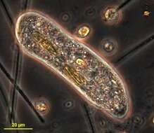
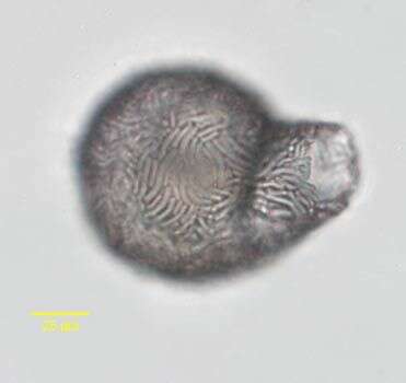
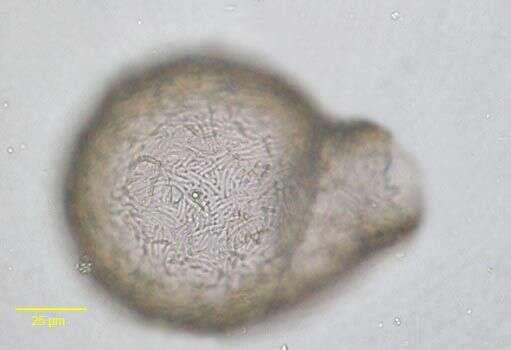
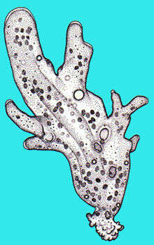
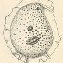
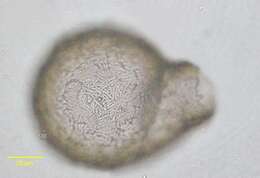
Test of the difflugiid amoeba, Lesquereusia epistomium (Penard, 1902).The test is composed of curved siliceous rods with occaisional interspersed quartz particles embedded in an organic matrix (sulfomucin complexed with proteins).The body of the test is more spherical and the neck more elongate than in the similar species L. spiralis.Collected from a freshwater pond near Boise, Idaho.Brightfield.
-
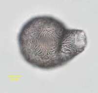
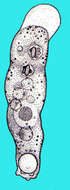
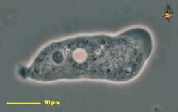
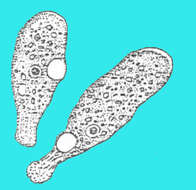
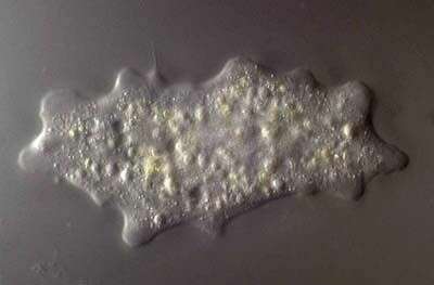
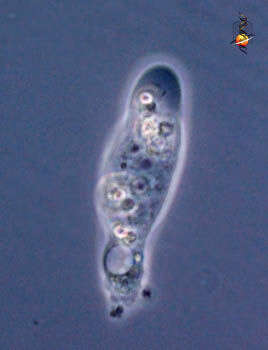
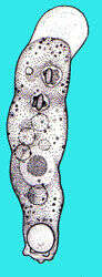
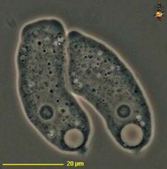
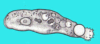
Trichospaherium is a marine amoeba that can occur in a spicule-covered form or as a naked form - as is illustrated here. the cone-shaped pseudopodia with terminal filaments are distinctive.
-
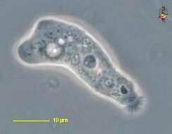
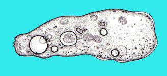
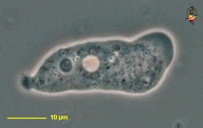
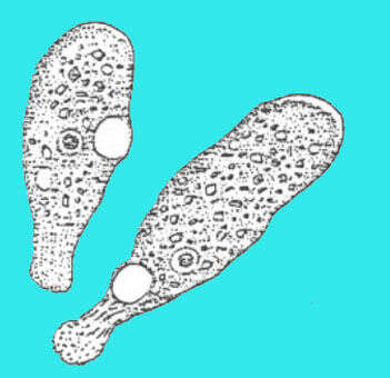
Marine amoeba, exists as a spicule-covered form and as a naked form. the second is illustrated here. The naked form used to be called Pontifex maximum before it became clear that the two forms are of the same species.
-
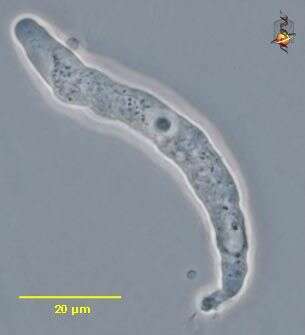
Hartmannella (heart-man-ella), a naked amoeba, limax (slug-like) body form, well developed hyaline cap, central nucleus and scrunched up uroidal region. Phase contrast.
-
Hartmannella (heart-man-ella), a naked amoeba, limax (slug-like) body form, cysts. Phase contrast.
-
Hartmannella (heart-man-ella), a naked amoeba, limax (slug-like) body form, well developed hyaline cap, central nucleus and scrunched up uroidal region. Phase contrast.
-
-
Cashia (cash-ee-a) - tentative identification - small limax (slug-shaped) amoeba, hyaline cap to right lacks inclusions, contractile vacuole is associated with the posterior end of the cell. Phase contrast.
-
-
Cashia. Cell observed in freshwater sediments in the vicinity of Broome, Western Australia in September 2003. This image was taken using phase contrast optics. This work was supported by the Australian Biological Resources Study.
-
Nolandella (no-lane-ell-a) is a small naked amoeba. Phase contrast micrograph.
-
-
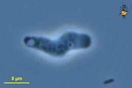
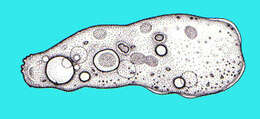
Saccamoeba (sack-a-me-ba), a monopodial naked free-living amoeba. With a lobose pseudopodium, usually progressing as a single pseudopodium (i.e. is monopodial). Hyaline cap absent or not well developed. Small uroid. These cells also with light-coloured contractile vacuoles and with nuclei with nucleoli. Phase contrast.
-
Saccamoeba (sack-a-me-ba), a monopodial naked free-living amoeba. With a lobose pseudopodium, usually progressing as a single pseudopodium (i.e. is monopodial). Hyaline cap absent or not well developed. Small uroid. This cell also with a light-coloured contractile vacuole and nucleus with nucleolus. Phase contrast.
-
-
-
-
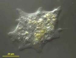
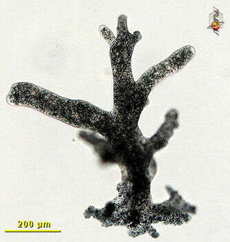
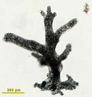
Darg ground illuminated image of an unconstrained cell. Its length is a little over half a millimeter. The cell is popypodial in that many broad rounded pseudopodia form at the same time. The cell is moving to the right. The posterior end of the cell has a rumpled appearance and is the uroid.
-
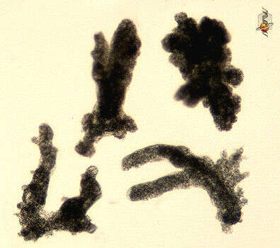
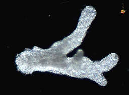
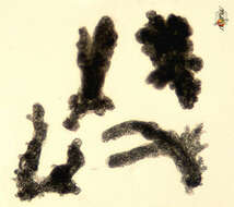
A smal collection of cells that are unconstrained and showing the different shapes that the amoebae may take. Stressed cells round up (upper right) and then begin to extend pseudopodia. Active cells are extended often producing many pseudopodia, and with the cells often becoming branched.
-
Bright field image of a moving amoeba. The cell is polypodial and has a number of rounded pseudopodia extending in the direction of movement (upwards in this image). The posterior end is rumpled and referred to as the uroid.
-
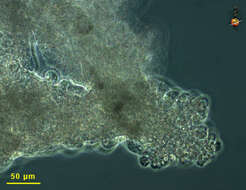
The posterior end of a moving cell has a rumpled appearance. this is the uroid. It forms as a result of the interactions between motility proteins such as actin and myosin. Phase contrast micrograph.
-
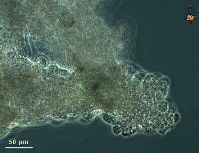
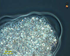
As many amoebae move, the front margin of the advancing psudopodia is filled with a transparent fluid. The cytoplasm and its contents are bound together in a more gelatinous cytoplasm. The transparent region is called the hyaline cap. Phase contrast micrograph.