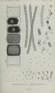
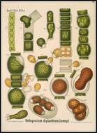
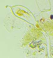
Summary.mw-parser-output table.commons-file-information-table,.mw-parser-output.fileinfotpl-type-information{border:1px solid #a2a9b1;background-color:#f8f9fa;padding:5px;font-size:95%;border-spacing:2px;box-sizing:border-box;margin:0;width:100%}.mw-parser-output table.commons-file-information-table>tbody>tr,.mw-parser-output.fileinfotpl-type-information>tbody>tr{vertical-align:top}.mw-parser-output table.commons-file-information-table>tbody>tr>td,.mw-parser-output table.commons-file-information-table>tbody>tr>th,.mw-parser-output.fileinfotpl-type-information>tbody>tr>td,.mw-parser-output.fileinfotpl-type-information>tbody>tr>th{padding:4px}.mw-parser-output.fileinfo-paramfield{background:#ccf;text-align:right;padding-right:0.4em;width:15%;font-weight:bold}.mw-parser-output.commons-file-information-table+table.commons-file-information-table,.mw-parser-output.commons-file-information-table+div.commons-file-information-table>table{border-top:0;padding-top:0;margin-top:-8px}@media only screen and (max-width:719px){.mw-parser-output table.commons-file-information-table,.mw-parser-output.commons-file-information-table.fileinfotpl-type-information{border-spacing:0;padding:0;word-break:break-word;width:100%!important}.mw-parser-output.commons-file-information-table>tbody,.mw-parser-output.fileinfotpl-type-information>tbody{display:block}.mw-parser-output.commons-file-information-table>tbody>tr>td,.mw-parser-output.commons-file-information-table>tbody>tr>th,.mw-parser-output.fileinfotpl-type-information>tbody>tr>td,.mw-parser-output.fileinfotpl-type-information>tbody>tr>th{padding:0.2em 0.4em;text-align:left;text-align:start}.mw-parser-output.commons-file-information-table>tbody>tr,.mw-parser-output.fileinfotpl-type-information>tbody>tr{display:flex;flex-direction:column}.mw-parser-output.commons-file-information-table+table.commons-file-information-table,.mw-parser-output.commons-file-information-table+div.commons-file-information-table>table{margin-top:-1px}.mw-parser-output.fileinfo-paramfield{box-sizing:border-box;flex:1 0 100%;width:100%}} Description: For background and links, please see: bibliodyssey.blogspot.com/2012/12/plant-anatomy-charts.html. Date: 25 August 2004, 15:21. Source: Oedogonium diplandrum, Juranyi. Author: Paul K from Sydney, Australia.
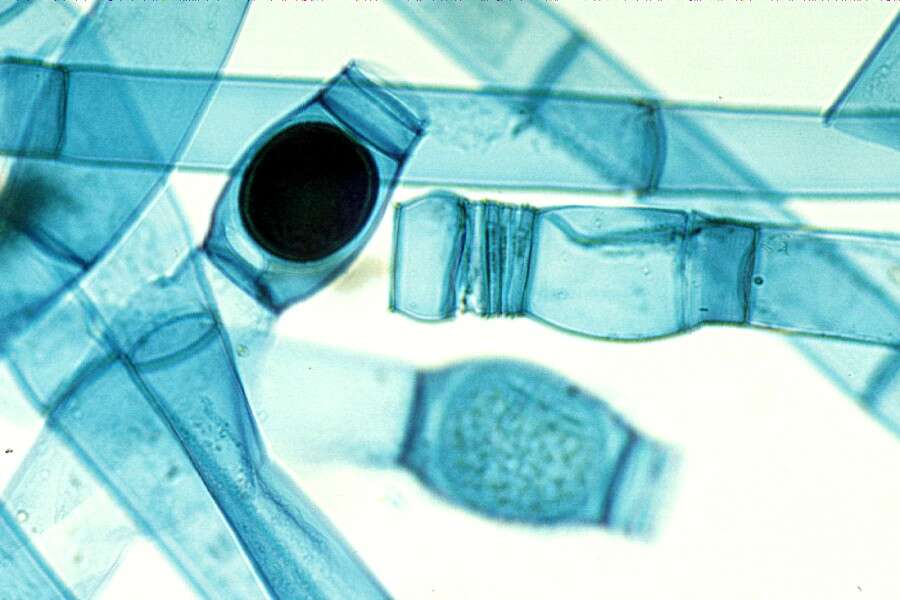
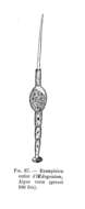
Description: Français : Oedogonium - dessin. Date: 27 November 2011. Source: Le Monde végétal ; Ernest Flammarion éditeur; 1907. Author: Gaston Bonnier.
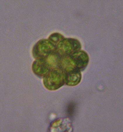
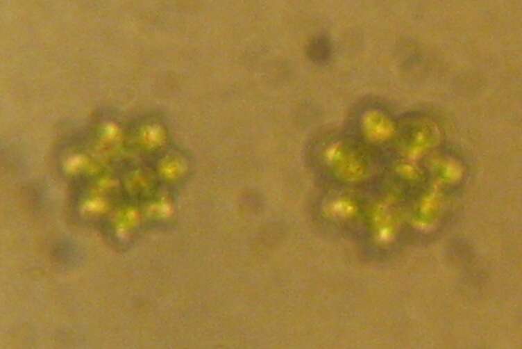
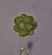
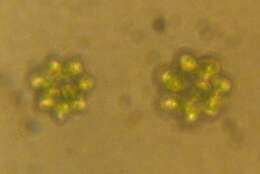
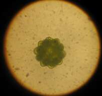
Description: Coenobium of Coelastrum sp. (A coenobium is a colony, containing a fixed number of cells, with little or no specialization). Eutrophic lake phytoplankton. Northern Poland. Date: 11 February 2007. Source: Own work. Author: http://commons.wikimedia.org/wiki/User:Panek. Permission(Reusing this file): 2.5.
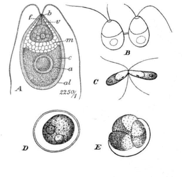
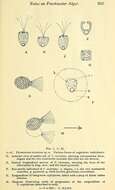
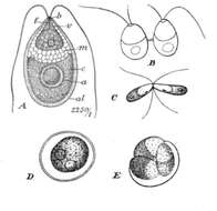
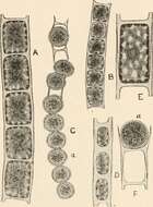
Description: English: 'Dunaliella salina' Teodor. A: Vegetative cell, B: Zoospores in cell division, C: Mating gametes, D: Ripe zygospore, E: Zygospore germination. Date: 1911. Source: Die natürlichen Pflanzenfamilien : Abt. 1a-Abt.1b. Euthallophyta, Unterabt. Schizophyta (Spaltpflanzen): Nebst ihren Gattungen und wichtigeren Arten, insbesondere den Nutzpflanzen By Adolf Engler, K Prantl Published by Engelmann, 1911, page 17. Author: Adolf Engler, K. Prantl.
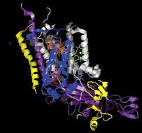
Description: English: The structure of cytochrome b6f from Chlamydomonas reinhardtii. Monomer. PDB id:1q90. Date: 9 August 2015. Source: Own work. Author: Эрг.
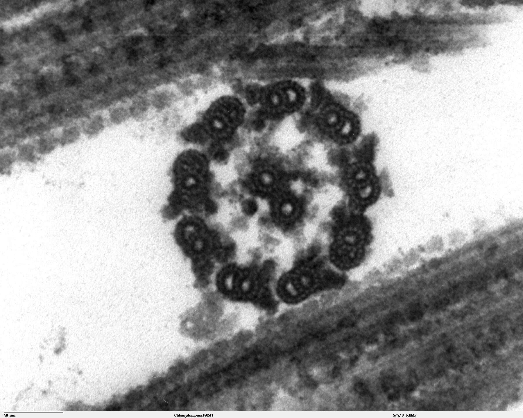
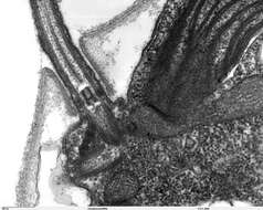
Description: Transmission electron microscope image, showing an example of green algae (Chlorophyta). Chlamydomanas reinhardtii is a unicellular flagellate used as a model system in molecular genetics work and flagellar motility studies. This image is a thin x-section cut through the isolated axoneme. Chlamydomonas flagella have the "9+2" structure characteristic of all eukaryotic cells. The axoneme has a central unit containing two single microtubules and nine peripheral doublet microtubules (known as the "9+2"). Dynein sidearms project from the A tubule of each doublet. Also visible in this image are the radial spokes and the inner sheath. Smith, E.F and P.A. Lefebvre (1996) "PF16 Encodes a Protein with Armadillo Repeats and Localizes to a Single Microtubule of the Central Apparatus in Chlamydomonas Flagella", J. Cell Biology, 132(3): 359-370 JEOL 100CX TEM. Source: http://remf.dartmouth.edu/imagesindex.htmlhttp://remf.dartmouth.edu/images/algaeTEM/source/14.html. Author: Elizabeth Smith, Louisa Howard, Erin Dymek. Permission(Reusing this file): PD.
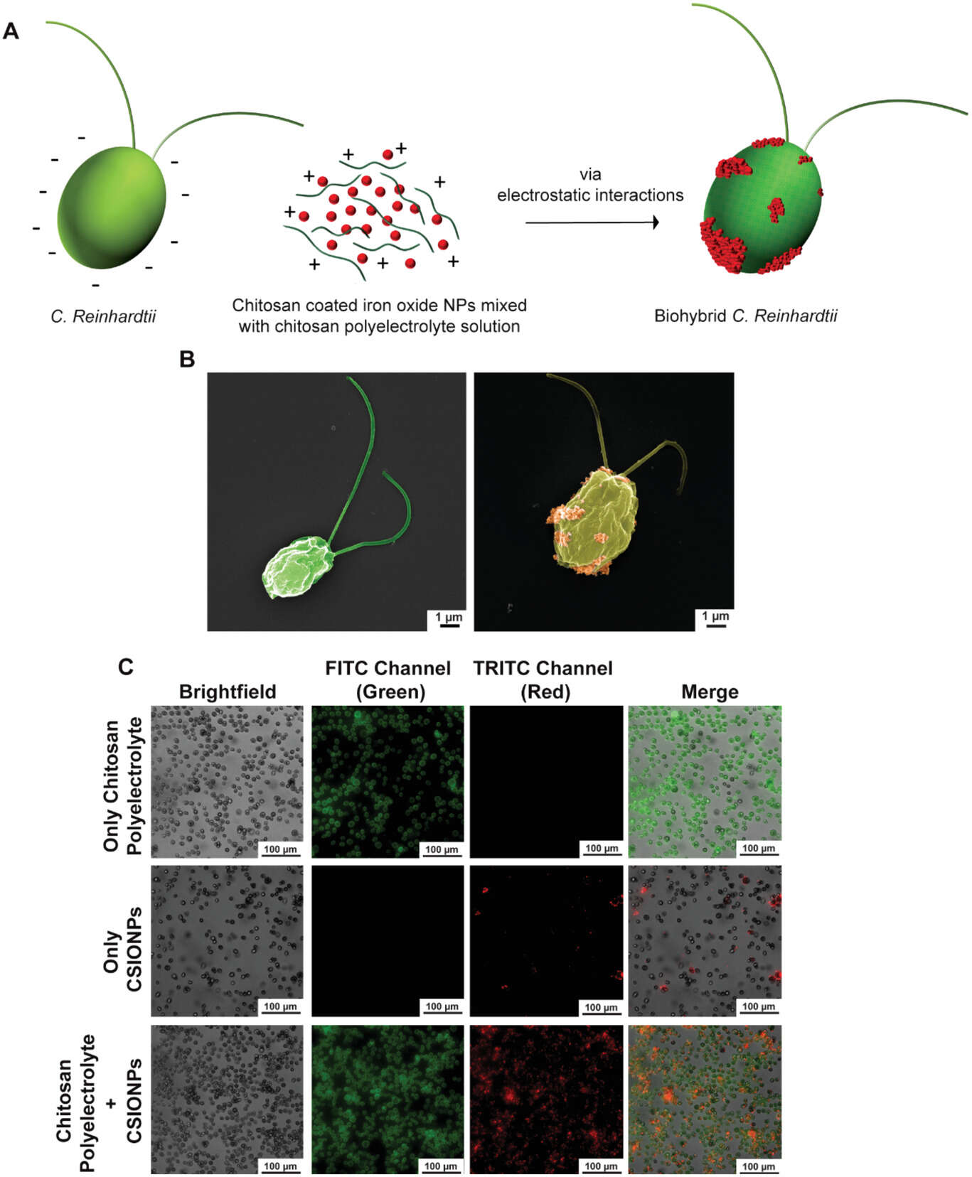
Mukrime Birgul Akolpoglu, Nihal Olcay Dogan, Ugur Bozuyuk, Hakan Ceylan, Seda Kizilel and Metin Sitti
Wikimedia Commons
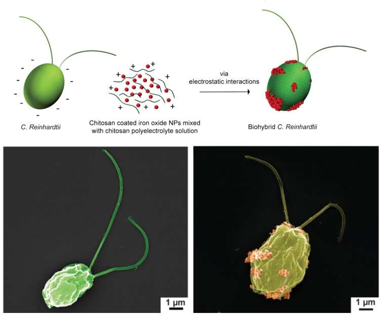
Description: English: Biohybrid Chlamydomonas reinhardtii microswimmers Top: Schematics of production steps for biohybrid C. reinhardtii. Bottom: SEM images of bare microalgae (left) and biohybrid microalgae (right) coated with chitosan-coated iron oxide nanoparticles (CSIONPs). Images were pseudocolored. A darker green color on the right SEM image represents chitosan coating on microalgae cell wall. Orange-colored particles represents CSIONPs. Date: 2 July 2020. Source: Extracted from this Commons file. Author: Mukrime Birgul Akolpoglu, Nihal Olcay Dogan, Ugur Bozuyuk, Hakan Ceylan, Seda Kizilel and Metin Sitti.
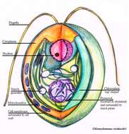
Description: English: Crossection of a Chlamydomonas reinhardtii algae cell, a 3D representation. Date: 18 September 2013. Source: Own work. Author: Ninghui Shi.
Ajay Vikram Singh, Vimal Kishore, Giulia Santomauro, Oncay Yasa, Joachim Bill, and Metin Sitti
Wikimedia Commons
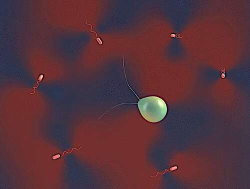
Description: English: Drawing of Chlamydomonas reinhardtii alga in coculture with Escherichia coli bacteria These two types of biological microswimmer (active colloid) populations exhibit opposite swimming behaviours: C. reinhardtii is a puller-type microswimmer while E. coli is a pusher-type microswimmer. Date: 28 April 2020. Source: [1]doi:10.1021/acs.langmuir.9b03665. Author: Ajay Vikram Singh, Vimal Kishore, Giulia Santomauro, Oncay Yasa, Joachim Bill, and Metin Sitti.
Description: English: Cross section of a Chlamydomonas reinhardtii algal cell, a 3D representation. Date: 18 September 2013. Source: Own work. Author: Ninghui Shi.
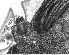
Description: Transmission electron microscope image, showing an example of green algae (Chlorophyta). Chlamydomanas reinhardtii is a unicellular flagellate used as a model system in molecular genetics work and flagellar motility studies. This image shows the flagellar apparatus, just after flagellar excision, which occurs at the transition zone(see area of flagella, with its fibers of the stellate structure). This image also shows components of the contractile vacuoles which are located just below the flagellar apparatus. JEOL 100CX TEM. Date: 7 October 2006. Source: Source and public domain notice at: http://remf.dartmouth.edu/imagesindex.htmlhttp://remf.dartmouth.edu/images/algaeTEM/source/11.html. Author: Elizabeth Smith, Louisa Howard, Erin Dymek (Dartmouth Electron Microscope Facility, Dartmouth College). Permission(Reusing this file): Released into the public domain.
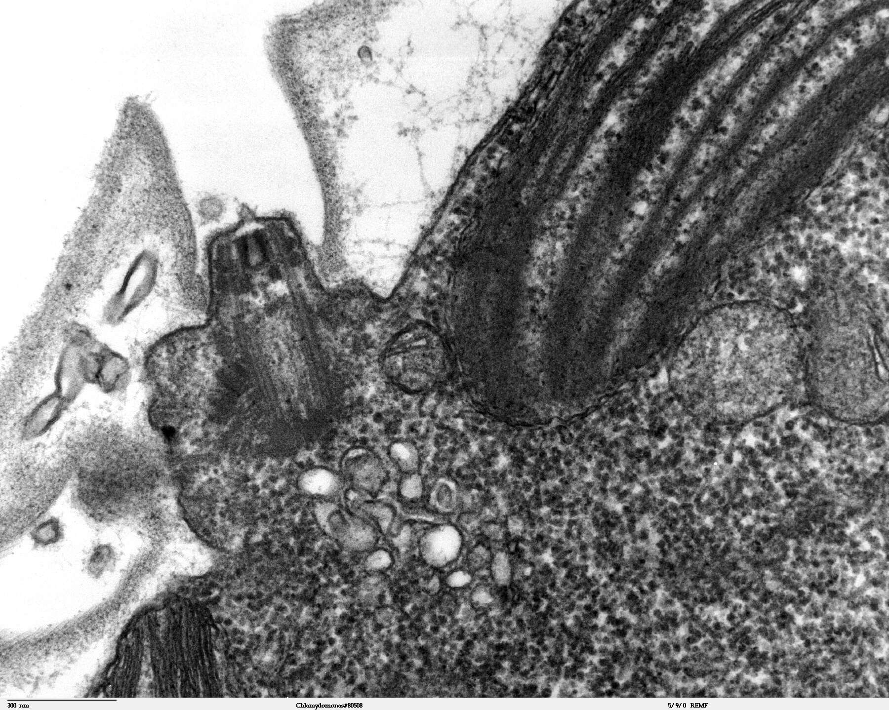
Description: Transmission electron microscope image, showing an example of green algae (Chlorophyta). Chlamydomanas reinhardtii is a unicellular flagellate used as a model system in molecular genetics work and flagellar motility studies. This image is a longitudinal section through a portion of the flagellar apparatus. In the cell apex are the basal body regions that are the anchoring sites for the flagella. This image shows that the two flagella form a V and they are connected at their bases by a transversely striated fibre. This connection is thought to play a part in the coordination of flagellar movement. Also visible is the transition region, with its fibers of the stellate structure. JEOL 100CX TEM. Source: http://remf.dartmouth.edu/imagesindex.htmlhttp://remf.dartmouth.edu/images/algaeTEM/source/10.html. Author: Elizabeth Smith, Louisa Howard, Erin Dymek. Permission(Reusing this file): PD.
Description: English: Schamatic of Chlamydomonas reinhardtii cell surface display forming. Starting from secretion to anchoring in the surface. Date: 2 June 2021. Source: Own work. Author: Candidomolino.
Mukrime Birgul Akolpoglu, Nihal Olcay Dogan, Ugur Bozuyuk, Hakan Ceylan, Seda Kizilel and Metin Sitti
Wikimedia Commons
Description: English: Biohybrid Chlamydomonas reinhardtii microswimmers A) Schematics of production steps for biohybrid C. reinhardtii. B) SEM images of bare microalgae (left) and biohybrid microalgae (right) coated with chitosan-coated iron oxide nanoparticles (CSIONPs). Images were pseudocolored. A darker green color on the right SEM image represents chitosan coating on microalgae cell wall. Orange-colored particles represents CSIONPs. C) Microscopy images of biohybrid microalgae treated with three different solutions: 1) microalgae coated with 5 µg mL−1< /sup> green-fluorescent chitosan polyelectrolyte solution (first row), 2) microalgae coated with 10 µg mL−1< /sup> red fluorescent CSIONPs (second row), and 3) microalgae coated with both CSIONPs (10 µg mL−1< /sup>) dispersed in chitosan polyelectrolyte solution (5 µg mL−1< /sup>) (third row. Date: 2 July 2020. Source: [1]doi:10.1002/advs.202001256. Author: Mukrime Birgul Akolpoglu, Nihal Olcay Dogan, Ugur Bozuyuk, Hakan Ceylan, Seda Kizilel and Metin Sitti.
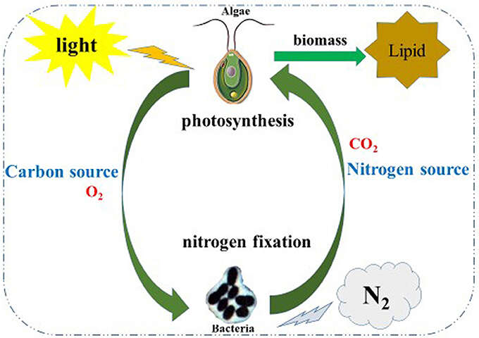
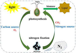
Description: English: Co-cultivation of an algae-bacterial system to improve biomass of and lipid production by algae In this co-system, algae (Chlamydomonas reinhardtii) and bacteria (Azotobacter chroococcum) could enhance the growth and biomass of each other through material exchange; algae supply carbohydrates and O2 by photosynthesis while A. chroococcum supply the nitrogen source and CO2 to algae by nitrogen fixation. Date: 29 April 2018, 23:30:52. Source: [1]doi:10.3389/fpls.2018.00741. Author: Lili Xu, Xianglong Cheng and Quanxi Wang.
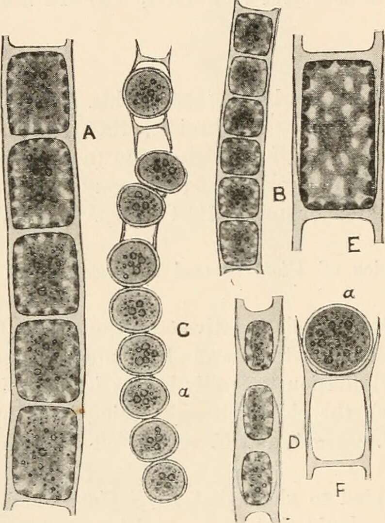
Description: English: Fig. 184. A, Microspora amœna (Kütz.) Lagerh. B and C, ? M. abbreriata (Rabenh.) Lagerh.; B, vegetative filament; C, filament with aplanospores (a). D, M. pachyderma (Wille) Lagerh. E, single vegetative cell of M. amœna var. crassior Hansg., showing the reticulated chloroplast. The indistinct blur in the centre of the cell indicates the position of the nucleus. F, fragment of filament of M. amœna with aplanospore (a). All x 520. Date: 1916. Source: https://www.flickr.com/photos/126377022@N07/14760704711/ page 301 of "Algæ. Vol. I. Myxophyceæ, Peridinieæ, Bacillarieæ, Chlorophyceæ, together with a brief summary of the occurrence and distribution of freshwat4er Algæ" (1916). Author: West, G. S. (George Stephen), 1876-1919.
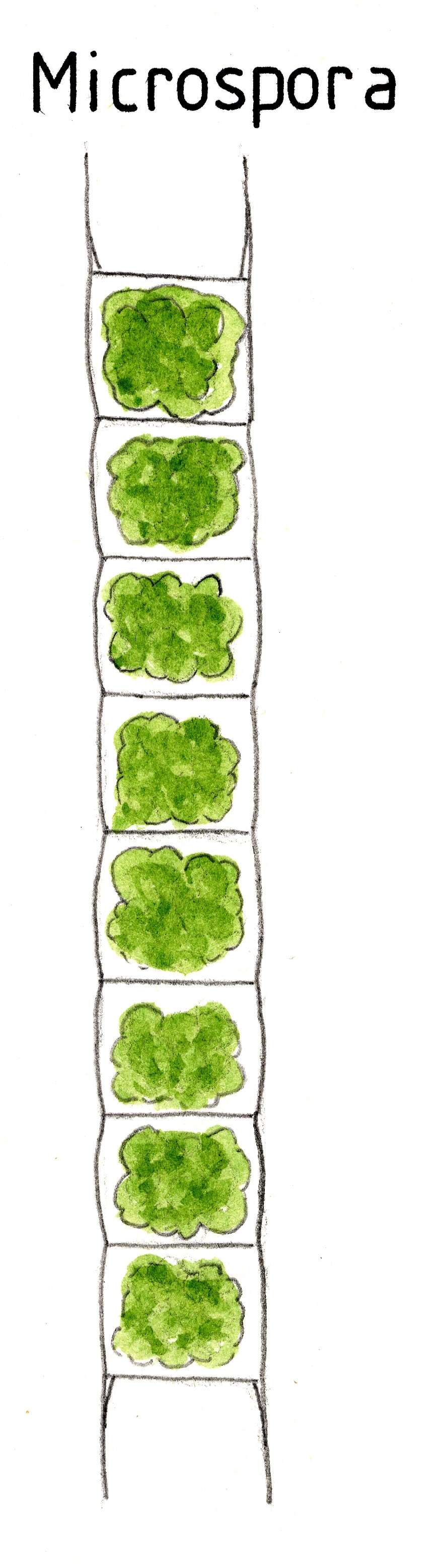
Pentecost, Allan [Artist] (2016) at Freshwater Biological Association [publisher]
Wikimedia Commons
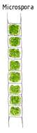
Description: English: Diagnostic Drawing: Microspora, an Ulothrix related genus. Date: 14 October 2008, 10:52:50. Source: Extract from http://www.environmentdata.org/archive/fbaia:2434 at http://www.environmentdata.org/ Agricultural & Environmental Data Archive (AEDA). Author: Pentecost, Allan [Artist] (2016) at Freshwater Biological Association [publisher].
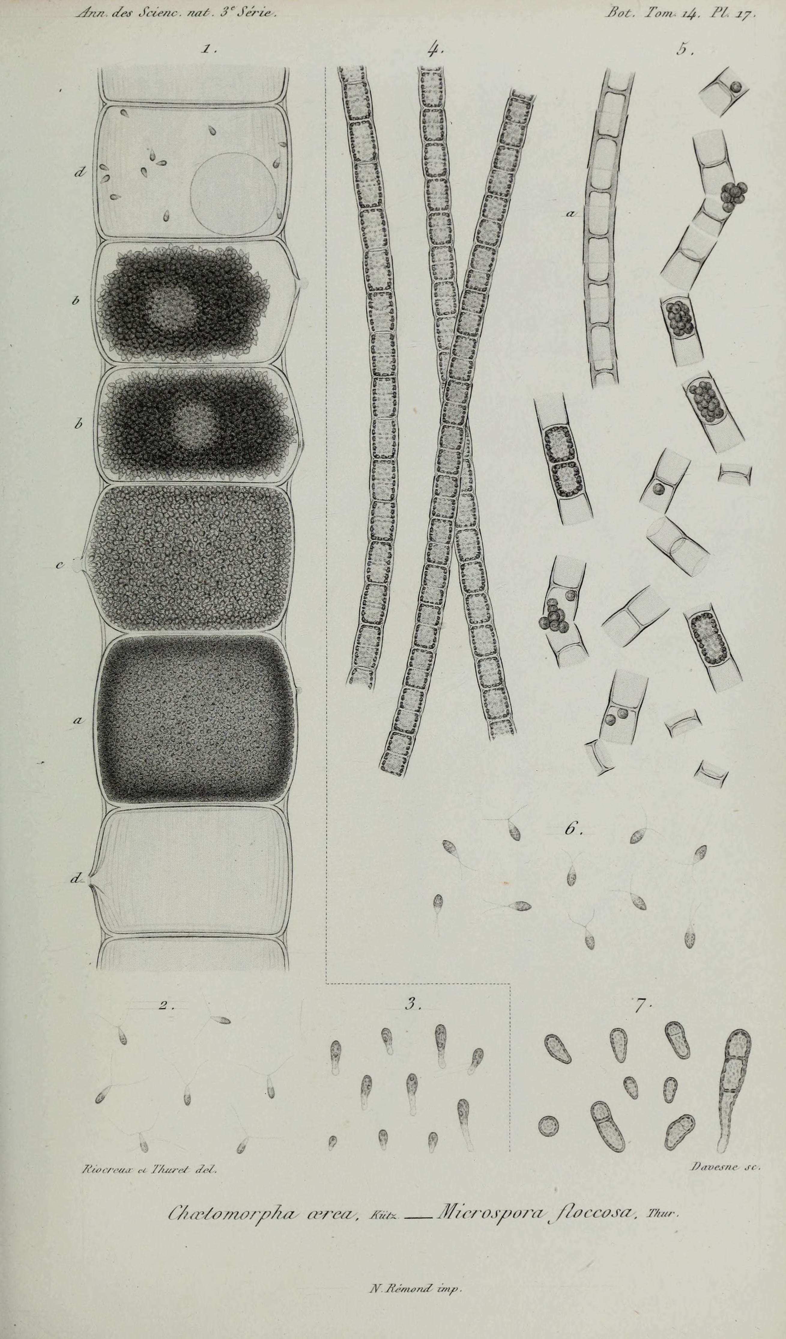
Description: English: Title: Annales des Sciences Naturelles Botaniques Identifier: annalesdesscienc3141850pa (find matches) Year: 1850 (1850s) Authors: Subjects: Publisher: Paris Contributing Library: Natural History Museum Library, London Digitizing Sponsor: BHL-SIL-FEDLINK View Book Page: Book Viewer About This Book: Catalog Entry View All Images: All Images From Book Click here to view book online to see this illustration in context in a browseable online version of this book. Text Appearing Before Image: y&isi. {/?s Sdeno. /m/-. 3eSr'/-iw /jfy. PL j/. Text Appearing After Image: T&ocretui ,■< TAuret ,M. Note About Images Please note that these images are extracted from scanned page images that may have been digitally enhanced for readability - coloration and appearance of these illustrations may not perfectly resemble the original work. Date: 1850. Source: https://www.flickr.com/photos/internetarchivebookimages/18408571515/. Author: Internet Archive Book Images. Permission (Reusing this file): At the time of upload, the image license was automatically confirmed using the Flickr API. For more information see Flickr API detail. Volume: Ser. 3, v. 14 (1850). Flickr tags: bookid:annalesdesscienc3141850pa bookyear:1850 bookdecade:1850 bookcentury:1800 bookpublisher:Paris bookcontributor:Natural_History_Museum_Library_London booksponsor:BHL_SIL_FEDLINK bookleafnumber:421 bookcollection:biodiversity BHL Collection BHL Consortium. Flickr posted date: 3 June 2015.


