Chlamydomanas reinhardtii Flagella 5 - TEM
Description:
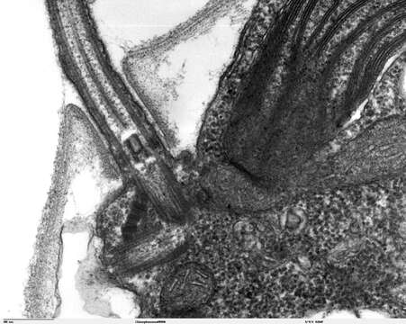
Description: Transmission electron microscope image, showing an example of green algae (Chlorophyta). Chlamydomanas reinhardtii is a unicellular flagellate used as a model system in molecular genetics work and flagellar motility studies. This image is a longitudinal section through a portion of the flagellar apparatus. In the cell apex are the basal body regions that are the anchoring sites for the flagella. This image shows that the two flagella form a V and they are connected at their bases by a transversely striated fibre. This connection is thought to play a part in the coordination of flagellar movement. Also visible is the transition region, with its fibers of the stellate structure. JEOL 100CX TEM. Source: http://remf.dartmouth.edu/imagesindex.html http://remf.dartmouth.edu/images/algaeTEM/source/10.html. Author: Elizabeth Smith, Louisa Howard, Erin Dymek. Permission(Reusing this file): PD.
Included On The Following Pages:
- Life (creatures)
- Cellular (cellular organisms)
- Eukaryota (eukaryotes)
- Archaeplastida (plants)
- Chloroplastida (green plants)
- Chlorophyta (chlorophytes)
- Chlorophyceae
- Chlamydomonadales
- Chlamydomonadaceae
- Chlamydomonas
- Chlamydomonas reinhardtii
This image is not featured in any collections.
Source Information
- license
- cc-publicdomain
- creator
- Elizabeth Smith, Louisa Howard, Erin Dymek
- original
- original media file
- visit source
- partner site
- Wikimedia Commons
- ID