-
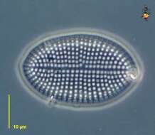
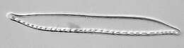
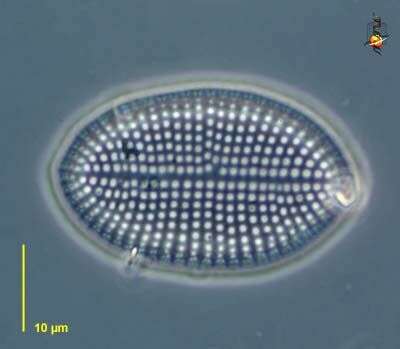
Cocconeis (cock-owe-neigh-us) one isolated valve seen from valve view, the perforations in this siliceous shell allow the cells which normally live within to exchange nutrients etc. with the outside world. Phase contrast micrograph.
-
-
-
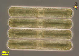
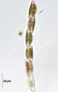
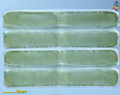
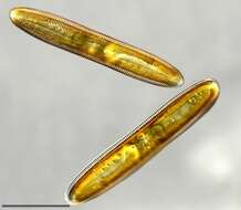
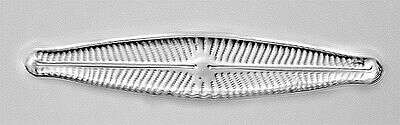
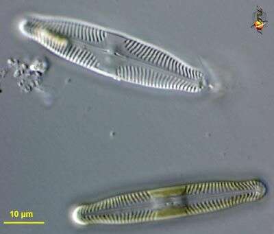
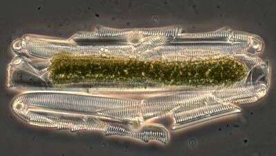
Pinnularia (pin-you-lair-ee-a). a pennate diatom, found either individually or in clusters as here. As with other diatoms with a siliceous cell wall. Nucleus central. Phase contrast.
-
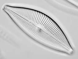
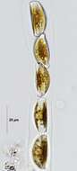
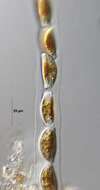
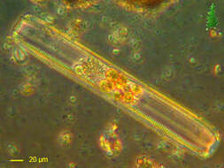
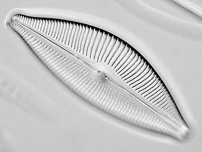
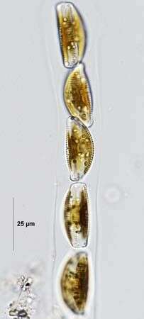
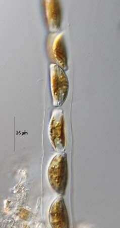
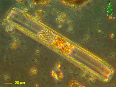
Cymbella tumida (Brébisson in Kützing) van Heurck 1882-1885. Frustules within gelatinous tube. Collected from detritus in a freshwater irrigation canal. Boise, Idaho. March 2009. Brightfield.
-
-
Pinnularia (pin-you-lair-ee-a). a pennate diatom, found either individually or in clusters as here. As with other diatoms with a siliceous cell wall. Nucleus central. Differential interference contrast.
-
Cymbella tumida (Brébisson in Kützing) van Heurck 1882-1885. Cymbella tumida (Brébisson in Kützing) van Heurck 1882-1885. Frustules within gelatinous tube. Collected from detritus in a freshwater irrigation canal. Boise, Idaho. March 2009. Brightfield.
-
-
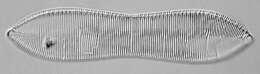
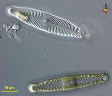
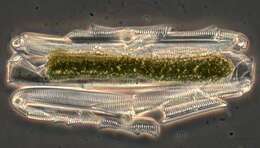
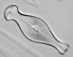
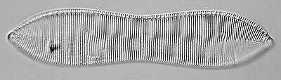
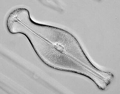
Pinnularia (pin-you-lair-ee-a) a pennate diatom, the upper cell is the empty siliceous wall or frustule, the lower cell is a living cell with brown-ish chloroplast and central nucleus. Diatoms are mostly identified and classified using the markings on the surface of the frustule. The central line is the raphe, and is associated with the gliding movements of the cell. Differential interference contrast. Material from Nymph Lake, thermal sites within Yellowstone National Park, photograph by Kathy Sheehan and David Patterson.
-
Cymbella tumida (Brébisson in Kützing) van Heurck 1882-1885. Frustules within gelatinous tube. Collected from detritus in a freshwater irrigation canal. Boise, Idaho. March 2009.DIC.
-
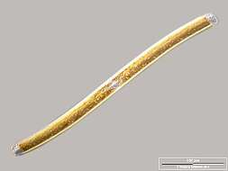
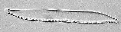
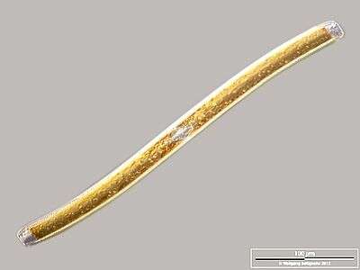
Cingular view. Scale bar indicates 100 µm. Sample from a wetland at the Pillersee (Tyrol, Austria). The image was built up using several photomicrographic frames with manual stacking technique. Images were taken using Zeiss Universal with Olympus C7070 CCD camera.Image under Creative Commons License V 3.0 (CC BY-NC-SA).
-
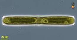
Pinnularia (pin-you-lair-ee-a), large pennate diatom. Margins reveal strengthening struts. The plastid has a browny-green colour. Phase contrast.
-
Cymbella tumida (Brébisson in Kützing) van Heurck 1882-1885. Frustules within gelatinous tube. Collected from detritus in a freshwater irrigation canal. Boise, Idaho. March 2009. Phase contrast.
-
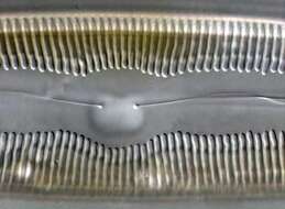
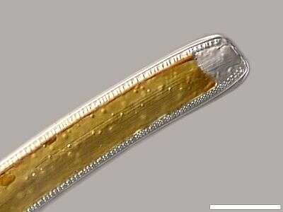
Detail: Apex in cingular view, displaying cingulum (central strae and the two canal raphes on the edges. Scale bar indicates 25 µm. Sample from a wetland at the Pillersee (Tyrol, Austria). The image was built up using several photomicrographic frames with manual stacking technique. Images were taken using Zeiss Universal with Olympus C7070 CCD camera.Image under Creative Commons License V 3.0 (CC BY-NC-SA).
-
The intricate frustule of the diatom Pinnularia. Frustules have ridges, grooves, and pores that are useful for identification of different diatoms. The two grooves in the center of the cell form the raphe and the raphe is used for propelling the diatom.
-
Cymbella tumida (Brébisson in Kützing) van Heurck 1882-1885. Frustule within gelatinous tube. Collected from detritus in a freshwater irrigation canal. Boise, Idaho. March 2009.DIC.
-
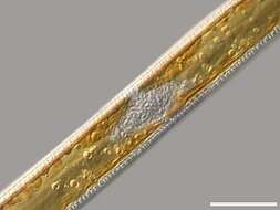
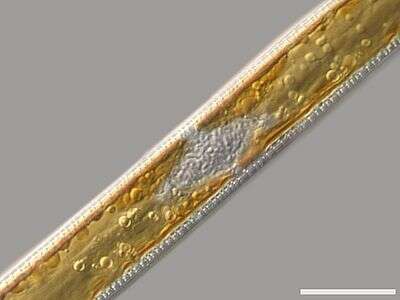
Detail: Center of the cell in cingular view with nucleus, cloroplasts with oil droplets (energy store) and the canal raphes. Scale bar indicates 25 µm. Sample from a wetland at the Pillersee (Tyrol, Austria). The image was built up using several photomicrographic frames with manual stacking technique. Images were taken using Zeiss Universal with Olympus C7070 CCD camera.Image under Creative Commons License V 3.0 (CC BY-NC-SA).
-
Diatom frustules are brittle and delicate. This Pinnularia frustule was broken when it was flattened between the microscope slide and the coverslip.
-
-
Specimen were selected from a sample of bottom sediments of a rain storage reservoir in Kiel (Schleswig-Holstein, Germany). The scale bar indicates 50 µm. Image was taken using Zeiss Universal with Olympus C7070 CCD camera.
-
Collected from Le Barron white cedar swamp on July 1, 2004.
-
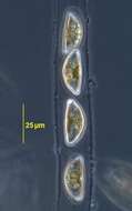
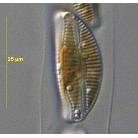
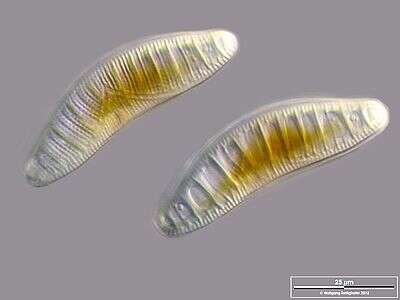
Cells in valvar view and in optical transversal section, showing shell reinforcement through transapical costae. Scale bar indicates 25 µm. Sample from Lake Constance near Bodman. Images were taken using Zeiss Universal with Olympus C7070 CCD camera.Image under Creative Commons License V 3.0 (CC BY-NC-SA).
-
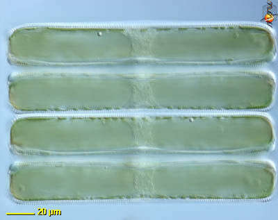
Pinnularia cell in valvar view (above) and an optical median cut in cingular projection showing the nucleus and oil droplets. Scale bar indicates 50 µm. Sample from sphagnum pond situated in the northern alpine region of Austria near Salzburg. Images were taken using Zeiss Universal with Olympus C7070 CCD camera.