-
-
-
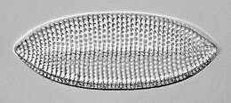
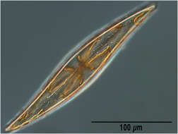
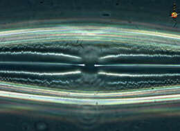
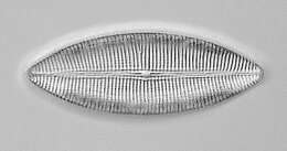
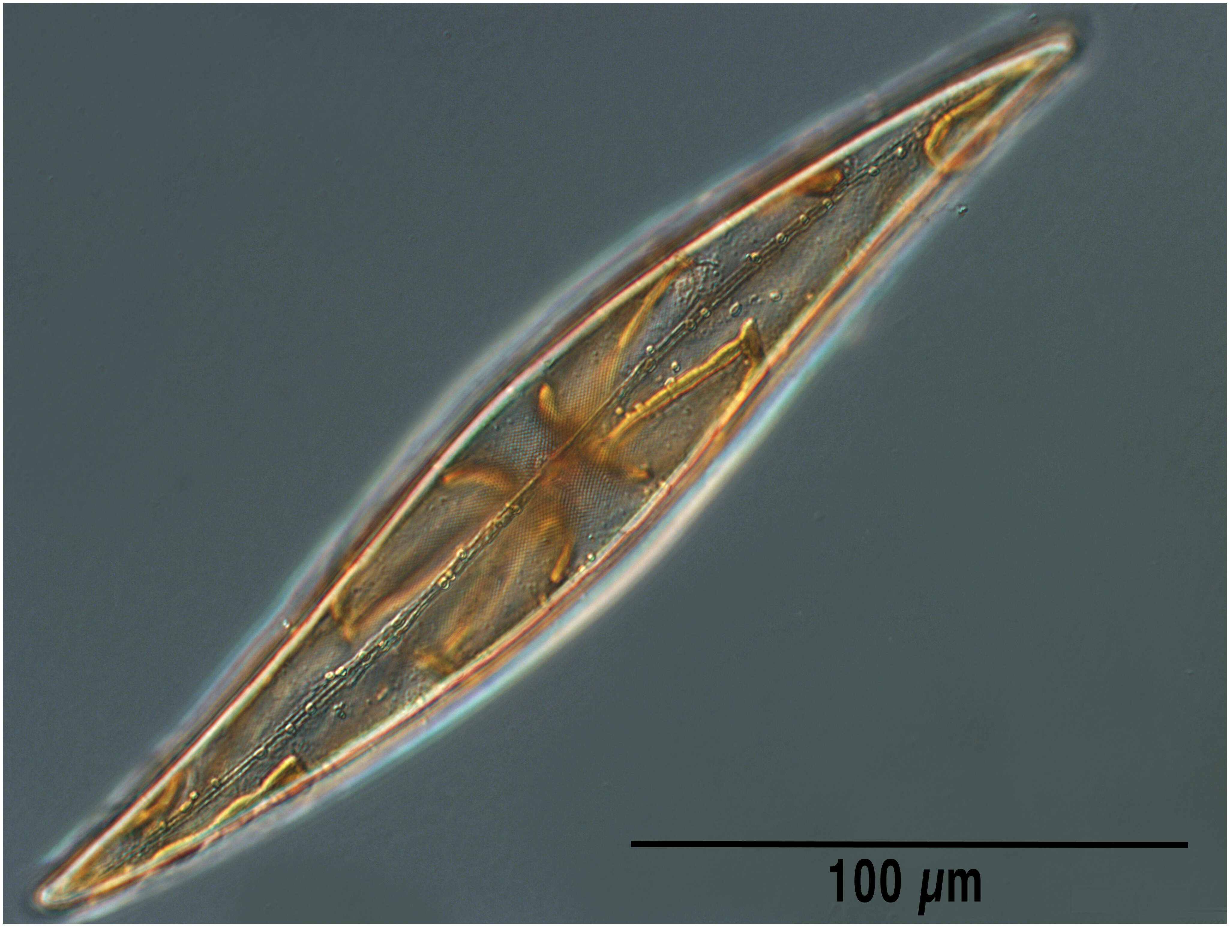
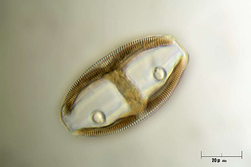
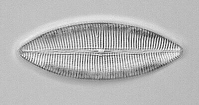
Pennate diatoms. The cells are enclosed in siliceous valves. There are typically two valves - a top one and a lower one, and they are joined together with fine bands or girdle strips. With plastids containing chlorophylls a and c (they are stramenopiles after all). Genera and species distinguished largely by the shape of the organism and the pattern of pores and sculptings of the siliceous shell or frustule. This pennate diatom is seen in valve view, there is an H shaped zone without sculptings in the frustule. Large plastids. The raphe is the two-part line running axially along the centre of the valve. Differential interference contrast.
-
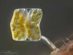
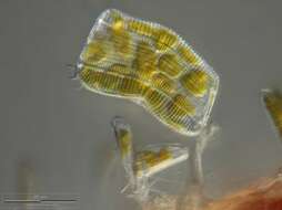
Two cells with stalk. Multi layer image using 20 frames generating depth of focus, stacked manually using Corel Photopaint. Collected from Bodden, the brackish waters lying between the isles of Hiddensee and Ruegen (German Baltic Sea). This image was taken using Zeiss Universal with Olympus C7070 CCD camera.
-
-
Material from the Netherlands.
-
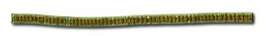
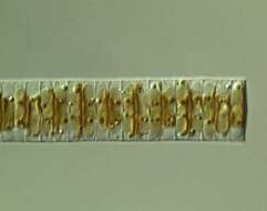
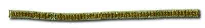
Filamentous diatom commonly found near the Tvarminne Zoological Station. Previously known as Achnantes taeniata.
-
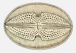
Navicula excavata.
-
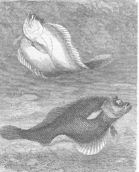
Dab (platessa Imanda).
-
FRom the Bay of Villefranche in March 2013
-
-
-
-
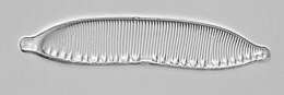
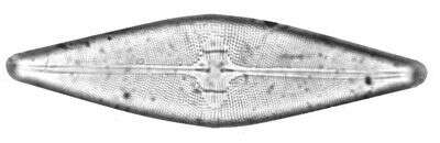
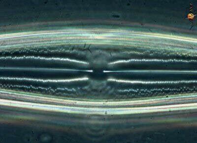
Centre of valve showing the inner termini of the raphe structure, phase contrast micrograph of empty frustule.
-
Single cell with stalk. Digital drawing using 18 frames generating depth of focus, stacked manually using Corel Photopaint. Scale bar indicates 25 µm. Collected from Bodden, the brackish waters lying between the isles of Hiddensee and Ruegen (German Baltic Sea). This image was taken using Zeiss Universal with Olympus C7070 CCD camera.
-
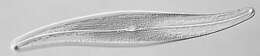
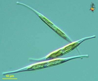
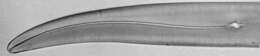
Cylindrotheca (sill-inn-dro-thee-ka) fusiformis, an elongate and slightly twisted pennate diatom (stramenopile), tends to move in a spiral motion, frustule not heavily silicifed and and can be deformed. Differential interference microscopy.
data on this strain.
-
Image from type material.
-
Detail showing plastids inside several cells at the end of a filament. Same filament as included in another picture in this collection.
-
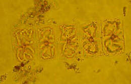
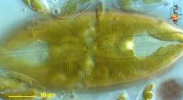
Valves are rectangular in girdle view and elliptical in valve view. Each cell has four ribbon like and folded chloroplasts, two along each side of the girdle.
-
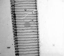
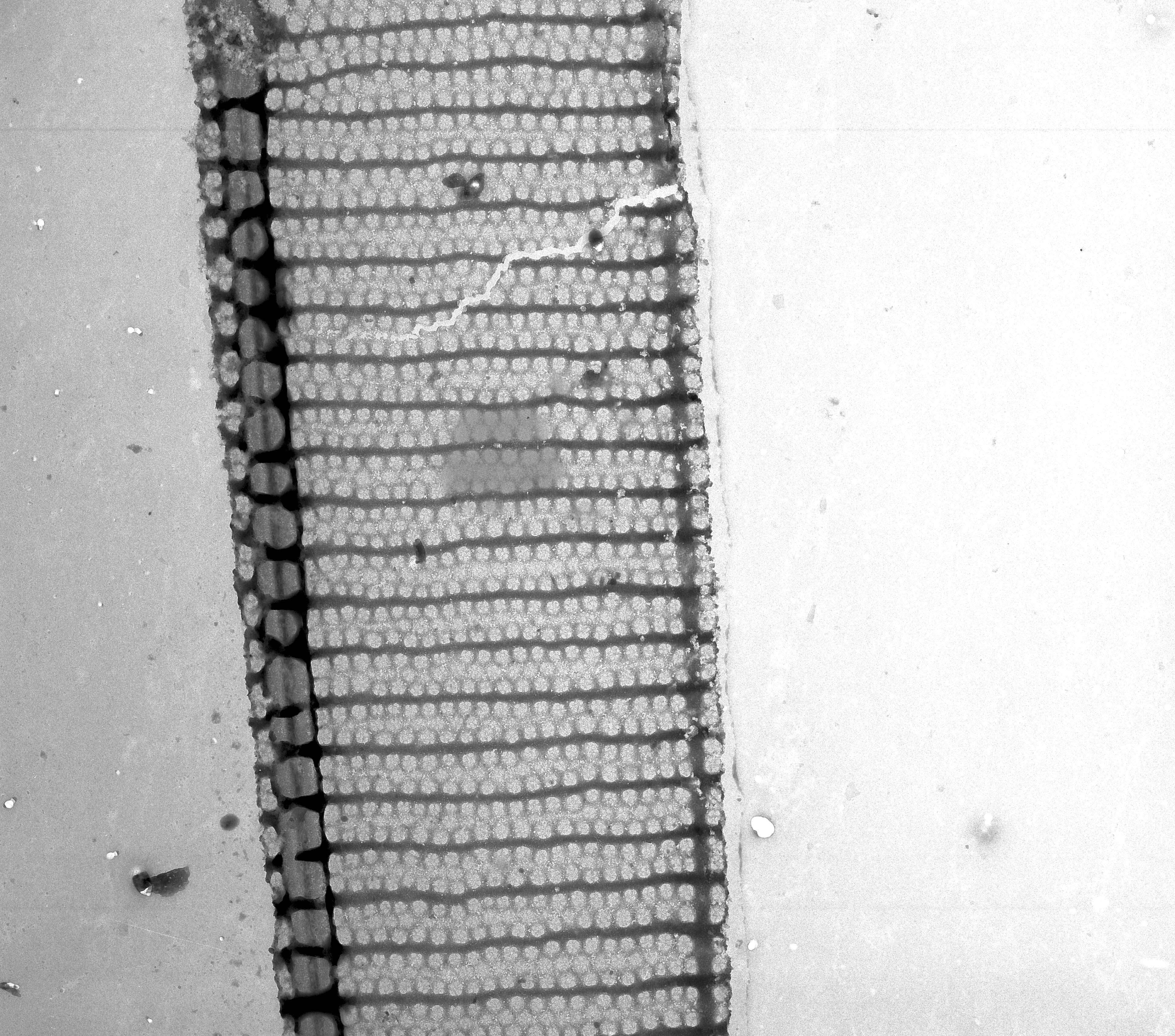
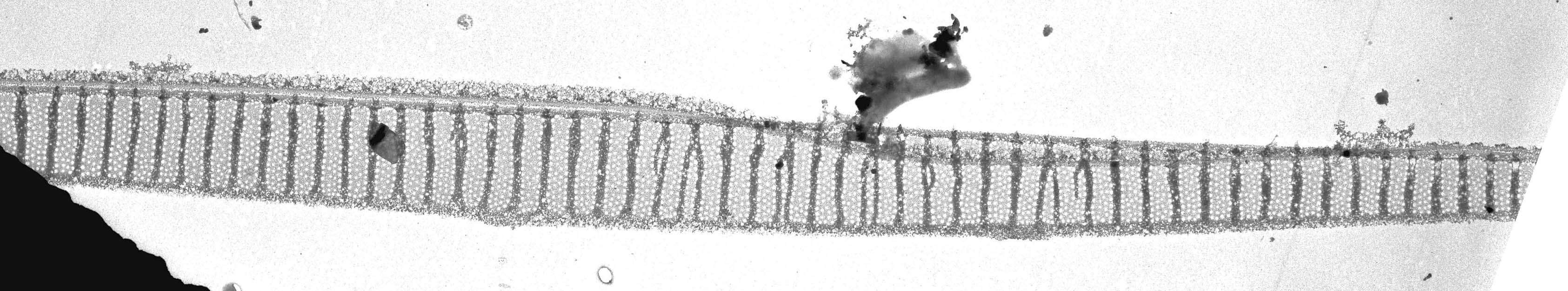
This EM isn't very good, but it's better than nothing.
-
-
-
-
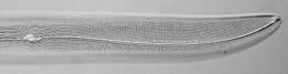
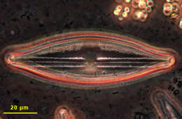
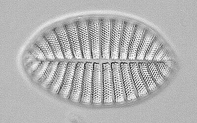
Pennate diatom, valve view of an empty frustule (shell). Phase contrast optics.