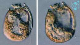
Sinophysis (sine-o-fi/fu-sis) ebriolum (Herdman) Balech 1956. The image on the left shows a cell in right lateral view. The cell is laterally compressed. The epicone is much smaller than the hypocone. The image on the right shows a cell in a mid-focal plane. The cells contain no plastids. The cells are thecate and have cingular lists.
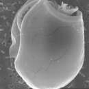
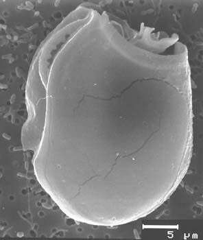
Sinophysis ebriolum, scanning electron microscope image. This image was taken by Mona Hoppenrath of a sample from Town Beach, Broome. This work was supported by the Australian Biological Resources Study.