-
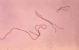
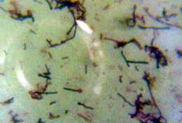
This 1976 Gram-stained photomicrograph depicted a number of chains of Gram-positive, anaerobic, coccoid (GPAC) Coprococcus eutactus bacteria. Bacteriologist commonly refer to these bacteria as Peptococci or Peptostreptococci.Created: 1976
-
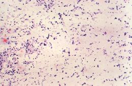
Magnified 1000X, this photomicrograph depicts a number of Gram-positive Eubacterium alactolyticum bacteria, which had been grown in Schaedler broth.Created: 1974
-
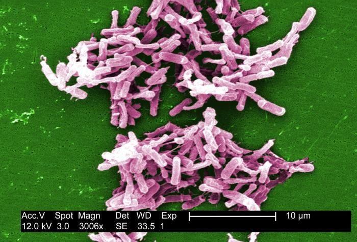
This micrograph depicts Gram-positive C. difficile bacteria from a stool sample culture obtained using a .1µm filter. See PHIL 6260 for a black and white version of this image.Created: 2004
-
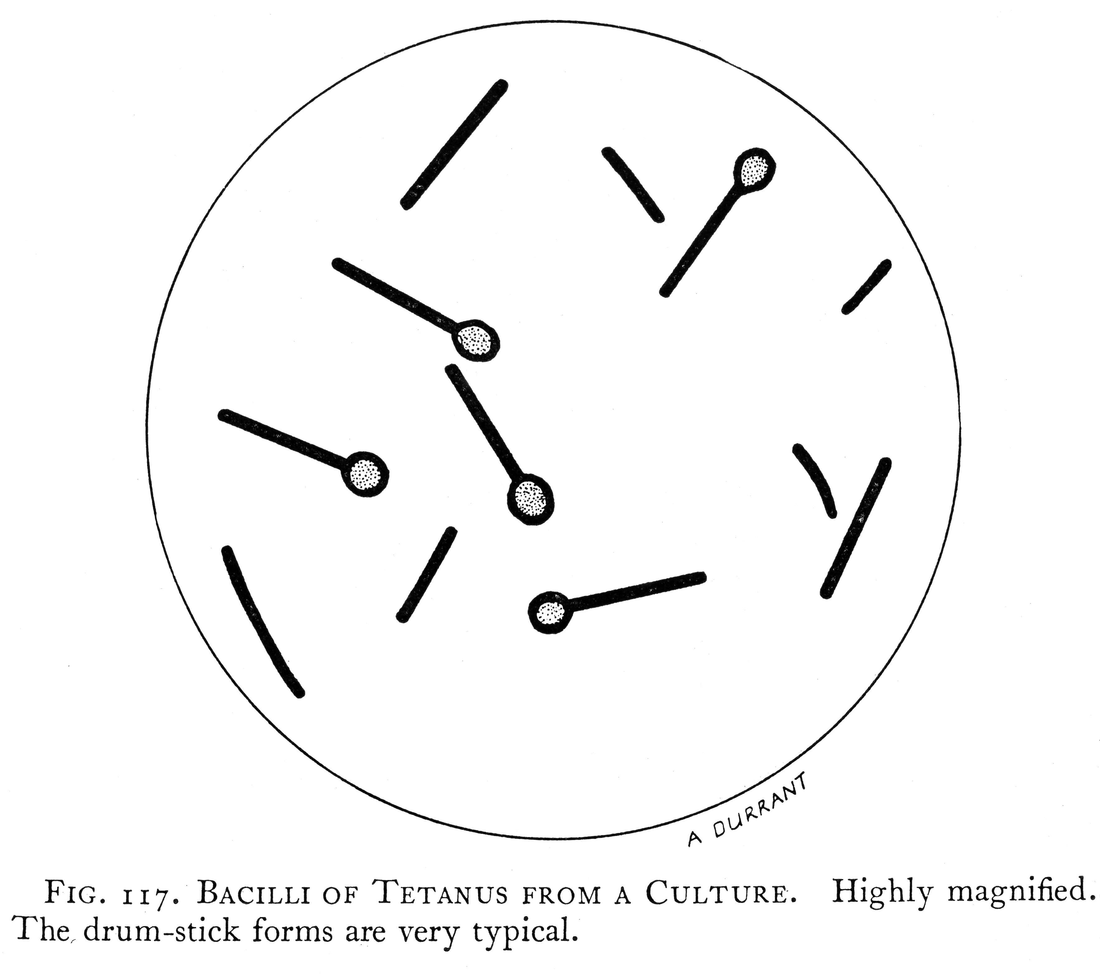
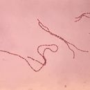
This micrograph depicts a group of Clostridium tetani bacteria, responsible for causing tetanus in humans.From
Wikimedia Commons
-
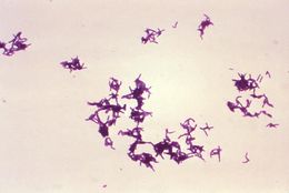
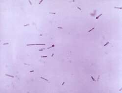
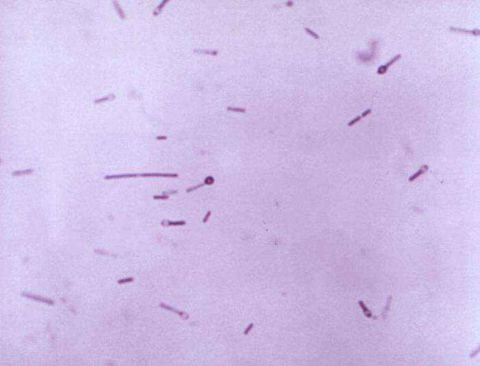
Magnified 1000X, this micrograph depicts numerous Peptostreptococcus sp. bacteria that were grown in Schaedlers broth, and stained using the Gram-stain technique.Created: 1974
-
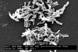
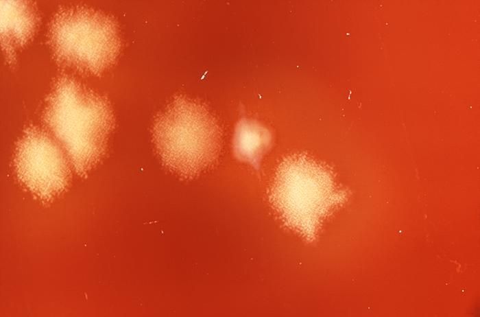
Under only a 2X magnification, this photograph revealed some of the details displayed atop a Petri dish culture of rod-shaped, Gram-positive Clostridium septicum bacteria.Created: 1971
-
This micrograph depicts Gram-positive C. difficile bacteria from a stool sample culture obtained using a .1µm filter. See PHIL 9999 for a colorized version of this image.Created: 2004
-
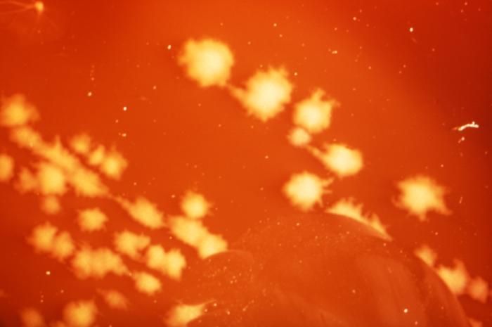
Under only a 2X magnification, this photograph revealed some of the details displayed atop a Petri dish culture of rod-shaped, Gram-positive Clostridium septicum bacteria.Created: 1971
-
Under only a 2X magnification, this photograph revealed some of the details displayed atop a Petri dish culture of rod-shaped, Gram-positive Clostridium septicum bacteria.Created: 1971
-
Under only a 2X magnification, this photograph revealed some of the details displayed atop a Petri dish culture of rod-shaped, Gram-positive Clostridium septicum bacteria.Created: 1971
-
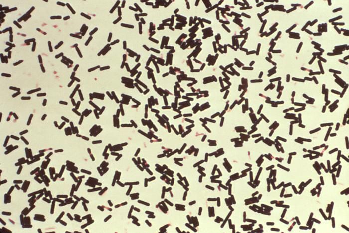
Magnified 1000X, this photomicrograph reveals numbers of Clostridium perfringens bacteria that had been grown in Schaedlers broth, and subsequently stained using Gram-stain.Created: 1974
-

Description: English: A photomicrograph of Clostridium botulinum bacteria. This is an illustration of a photomicrograph of Clostridium botulinum stained with Gentian violet. The bacterium C. botulinum produces a nerve toxin, which causes the rare, but serious paralytic illness Botulism. Italiano: Il botulino, un batterio che produce una tossina mortale. Date: 1979. Source: : This media comes from the
Centers for Disease Control and Prevention's
Public Health Image Library (PHIL), with identification number
#2107. Note: Not all PHIL images are public domain; be sure to check copyright status and credit authors and content providers.
العربية |
Deutsch |
English |
македонски |
slovenščina |
+/−. Author: Content Providers: CDC. Permission(
Reusing this file): English: None - This image is in the public domain and thus free of any copyright restrictions. As a matter of courtesy we request that the content provider be credited and notified in any public or private usage of this image. Other versions:
PNG version.
-
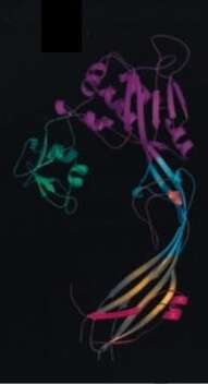
Description: English: Model of Aerolysin. Date: 2004. Source: Melton JA, Parker MW, Rossjohn J, Buckley JT and Tweten RK (2004). The identification and structure of the membrane-spanning domain of the clostridium septicum alpha toxin. The Journal of Biological Chemistry. 279(14): 14315-14322. Author: Jody A. Melton, Michael W. Parker, Jamie Rossjohn, J. thomas Buckley and Rodney K. Tweten.
-
-
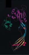
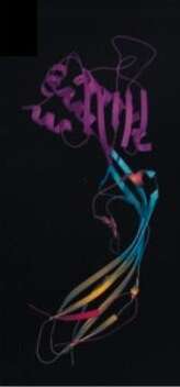
Description: English: Model of Alpha-toxin. Date: 2004. Source: Melton JA, Parker MW, Rossjohn J, Buckley JT and Tweten RK (2004). The identification and structure of the membrane-spanning domain of the clostridium septicum alpha toxin. The Journal of Biological Chemistry. 279(14): 14315-14322. Author: Jody A. Melton, Michael W. Parker, Jamie Rossjohn, J. thomas Buckley and Rodney K. Tweten.
-
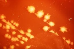
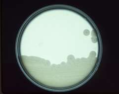
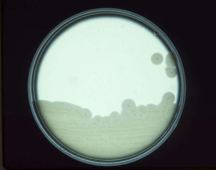
Description: English: This image depicts a Petri dish culture plate, which contained a growth medium of egg yolk agar that had been inoculated with the anaerobic bacterium, Clostridium perfringens. Date: 1973. Source: : This media comes from the
Centers for Disease Control and Prevention's
Public Health Image Library (PHIL), with identification number
#3872. Note: Not all PHIL images are public domain; be sure to check copyright status and credit authors and content providers.
العربية |
Deutsch |
English |
македонски |
slovenščina |
+/−. Author: CDC. Permission(
Reusing this file): This image is in the public domain and thus free of any copyright restrictions. As a matter of courtesy we request that the content provider be credited and notified in any public or private usage of this image.
-
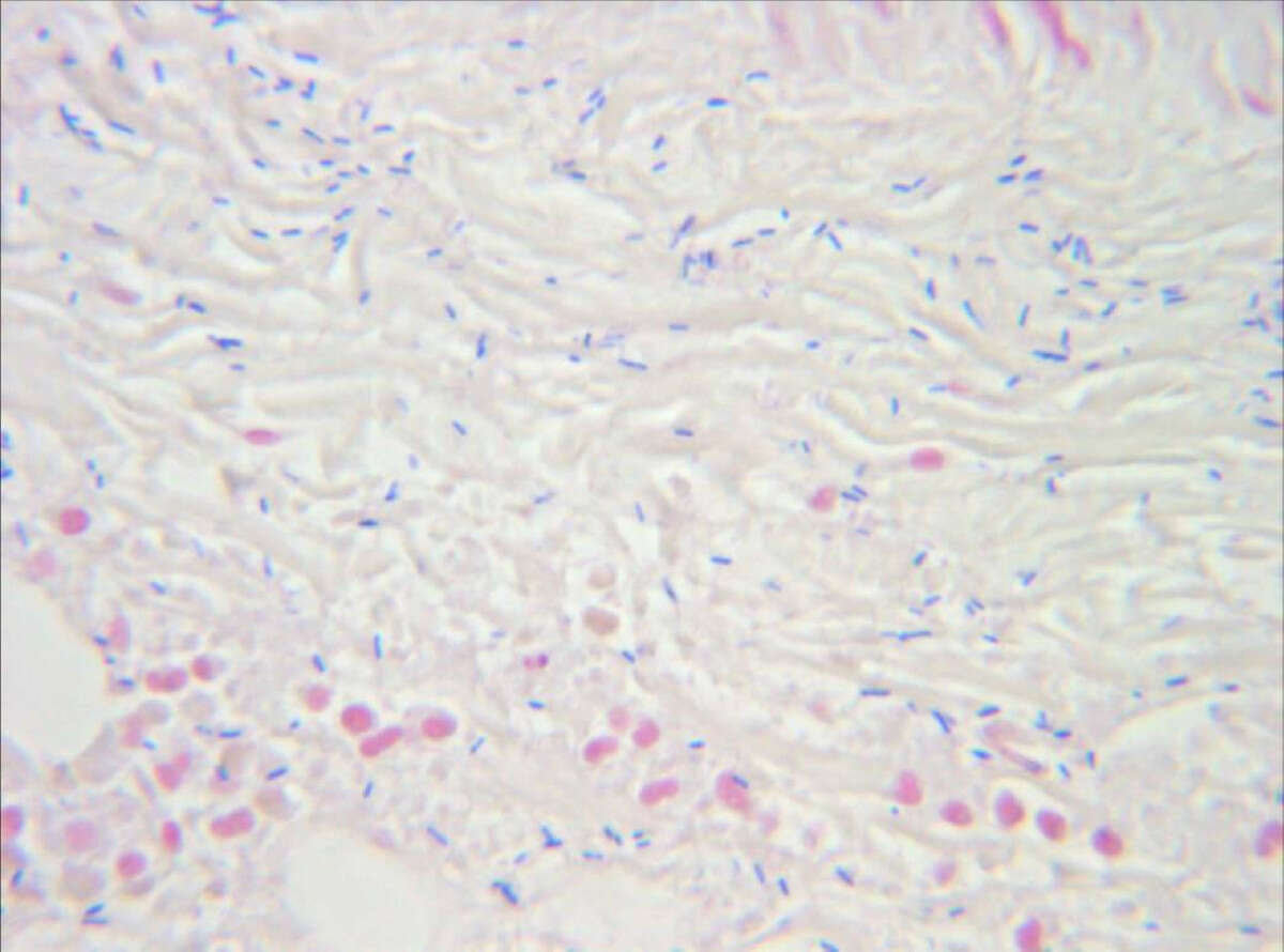
Description: English: Postoperative pathologic findings of the muscle tissue obtained during surgical removal. Gram-stain, Zoom 1000×. Identification of gram-positive, rod-shaped, anaerobic, spore-forming bacteria in the infected muscle tissue. The result is highly compatible to an infection with clostridium perfringens. Date: Published: 20 Oct 2008. Source:
Diagnosis and misdiagnosis of necrotizing soft tissue infections: three case reports. Cases J 2008, 1:252. doi: 10.1186/1757-1626-1-252. Author: Engelbert Schröpfer, Stephan Rauthe and Thomas Meyer.
-

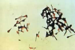
Description: English: This photomicrograph reveals Clostridium perfringens grown in Schaedler’s broth using Gram-stain. Clostridium perfringens is a spore-forming, heat-resistant bacterium that can cause foodborne disease. The spores persist in the environment, and often contaminate raw food materials. These bacteria are found in mammalian feces, and soil. Polski: Mikrofotografia laseczek Clostridium perfringens. Date: 1974. Source: : This media comes from the
Centers for Disease Control and Prevention's
Public Health Image Library (PHIL), with identification number
#2995. Note: Not all PHIL images are public domain; be sure to check copyright status and credit authors and content providers.
English |
Slovenščina |
+/−. Author: Content Providers(s): CDC/Don Stalons. Permission (
Reusing this file): PD-USGov-HHS-CDC English: None - This image is in the public domain and thus free of any copyright restrictions. As a matter of courtesy we request that the content provider be credited and notified in any public or private usage of this image.
-
-

Description: English: This micrograph depicts a group of Clostridium tetani bacteria, responsible for causing tetanus in humans. Tetanus is an acute, often fatal, disease caused by an exotoxin produced by C. tetani. It is characterized by generalized rigidity and convulsive spasms of skeletal muscles, usually involving the jaw (lockjaw) and neck, then becoming generalized. Deutsch: Eine Gruppe Clostridium tetani. 日本語: 破傷風菌. 顕微鏡で見た破傷風菌. Date: 1995. Source: : This media comes from the
Centers for Disease Control and Prevention's
Public Health Image Library (PHIL), with identification number
#6372. Note: Not all PHIL images are public domain; be sure to check copyright status and credit authors and content providers.
English |
Slovenščina |
+/−. Author: Content Providers(s): CDC. Permission (
Reusing this file): PD-USGov-HHS-CDC English: None - This image is in the public domain and thus free of any copyright restrictions. As a matter of courtesy we request that the content provider be credited and notified in any public or private usage of this image. Other versions:
png version.
-
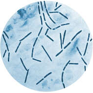
Description: English: Gram stain showing Clostridium tetani with terminal round spore. Date: Unknown date. Source: Own work. Author:
Microrao.
-
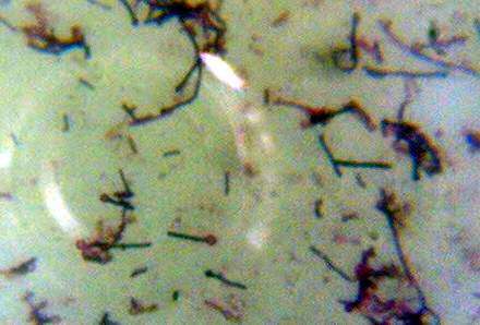
Description: English: Under a magnification of 956X, this photomicrograph of a Gram-stained specimen, depicted numbers of Gram-positive, Clostridium tetani bacteria, which had been cultivated on a blood agar plate (BAP), and incubated over a 48 hour time period. Note that several of these organisms had entered their endospore phase, assuming the characteristic tennis racket-type morphology. Date: 1965. Source:
https://phil.cdc.gov/Details.aspx?pid=12056. Author: CDC/ Dr. Holdeman.
-
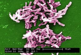
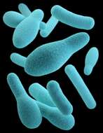
Description: English: This illustration depicts a three-dimensional (3D), computer-generated image of a group of anaerobic, spore-forming, Clostridium sp. organisms. The artistic recreation was based upon scanning electron microscopic (SEM) imagery. Note that a number of these organisms had assumed a safety-pin, or club-shape, indicating that they had entered what is known as an endospore phase, which is a tougher, dormant phase, resistant to heat and UV-radiation. Date: 2016. Source: : This media comes from the
Centers for Disease Control and Prevention's
Public Health Image Library (PHIL), with identification number
#21911. Note: Not all PHIL images are public domain; be sure to check copyright status and credit authors and content providers.
العربية |
Deutsch |
English |
македонски |
slovenščina |
+/−. Author: James Archer, Jennifer Oosthuizen.
-
Stefan Kircher, Rupert Wössner, Hans-Konrad Müller-Hermelink, Hans-Ullrich Völker.
Wikimedia Commons
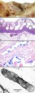
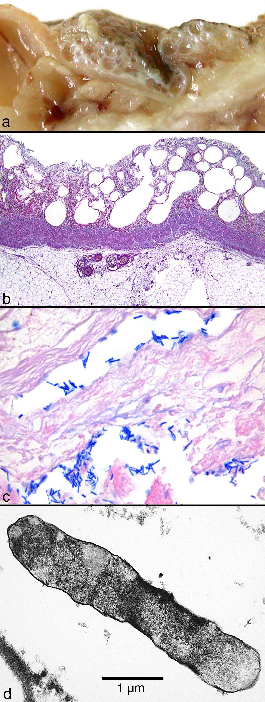
Description: English: Macroscopic, histological and ultrastructural assessment of small intestine tissue. (a) Macroscopic picture of the oedematous intestinal wall with multiple submucosal and subserosal cysts. (b) Histological picture of the intestinal mucosa with areactive necrosis. (c) Gram stain of cysts with large rod-shaped bacteria. (d) Electron microscopic picture of a bacterium found in a submucosal cyst. Date: Published: 24 July 2008. Source:
Lethal pneumatosis coli in a 12-month-old child caused by acute intestinal gas gangrene after prolonged artificial nutrition: a case report. Journal of Medical Case Reports. 2008;2:238. doi:10.1186/1752-1947-2-238. Author: Stefan Kircher, Rupert Wössner, Hans-Konrad Müller-Hermelink, Hans-Ullrich Völker. Permission(
Reusing this file): This is an Open Access article distributed under the terms of the Creative Commons Attribution License (
https://creativecommons.org/licenses/by/2.0), which permits unrestricted use, distribution, and reproduction in any medium, provided the original work is properly cited.


