Die Familie Hantaviridae aus der Ordnung der Bunyavirales umfasst neben wenigen Spezies der Gattungen Loanvirus, Mobatvirus und Thottimvirus vor allem zahlreiche Arten der Gattung Orthohantavirus: unter anderem die humanpathogenen Arten Hantaan-Virus (HTNV), Puumala-Virus (PUUV), Dobrava-Belgrad-Virus (DOBV), Seoul-Virus (SEOV), Sin-Nombre-Virus (SNV) und Andes-Virus (ANDV). Diese behüllten Einzel-Strang(−)-RNA-Viren [ss(−)RNA] verursachen je nach Virustyp verschiedene Erkrankungen. Dazu zählen schwere Lungenerkrankungen (Pneumonie), ein akutes Nierenversagen (nephrotisches Syndrom) oder hämorrhagische Fiebererkrankungen. Die Viren sind weltweit verbreitet und treten auch in Mitteleuropa auf. Sie werden durch den Kot oder Urin infizierter Nagetiere (Mäuse und Ratten), der als Staub eingeatmet wird, auf den Menschen übertragen. Die infizierten Nagetiere selbst zeigen keine Krankheitssymptome. Die menschlichen Erkrankungen verlaufen unterschiedlich schwer. Während die in Mitteleuropa auftretenden Puumala-Virus-Infektionen in weniger als 1 Prozent der klinisch auffälligen Fälle tödlich verlaufen, beträgt die Letalität bei Infektionen mit dem in Ostasien auftretenden Hantaan-Virus und mit dem auf dem Balkan zu findenden Dobrava-Virus bis zu 15 Prozent und bei den amerikanischen Hantaviren (Sin-Nombre-Virus, Andes-Virus und andere) etwa 30–40 Prozent.[3]
Der Name Hanta geht auf den Fluss Hantan in Südkorea zurück, an dem in den 1950er-Jahren während des Koreakrieges mehr als 3.000 amerikanische Soldaten an einem ungewöhnlich starken Fieber häufig mit einem anschließenden Nierenversagen erkrankten. Erst 1977 gelang es Ho Wang Lee und anderen, das bis dahin unbekannte Hantaan-Virus (HTNV) zu isolieren.[4] Auf diese Historie geht die Bezeichnung Korea-Fieber für durch humanpathogene Orthohantaviren verursachte Erkrankungen (englisch Hantavirus hemorrhagic fever with renal syndrome, HFRS) zurück.
Die Übertragung geschieht durch verschiedene Nager, die mit dem Speichel, den Fäkalien und dem Urin (Virurie) große Mengen an Erregern ausscheiden. Bei den Nagern sind vor allem Mäuse, in Deutschland besonders die Rötelmaus, als Überträger festgestellt, die jedoch selbst nicht erkranken, auch wenn sie, einmal infiziert, lebenslang infektiös bleiben.[4] Die Übertragung auf den Menschen erfolgt sowohl durch Kontaktinfektion als auch durch orale, überwiegend jedoch durch respiratorische Aufnahme der Erreger durch die Atemwege, seltener durch Nagetierbisse.[5] Eine typische Situation, bei der eine Übertragung mit relativ hoher Wahrscheinlichkeit erfolgen kann, ist beispielsweise das Ausfegen einer im Winter nicht genutzten Hütte im Frühjahr. Generell sind vor allem Personen gefährdet, die in der Land- und Forstwirtschaft arbeiten oder sich viel in der Natur aufhalten. In Asien sind häufig Reisbauern betroffen. Eine Mensch-zu-Mensch-Übertragung ist nur bei einem Ausbruch in Südamerika 1996 beschrieben worden.[6][7]
Die Inkubationszeit beträgt je nach Virustyp zwischen 5 und 60 Tagen.
Bei den europäischen und asiatischen Hantaviren stehen eine Nierenschädigung, die bis zum (meist reversiblen) akuten Nierenversagen führen kann, sowie eine fieberhafte Erkrankung mit Störung der Blutgerinnung und Blutungsneigung im Vordergrund. Der Symptomkomplex wird oft als „hämorrhagisches Fieber mit renalem Syndrom“ (HFRS) bezeichnet. In Europa kommen das Dobrava-Belgrad-Virus und Puumala-Virus vor, die leichte bis mittelschwere Verläufe verursachen. Als Überträger sind für das Puumala-Virus die Rötelmaus, für das Dobrava-Virus die Brandmaus und die Gelbhalsmaus identifiziert worden.[3] Die durch das auch in Deutschland und Skandinavien häufig vorkommende Puumala-Virus ausgelöste Nierenschädigung wird auch als Nephropathia epidemica[8] (NE) bezeichnet. Dabei treten sehr selten Blutungen auf.[5]
Klinisch äußert sich die Infektion mit abrupt einsetzendem Fieber, Kopfschmerzen, Gliederschmerzen, Blutdruckabfall, neurologischen Symptomen (Sehstörungen) und Zeichen der Nierenschädigung. Labormedizinisch kommt es zu Blutbildveränderungen (Thrombozytopenie), zu einem Anstieg von Serum-Kreatinin, zu einer Mikrohämaturie, zu einer Proteinurie und zu einem Rückgang der glomerulären Filtrationsrate. Akute Glaukomanfälle, eine Beteiligung des zentralen Nervensystems (ZNS), Myokarditiden und intestinale (den Darm betreffende) Blutungen können als Komplikationen auftreten. Die Erkrankung heilt meist folgenlos aus.[3][9]
Die durch die südosteuropäische Variante des Dobrava-Virus hervorgerufenen HFRS-Erkrankungen neigen zu signifikant schwereren Verläufen und weisen eine Letalität von bis zu 12 % auf. Ähnliches gilt für das durch den Hantaan-Genotyp verursachte ostasiatische Erkrankungsbild, bei dem die Todesrate etwa 15 % beträgt.[10]
Erkrankungen durch Hantaviren müssen bei schwerem Verlauf im Krankenhaus behandelt werden, andernfalls kann die Erkrankung zum Tode führen. Eine Anämie kann Monate fortdauern. Der Nachweis von Hantaviren oder einer Hantavirus-Infektion ist in Deutschland seit dem 1. Januar 2001 meldepflichtig.[5]
Bei den auf dem amerikanischen Doppelkontinent vorkommenden Hantaviren stehen in der Regel nicht die Nierenschädigung oder ein hämorrhagisches Fieber im Vordergrund, sondern eine schwere Lungenentzündung (Pneumonie) mit Lungenödem (Hantavirus-assoziiertes pulmonales Syndrom, abgekürzt HPS, synonym auch Hantavirus cardiopulmonary syndrome, HCPS). Die Erstbeschreibung dieser Erkrankung erfolgte im Jahr 1993, nachdem im Grenzgebiet der vier Bundesstaaten New Mexico, Utah, Arizona und Colorado (einer Region, die als „Four Corners“ bekannt ist) mehrere Fälle einer schweren Lungenentzündung bei Navajo-Indianern auftraten, von denen einige tödlich endeten.[11] Die Umstände und die Epidemiologie dieses Ausbruchs wiesen auf eine infektiöse Ursache der Lungenentzündung hin. Wissenschaftler der Centers for Disease Control and Prevention (CDC) konnten in der Folge nachweisen, dass die Erkrankung durch ein neues, bis dahin nicht bekanntes Hantavirus verursacht wurde. Das Virus wurde sowohl am CDC als auch am United States Army Medical Research Institute of Infectious Diseases (USAMRIID) isoliert und erhielt zunächst den Namen Muerto Canyon virus, später geändert in Sin Nombre virus (SNV, „namenloses Virus“).[11]
Da bekannt war, dass Hantaviren durch Nagetiere übertragen werden, wurde eine umfangreiche Suche nach dem Überträger dieser lokalen Hantavirus-Epidemie in der betroffenen Region eingeleitet. Zahlreiche Nagetiere wurden in Fallen gefangen und untersucht. Dabei stellte sich heraus, dass Weißfußmäuse (Peromyscus maniculatus, englisch deer mouse) die Hauptüberträger dieses neu entdeckten Hantavirus waren. Diese Neuweltmäuse leben häufig in der Nähe menschlicher Ansiedlungen oder auch direkt in älteren Häusern. Untersuchungen noch im selben Jahr 1993 ergaben, dass auch andere verwandte Hantaviren ein ähnliches Krankheitsbild auslösen können. Bei zwei Personen aus Louisiana und aus Florida mit HPS wurden zwei neue Hantaviren entdeckt, das später so benannte Bayou virus und das Black Creek Canal virus. Als Überträger wurden Reisratten (Oryzomys) bzw. Baumwollratten (Sigmodon hispidus) identifiziert.
Bei den nordamerikanischen Hantaviren ("new world hanta viruses") und besonders beim Sin-Nombre-Virus beträgt die Inkubationszeit zwei bis vier Wochen, gefolgt von einer Prodromalphase. Dabei tritt ein hohes Fieber auf, es kommt zu Muskelschmerzen, zu starken Kopf- und Nackenschmerzen, zu einem allgemeinen Krankheitsgefühl, zu einer Körperschwäche, zu Bauchschmerzen, zu Übelkeit und Erbrechen und gelegentlich zu einem Durchfall. Es entwickeln sich eine schwere Leukozytose mit Bandämie und eine Thrombozytopenie. Nieren und Leber sind typischerweise nicht involviert. Abrupt kann es nach drei bis fünf Tagen zu einer Verschlechterung und zu einer Progression in die "kardiopulmonale Phase" mit Atemnot, nichteitriger Sekretion, bis hin zum akuten Lungenversagen und myokardialen Dekompensation kommen, die mit einer Letalität von 40 % verbunden ist. Sehr früh werden spezifische Antikörper gebildet, neben IgM auch innerhalb weniger Tage IgG-Antikörper.[12]
Später wurden weitere Hantaviren bei Patienten mit Lungensymptomen in Argentinien, Brasilien, Kanada, Chile, Paraguay und Uruguay beschrieben.[11] Im Südkegel Südamerikas gilt die Langschwanz-Zwergreisratte Oligoryzomys longicaudatus als Hauptüberträger von Hantaviren, die je etwa in der Hälfte der Erkrankungsfälle die fiebrige Nierenform (HFRS) und die sehr gefährliche Herz-Lungenform (HCPS) der Hantavirenkrankheit verursachen.[13] Der dominierende Erreger ist das 1998 in Argentinien identifizierte Andes-Virus (ANDV), das neben HPS/HCPS häufiger als das Sin-Nombre-Virus auch HFRS hervorruft.[14][15][16]
Die Diagnose einer Hantavirus-Infektion wird zum einen aufgrund des typischen Krankheitsbildes (siehe oben) und zum anderen aufgrund von spezifischen Laborwerten gestellt. Die direkte Erregerisolation ist im Tierversuch und in Zellkulturen zu Krankheitsbeginn möglich, aber ungebräuchlich. Der serologische Nachweis (d. h. Nachweis von Antikörpern gegen das Virus im Blut) wird im Immunfluoreszenztest und mittels ELISA erbracht. IgM-Antikörper sind nur einige Wochen nachweisbar, wohingegen die 14 Tage nach Krankheitsbeginn auftretenden IgG-Antikörper jahrelang bestehen bleiben können.
Die Therapie ist vor allem symptomatisch und unterstützend mit ausreichender Flüssigkeitszufuhr, intensivmedizinischer Versorgung und Intubation bei Ausbildung eines akuten Lungenversagens bzw. mit einer Dialyse bei akuter Niereninsuffizienz.
Das antiviral wirksame Ribavirin zeigt im Laborversuch zwar eine gute Aktivität gegen Hantaviren, die therapeutischen Ergebnisse sind aber uneindeutig und die schweren Nebenwirkungen führen dazu, dass es in der Regel nicht verwendet wird.[12]
Eine Impfung gegen Hantaviren befindet sich erst im Entwicklungsstadium.[17][18][19][20]
Zur Infektionsverhütung können Nagetiere im Umfeld menschlicher Siedlungen bekämpft werden. Ist ein Kontakt nicht zu vermeiden, zum Beispiel beim Reinigen befallener Bereiche, wird empfohlen, Nagetierkadaver nur mit Einmalhandschuhen zu entfernen.[21] Ein Anfeuchten betroffener Flächen, beispielsweise in Schuppen oder Scheunen, vermindert das Aufwirbeln von Staub. Bei Reinigungsarbeiten sollten Mundschutz und Handschuhe getragen werden. Auch dicht schließende Mülleimer sowie regelmäßiges Reinigen und Lüften gehören zu den vom Robert Koch-Institut empfohlenen Vorbeugemaßnahmen.[4] Verschmutzte Flächen sollten nach dem Reinigen desinfiziert werden.[22]
Europa (NE)
NE = Nephropathia epidemica
HFRS = Hämorrhagisches Fieber mit renalem Syndrom
HPS = Hantavirus-assoziiertes pulmonales Syndrom
Hantaviren sind weltweit verbreitet. Für die meisten Länder existieren aber keine genauen epidemiologischen Kennzahlen. Einen Eindruck vermittelt die nebenstehende Karte. Es ist davon auszugehen, dass viele Hantavirus-Infektionen, insbesondere in Ländern mit weniger entwickeltem Gesundheitssystem, nicht erkannt werden, da z. B. nicht an die Möglichkeit einer infektiösen Ursache gedacht wird.[3][5]
In China wurde am 24. März 2020 ein Todesfall und weitere Verdachtsfälle in der Provinz Yunnan gemeldet.[24]
Die höchsten Fallzahlen werden aus Schweden und Finnland gemeldet. In Mitteleuropa sind beispielsweise einige Regionen in Niedersachsen, Hessen, Bayern und Baden-Württemberg sowie in Österreich Teile der Steiermark als Endemiegebiete für das Puumala-Virus bekannt, während in Norddeutschland, vor allem aber in Ost- und Südosteuropa, das Dobrava-Belgrad-Virus endemisch ist. Besonders im Frühjahr kann es dadurch zu Erkrankungen mit plötzlichem Nierenversagen kommen.
Da Hantavirus-Erkrankungen in Deutschland erst seit 2001 meldepflichtig sind, liegen für die Erkrankungsraten aus früheren Jahren keine verlässlichen Daten vor. Etwa 1–2 Prozent der Bevölkerung weisen Hantavirus-spezifische Antikörper auf.[3] Die Zahl der gemeldeten Erkrankungen liegt jedoch weit darunter, was zum einen darauf hinweist, dass die Infektion häufig ohne klinische Symptome abläuft. Zum anderen wird aber auch bei entsprechenden klinischen Symptomen (Nierenschaden) nicht immer an eine infektiöse Ursache gedacht. Mit 2017 gemeldeten Fällen gehörten Hantavirus-Infektionen im Jahr 2010 zu den fünf häufigsten meldepflichtigen Viruserkrankungen in Deutschland (nach Noroviren (140519), Rotaviren (54051), Hepatitis C (5301) und Influenza (3468)). Auffällig sind die jährlich großen Unterschiede der Erkrankungsfälle: Während in einigen Jahren weit über 1000 Fälle gemeldet wurden, lagen die Fallzahlen in anderen Jahren zum Teil nur bei wenigen Hundert. Im Jahr 2006 wurden sogar nur 72 Erkrankungen gemeldet. 2016 wurden bundesweit 282 Fälle erfasst, 2017 hingegen 1731.[25][26] Auch regionale Unterschiede bestehen: Hohe Fallzahlen wurden 2007 und 2010 aus Baden-Württemberg von der Schwäbischen Alb gemeldet,[4] aber auch aus dem Bayerischen Wald, aus dem Spessart, aus Köln und aus dem Münsterland. Es wird ein Zusammenhang mit dem Vorkommen von Buchenwäldern vermutet, da sich die Rötelmaus von Bucheckern ernährt.[4] Im Frühjahr 2021 ist von einer Gruppe Wissenschaftler um Jörg Hoffmann in Deutschland die erste Infektion von Menschen mit einem Tula-Virus berichtet worden.[27] Tula-Viren, die zu den Hanta-Viren zählen, werden wahrscheinlich durch Feldmäuse übertragen und haben bei dem betroffenen 21-Jährigen zu einem akuten Nierenversagen geführt.[28]
*) nur klinisch-labordiagnostische Fälle
In Österreich ist das Puumala-Virus das am häufigsten nachgewiesene Hantavirus.[30] In den Jahren 2004 bis 2011 wurden landesweit durchschnittlich 20 Infektionen bekannt, allerdings mit großen Unterschieden zwischen einzelnen Jahren. So gab es 2007 knapp 80 Infektionen. Im Jahr 2012 zeigte sich wie auch in Deutschland eine ungewöhnliche Häufung von Hantavirus-Infektionen; in den ersten sieben Monaten wurden schon 180 Fälle gezählt. Die Letalität wird mit 0,2 % angegeben.[31] Im Jahr 2011 wurde erstmals auch eine vermutlich in Österreich erworbene Infektion mit Dobravaviren diagnostiziert, die schwerere Krankheitsverläufe auslösen.[30] Die meisten der zwischen 1993 und 2010 dokumentierten Hantavirusinfektionen ereigneten sich in der Steiermark, in Kärnten (vor allem im Bezirk Wolfsberg), im südlichen Burgenland und im Bezirk Rohrbach in Oberösterreich. Aus den Bundesländern Tirol und Vorarlberg waren dagegen 2012 noch keine Fälle bekannt.[30]
In der Schweiz sind Hantavirus-Infektionen bisher nur als Einzelfälle beschrieben worden.[32]
Es liegen keine Daten zu in Luxemburg erworbenen Hantavirus-Infektionen vor.
In den Vereinigten Staaten wurden zwischen 1993 und 2011 insgesamt 587 Fälle von Hantavirus-assoziiertem pulmonalem Syndrom registriert, wovon im Durchschnitt 37 % tödlich verliefen.[33] Ganz überwiegend sind die Bundesstaaten im Südwesten betroffen.
Im Sommer 2012 kam es zu einer Reihe von HPS-Erkrankungen mit drei Todesfällen bei Touristen, die im kalifornischen Yosemite-Nationalpark übernachtet hatten.[34] Der genaue Ort der Infektion konnte bislang nicht ermittelt werden: Nachdem zunächst Nagetiere im Umfeld von Zeltkabinen verdächtigt wurden, trat ein weiterer Fall in einem anderen Teil des Parks auf. Daraufhin wurden im September 2012 alle Parkbesucher der Sommersaison gewarnt und aufgefordert, beim Auftreten von Symptomen einer Lungenerkrankung sofort ärztliche Hilfe zu suchen und auf den Parkbesuch hinzuweisen.[35][36]
Aus verschiedenen süd- und mittelamerikanischen Staaten sind Hantavirus-Infektionen dokumentiert. Hierzu zählen Argentinien, Bolivien, Brasilien, Chile, Ecuador, Paraguay, Panama, Uruguay und Venezuela.[7] In Mexiko, Kolumbien und Costa Rica wurden Hantaviren gefunden, die dem Sin-Nombre-Virus ähneln, aber beim Menschen anscheinend keine Erkrankung auslösen.[7] Die meisten Fälle traten in Brasilien auf. Die Letalität des Hantavirus-assoziierten pulmonalen Syndroms entsprach dort mit etwa 37 % derjenigen in den Vereinigten Staaten.[37]
In Argentinien und Chile dominiert das 1998 identifizierte Andes-Virus (ANDV), das sowohl die fiebrige Nieren-Form (HFRS) als auch die gefährliche Herz-Lungen-Form (HCPS) der Hantavirenkrankheit verursacht. In Chile wurden zwischen 1993 und 2001 204 HCPS-Erkrankungsfälle gezählt, von denen anfänglich mehr als die Hälfte, zuletzt etwa ein Drittel tödlich verliefen. In Argentinien, wo das Andes-Virus im Anschluss an die schweren HPS-Ausbrüche der Jahre 1995 und 1996 erstmals isoliert wurde, soll auch die Übertragbarkeit des Virus von Mensch zu Mensch beobachtet worden sein.[7][14][15][16]
Nach ICTV mit Stand Herbst 2018 gliedert sich die Familie Hantaviridae wie folgt:
Die folgenden Viren der Gattung Orthohantavirus ohne Zuordnung einer Spezies werden vom ICTV wegen zu schlechter Datenlage nicht mehr gelistet:
Die folgenden Hantaviren von Fledermäusen wurden von ICTV noch nicht aufgenommen:[46][47]
Weitere Vorschläge:
In Deutschland ist der direkte oder indirekte Nachweis von Hantaviren namentlich meldepflichtig nach des Infektionsschutzgesetzes, soweit der Nachweis auf eine akute Infektion hinweist. Meldepflichtig sind vor allem die Leitungen der Labore usw. ( IfSG).
Hanta-Virus-Infektionen sind in Österreich gemäß Abs. 1 Nummer 2 Epidemiegesetz 1950 bei Erkrankung und Tod anzeigepflichtig. Zur Anzeige verpflichtet sind unter anderen Ärzte und Labore ( Epidemiegesetz).
In der Schweiz ist der positive und negative laboranalytische Befund zu einem Hanta-Virus meldepflichtig und zwar nach dem Epidemiengesetz (EpG) in Verbindung mit der Epidemienverordnung und der Verordnung des EDI über die Meldung von Beobachtungen übertragbarer Krankheiten des Menschen. Meldepflichtig ist das untersuchende Labor. Zudem ist die Erkrankung Hanta-Fieber meldepflichtig nach den genannten Normen und der genannten Verordnung des EDI. Meldepflichtig sind Ärzte, Spitäler usw.
Die Familie Hantaviridae aus der Ordnung der Bunyavirales umfasst neben wenigen Spezies der Gattungen Loanvirus, Mobatvirus und Thottimvirus vor allem zahlreiche Arten der Gattung Orthohantavirus: unter anderem die humanpathogenen Arten Hantaan-Virus (HTNV), Puumala-Virus (PUUV), Dobrava-Belgrad-Virus (DOBV), Seoul-Virus (SEOV), Sin-Nombre-Virus (SNV) und Andes-Virus (ANDV). Diese behüllten Einzel-Strang(−)-RNA-Viren [ss(−)RNA] verursachen je nach Virustyp verschiedene Erkrankungen. Dazu zählen schwere Lungenerkrankungen (Pneumonie), ein akutes Nierenversagen (nephrotisches Syndrom) oder hämorrhagische Fiebererkrankungen. Die Viren sind weltweit verbreitet und treten auch in Mitteleuropa auf. Sie werden durch den Kot oder Urin infizierter Nagetiere (Mäuse und Ratten), der als Staub eingeatmet wird, auf den Menschen übertragen. Die infizierten Nagetiere selbst zeigen keine Krankheitssymptome. Die menschlichen Erkrankungen verlaufen unterschiedlich schwer. Während die in Mitteleuropa auftretenden Puumala-Virus-Infektionen in weniger als 1 Prozent der klinisch auffälligen Fälle tödlich verlaufen, beträgt die Letalität bei Infektionen mit dem in Ostasien auftretenden Hantaan-Virus und mit dem auf dem Balkan zu findenden Dobrava-Virus bis zu 15 Prozent und bei den amerikanischen Hantaviren (Sin-Nombre-Virus, Andes-Virus und andere) etwa 30–40 Prozent.
Der Name Hanta geht auf den Fluss Hantan in Südkorea zurück, an dem in den 1950er-Jahren während des Koreakrieges mehr als 3.000 amerikanische Soldaten an einem ungewöhnlich starken Fieber häufig mit einem anschließenden Nierenversagen erkrankten. Erst 1977 gelang es Ho Wang Lee und anderen, das bis dahin unbekannte Hantaan-Virus (HTNV) zu isolieren. Auf diese Historie geht die Bezeichnung Korea-Fieber für durch humanpathogene Orthohantaviren verursachte Erkrankungen (englisch Hantavirus hemorrhagic fever with renal syndrome, HFRS) zurück.
Ο χανταϊός είναι ιός της οικογένειας των Χανταϊών. Ο ιός μπορεί να μεταδοθεί από το ποντίκι στον άνθρωπο εφόσον ο άνθρωπος καταναλώσει το σωματικό υγρό του ποντικιού. Ακόμη μπορεί να μεταδοθεί μέσω της έκθεσης σε ούρα, περιττώματα ή σάλιο τρωκτικών που φέρουν τον ιό ή από δάγκωμα ποντικιού. Η περίοδος επώασης φτάνει μέχρι και τις 8 εβδομάδες.[1]
Τα συμπτώματα του χανταϊού είναι συνήθως πυρετός, πονοκέφαλος, βήχας και δύσπνοια.[1]
Οι μολύνσεις ανθρώπων με χανταϊό σχετίζονται σχεδόν εξ ολοκλήρου με την ανθρώπινη επαφή με περιττώματα τρωκτικών, ωστόσο, το 2005 και το 2019, αναφέρθηκε μετάδοση του ιού των Άνδεων από άνθρωπο σε άνθρωπο στη Νότια Αμερική.[2]
Το όνομα του χανταϊού προέρχεται από τον ποταμό Χαντάν στην Νότια Κορέα όπου καταγράφηκε ένα ξέσπασμα.[3] Ο Χο-Βανγκ Λι απομόνωσε τον ιό το 1976.
Ο χανταϊός είναι ιός της οικογένειας των Χανταϊών. Ο ιός μπορεί να μεταδοθεί από το ποντίκι στον άνθρωπο εφόσον ο άνθρωπος καταναλώσει το σωματικό υγρό του ποντικιού. Ακόμη μπορεί να μεταδοθεί μέσω της έκθεσης σε ούρα, περιττώματα ή σάλιο τρωκτικών που φέρουν τον ιό ή από δάγκωμα ποντικιού. Η περίοδος επώασης φτάνει μέχρι και τις 8 εβδομάδες.
Τα συμπτώματα του χανταϊού είναι συνήθως πυρετός, πονοκέφαλος, βήχας και δύσπνοια.
Οι μολύνσεις ανθρώπων με χανταϊό σχετίζονται σχεδόν εξ ολοκλήρου με την ανθρώπινη επαφή με περιττώματα τρωκτικών, ωστόσο, το 2005 και το 2019, αναφέρθηκε μετάδοση του ιού των Άνδεων από άνθρωπο σε άνθρωπο στη Νότια Αμερική.
Το όνομα του χανταϊού προέρχεται από τον ποταμό Χαντάν στην Νότια Κορέα όπου καταγράφηκε ένα ξέσπασμα. Ο Χο-Βανγκ Λι απομόνωσε τον ιό το 1976.

ऑर्थो हॅन्टा विषाणू (किंवा हंताविषाणू (हंता व्हायरस) हा एक एकल, आच्छादित, नकारात्मक अर्थाने आरएनए व्हायरस आहे. हे विषाणू सामान्यपणे उंदीरांना संक्रमित करतात परंतु त्यांच्यात आजार उद्भवत नाहीत. [१] उंदीर मूत्र, लाळ किंवा विष्ठ यांच्या संपर्कातून मानवांना हंताविषाणूची लागण होऊ शकते.
वर्गीकरण
हंता व्हायरस बन्याव्हायरस आहे. बन्याव्हायरस ऑर्डर बारा कुटुंबांमध्ये विभागली आहे. या ऑर्डरच्या सर्व सदस्यांप्रमाणेच हंताव्हायरसमध्ये तीन नकारात्मक अर्थाने, एकल-अडकलेल्या आरएनए सेगमेंट्सचा समावेश जनुकांमध्ये असतो म्हणून नकारात्मक अर्थाने आरएनए व्हायरस वर्गीकृत केले जातात. इतर बुनियाविरलेस कुटुंबातील सदस्य सामान्यत: आर्थ्रोपॉड-जनित विषाणू असतात,[२] परंतु मानवांमध्ये प्रामुख्याने एरोसोलिज्ड मलमूत्र किंवा उंदीर चाव्याव्दारे श्वास माध्यमातून हंता व्हायरस मानवांमध्ये संक्रमित केले जातात.
१) हंता व्हायरस कारणे, लक्षणे, प्रसार, प्रतिबंध, उपचार
|date= मधील दिनांक मूल्ये तपासा (सहाय्य) ऑर्थो हॅन्टा विषाणू (किंवा हंताविषाणू (हंता व्हायरस) हा एक एकल, आच्छादित, नकारात्मक अर्थाने आरएनए व्हायरस आहे. हे विषाणू सामान्यपणे उंदीरांना संक्रमित करतात परंतु त्यांच्यात आजार उद्भवत नाहीत. उंदीर मूत्र, लाळ किंवा विष्ठ यांच्या संपर्कातून मानवांना हंताविषाणूची लागण होऊ शकते.
Àdàkọ:Infobox medical condition (new)
Hantavirus tàbí orthohantavirus jẹ́ kòkòrò ẹ̀ràn àìfojúrí tí ó máa ń jẹyọ lára eku.[3] Ó jẹ́ ẹ̀ràn kòkòrò àìfojúrí tí ó máa ṣẹ̀yọ lára eku, ṣùgbọ́n kìí ṣe wọ́n ní ìjàmbá kankan.[3] Ènìyàn lè kó ẹ̀ràn kòkòrò àìfojúrí, Hantavirus láti ara ìtọ̀, irọ́ tàbí imìgbẹ́ eku. Èyí lè fà àìsàn ìbà tí wọ́n ń pè ní hantavirus hemorrhagic fever with renal syndrome (HFRS), tàbí èyí tí wọ́n ń pè ní hantavirus pulmonary syndrome (HPS), tí gbogbo ènìyàn mọ̀ sí hantavirus cardiopulmonary syndrome (HCPS),[4] àwọn àìsàn mìíràn tí ẹ̀ràn kòkòrò àìfojúrí yìí máa ń fà, kò ì tí ì ṣẹ̀yọ lára ènìyàn. [5] HPS (HCPS) yìí jẹ́ àrùn tí ó máa ń jẹ mọ́ èémí nípa fífa òórùn ìgbẹ́ tàbí ìtọ̀ eku tí ó ti ní ẹ̀ràn kòkòrò àìfojúrí Hantavirus símú."[4]
Ènìyàn lè ko àgbákò àrùn tí ẹ̀ràn kòkòrò àìfojúrí Hantavirus máa ń fà bí ènìyàn bá fara kan ìgbẹ́ eku, ṣùgbọ́n lọ́dún 2005, ìròyìn gbé e pé ẹni kan kó àrùn náà lára ẹlòmíràn ní Gúúsù Amẹ́ríkà (South America).[5]
Wọ́n ṣẹ̀dá orúkọ yìí, Hantavirus láti ara odò kan tí wọ́n ń pè ní Hanta lórílẹ̀-èdè South Korea, níbi tí àrùn náà ti kọ́kọ́ ṣẹ́yọ ,[6]
Àdàkọ:Infobox medical condition (new)
Hantavirus tàbí orthohantavirus jẹ́ kòkòrò ẹ̀ràn àìfojúrí tí ó máa ń jẹyọ lára eku. Ó jẹ́ ẹ̀ràn kòkòrò àìfojúrí tí ó máa ṣẹ̀yọ lára eku, ṣùgbọ́n kìí ṣe wọ́n ní ìjàmbá kankan. Ènìyàn lè kó ẹ̀ràn kòkòrò àìfojúrí, Hantavirus láti ara ìtọ̀, irọ́ tàbí imìgbẹ́ eku. Èyí lè fà àìsàn ìbà tí wọ́n ń pè ní hantavirus hemorrhagic fever with renal syndrome (HFRS), tàbí èyí tí wọ́n ń pè ní hantavirus pulmonary syndrome (HPS), tí gbogbo ènìyàn mọ̀ sí hantavirus cardiopulmonary syndrome (HCPS), àwọn àìsàn mìíràn tí ẹ̀ràn kòkòrò àìfojúrí yìí máa ń fà, kò ì tí ì ṣẹ̀yọ lára ènìyàn. HPS (HCPS) yìí jẹ́ àrùn tí ó máa ń jẹ mọ́ èémí nípa fífa òórùn ìgbẹ́ tàbí ìtọ̀ eku tí ó ti ní ẹ̀ràn kòkòrò àìfojúrí Hantavirus símú."
Ènìyàn lè ko àgbákò àrùn tí ẹ̀ràn kòkòrò àìfojúrí Hantavirus máa ń fà bí ènìyàn bá fara kan ìgbẹ́ eku, ṣùgbọ́n lọ́dún 2005, ìròyìn gbé e pé ẹni kan kó àrùn náà lára ẹlòmíràn ní Gúúsù Amẹ́ríkà (South America).
Wọ́n ṣẹ̀dá orúkọ yìí, Hantavirus láti ara odò kan tí wọ́n ń pè ní Hanta lórílẹ̀-èdè South Korea, níbi tí àrùn náà ti kọ́kọ́ ṣẹ́yọ ,
Orthohantavirus is a genus of single-stranded, enveloped, negative-sense RNA viruses in the family Hantaviridae within the order Bunyavirales.[3] Members of this genus may be called orthohantaviruses or simply hantaviruses.
Orthohantaviruses typically cause chronic asymptomatic infection in rodents.[3][4] Humans may become infected with hantaviruses through contact with rodent urine, saliva, or feces. Some strains cause potentially fatal diseases in humans, such as hantavirus hemorrhagic fever with renal syndrome (HFRS), or hantavirus pulmonary syndrome (HPS), also known as hantavirus cardiopulmonary syndrome (HCPS),[5] while others have not been associated with known human disease (e.g. Prospect Hill virus).[6] HPS (HCPS) is a "rare respiratory illness associated with the inhalation of aerosolized rodent excreta (urine and feces) contaminated by hantavirus particles."[5]
Human infections of hantaviruses have almost entirely been linked to human contact with rodent excrement; however, in 2005 and 2019, human-to-human transmission of the Andes virus was reported in South America.[7]
Orthohantaviruses are named for the Greek word ortho- meaning "straight" or "true" and for the Hantan River in South Korea, where the first member species (Hantaan virus) was identified and isolated in 1976 by Ho Wang Lee.[8][9]
Hantavirus infections in humans are associated with two diseases: hemorrhagic fever with renal syndrome (HFRS) and hantavirus pulmonary syndrome (HPS), caused by Old World and New World hantaviruses, respectively. A common feature of the two diseases is increased vascular permeability, which causes hypotension, thrombocytopenia, and leukocytosis. The pulmonary illness is the more fatal of the two, whereas the hemorrhagic fever is much more common. Treatment for both is primarily supportive as there is no specific treatment for hantavirus infections.[10] While many hantaviruses cause either of the two diseases, some are not known to cause illness, such as the Prospect Hill orthohantavirus.[11]
Hemorrhagic fever with renal syndrome (HFRS) is caused chiefly by hantaviruses in Asia and Europe. Clinical presentation varies from subclinical to fatal, depending on the virus. After an incubation period of 2–4 weeks, the typical illness starts with non-specific symptoms such as high fever, chills, headache, backache, abdominal pains, nausea, and vomiting. After the initial period, bleeding under the skin begins, often paired with low blood pressure, followed by further internal bleeding throughout the body. Renal dysfunction leading to further health issues begins thereafter, which may cause death.[10] A more mild form of HFRS that occurs in Europe is called "nephropathia epidemica" (NE).[12] Trench nephritis during World War I is now thought to have been HFRS.
Hantavirus pulmonary syndrome (HPS), also called hantavirus cardiopulmonary syndrome (HCPS), is usually caused by hantaviruses in the Americas. Its incubation period ranges from 16 to 24 days. Illness initially shows similar symptoms as HFRS. After a few days of non-specific symptoms, sudden onset of progressive, or productive, coughing, shortness of breath, and elevated heart rate occur due to fluid buildup in the lungs. These symptoms are accompanied by impairment of lymphoid organs. Death from cardiovascular shock may occur rapidly after the appearance of severe symptoms.[10][11] While HCPS is typically associated with New World hantaviruses, the Puumala orthohantavirus in Europe has also caused the syndrome on rare occasions.[12]
Hantaviruses are transmitted by contact with the bodily fluids of rodents, particularly from saliva from bites and especially from inhalation of viral particles from urine and feces in aerosols. The manner of transmission is the same for both diseases caused by hantaviruses. Among the HCPS-causing hantaviruses is the Andes orthohantavirus, which is the only hantavirus confirmed to be capable of spreading from person to person, though this is rare.[10][11]
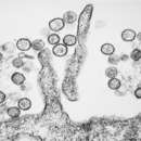
Hantavirus virions are about 120–160 nm in diameter. The lipid bilayer of the viral envelope is about 5 nm thick and is embedded with viral surface proteins to which sugar residues are attached. These glycoproteins, known as Gn and Gc, are encoded by the M segment of the viral genome. They tend to associate (heterodimerize) with each other and have both an interior tail and an exterior domain that extends to about 6 nm beyond the envelope surface.
Inside the envelope are the nucleocapsids. These are composed of many copies of the nucleocapsid protein N, which interact with the three segments of the viral genome to form helical structures. The virally encoded RNA polymerase is also found in the interior. By mass, the virion is greater than 50% protein, 20–30% lipid, and 2–7% carbohydrate. The density of the virions is 1.18 g/cm3. These features are common to all members of the family Hantaviridae.
The genome of hantaviruses is negative-sense, single-stranded RNA. Their genomes are composed of three segments: the small (S), medium (M), and large (L) segments. The S segment, 1-3 kilobases (kb) in length, encodes for the nucleocapsid (N) protein. The M segment, 3.2-4.9 kb in length, encodes a glycoprotein precursor polyprotein that is co-translationally cleaved into the envelope glycoproteins Gn and Gc, alternatively called G1 and G2. The L segment, 6.8–12 kb in length, encodes the L protein which functions primarily as the viral RNA-dependent RNA polymerase used for transcription and replication.[11][13]
Within virions, the genomic RNAs of hantaviruses are thought to complex with the N protein to form helical nucleocapsids, the RNA component of which circularizes due to sequence complementarity between the 5' and 3' terminal sequences of genomic segments.
As with other Bunyavirales, each of the three segments has a consensus 3'-terminal nucleotide sequence (AUCAUCAUC), which is complementary to the 5'-terminal sequence and is distinct from those of the other four genera in the family.[14] These sequences appear to form panhandle structure which seem likely to play a role in replication and encapsidation facilitated by binding with the viral nucleocapsid (N) protein.[15] The large segment is 6530–6550 nucleotides (nt) in length, the medium is 3613–3707 nt in length and the small is 1696–2083 nt in length.
No nonstructural proteins are known, unlike the other genera in this family. At the 5' and 3' of each segment are short noncoding sequences: the noncoding segment in all sequences at the 5' end is 37–51 nt. The 3' noncoding regions differ: L segment 38–43 nt; M segment 168–229 nt; and S segment 370–730 nt. The 3' end of the S segment is conserved between the genera suggesting a functional role.
Viral entry into host cells initiates by binding to surface cell receptors. Integrins are considered to be the main receptors for hantaviruses in vitro, but complement decay-accelerating factor (DAF) and globular heads of complement C1q receptor (gC1qR) have mediated attachment in cultured cells too. Entry may proceed through a number of possible routes, including clathrin-dependent endocytosis, clathrin-independent receptor-mediated endocytosis, and micropinocytosis. Viral particles are then transported to late endosomes. Gc-mediated membrane fusion with the endosomal membrane, triggered by low pH, releases the nucleocapsid into the cytoplasm.[13]
After the release of the nucleocapsids into cytoplasm, the complexes are targeted to the endoplasmic reticulum–Golgi intermediate compartments (ERGIC) through microtubular-associated movement resulting in the formation of viral factories at ERGIC.
These factories then facilitate transcription and subsequent translation of the viral proteins. Transcription of viral genes must be initiated by association of the L protein with the three nucleocapsid species. In addition to transcriptase and replicase functions, the viral L protein is also thought to have an endonuclease activity that cleaves cellular messenger RNAs (mRNAs) for the production of capped primers used to initiate transcription of viral mRNAs. As a result of this cap snatching, the mRNAs of hantaviruses are capped and contain nontemplated 5'-terminal extensions.[16]
The G1 (or Gn) and G2 (Gc) glycoproteins form hetero-oligomers and are then transported from the endoplasmic reticulum to the Golgi complex, where glycosylation is completed. The L protein produces nascent genomes by replication via a positive-sense RNA intermediate. Hantavirus virions are believed to assemble by association of nucleocapsids with glycoproteins embedded in the membranes of the Golgi, followed by budding into the Golgi cisternae. Nascent virions are then transported in secretory vesicles to the plasma membrane and released by exocytosis.
The pathogenesis of hantavirus infections is unclear as there is a lack of animal models to describe it (rats and mice do not seem to acquire severe disease). While the primary site of viral replication in the body is not known, in HFRS the main effect is in the blood vessels while in HPS most symptoms are associated with the lungs. In HFRS, there are increased vascular permeability and decreased blood pressure due to endothelial dysfunction and the most dramatic damage is seen in the kidneys, whereas in HPS, the lungs, spleen, and gall bladder are most affected. Early symptoms of HPS tend to present similarly to the flu (muscle aches, fever and fatigue) and usually appear around 2 to 3 weeks after exposure. Later stages of the disease (about 4 to 10 days after symptoms start) include difficulty breathing, shortness of breath and coughing.[17]
Findings of significant congruence between phylogenies of hantaviruses and phylogenies of their rodent reservoirs have led to the theory that rodents, although infected by the virus, are not harmed by it because of long-standing hantavirus–rodent host coevolution,[18][19] although findings in 2008 led to new hypotheses regarding hantavirus evolution.[20][21]
Various hantaviruses have been found to infect multiple rodent species, and cases of cross-species transmission (host switching) have been recorded.[22][23][24] Additionally, rates of substitution based on nucleotide sequence data reveal that hantavirus clades and rodent subfamilies may not have diverged at the same time.[21][25] Furthermore, as of 2007 hantaviruses have been found in multiple species of non-rodent shrews and moles.[21][26][27][28]
Taking into account the inconsistencies in the theory of coevolution, it was proposed in 2009 that the patterns seen in hantaviruses in relation to their reservoirs could be attributed to preferential host switching directed by geographical proximity and adaptation to specific host types.[21] Another proposal from 2010 is that geographical clustering of hantavirus sequences may have been caused by an isolation-by-distance mechanism.[24] Upon comparison of the hantaviruses found in hosts of orders Rodentia and Eulipotyphla, it was proposed in 2011 that the hantavirus evolutionary history is a mix of both host switching and codivergence and that ancestral shrews or moles, rather than rodents, may have been the early original hosts of ancient hantaviruses.[26]
A Bayesian analysis in 2014 suggested a common origin for these viruses ~2000 years ago. The association with particular rodent families appears to have been more recent. The viruses carried by the subfamilies Arvicolinae and Murinae originated in Asia 500–700 years ago. These subsequently spread to Africa, Europe, North America and Siberia possibly carried by their hosts. The species infecting the subfamily Neotominae evolved 500–600 years ago in Central America and then spread toward North America. The species infecting Sigmodontinae evolved in Brazil 400 years ago. Their ancestors may have been a Neotominae-associated virus from northern South America.[29]
The evolution of shrew-borne hantaviruses appears to have involved natural occurrences of homologous recombination events and the reassortment of genome segments.[30] The evolution of Tula orthohantavirus carried by the European common vole also appears to have involved homologous recombination events.[31]
Orthohantaviruses belong to the family Hantaviridae and members of both the genus and the family are called hantaviruses. The genus also belongs to the subfamily Mammantavirinae, the mammalian hantaviruses, with three other genera. Orthohantaviruses specifically are mammalian hantaviruses that are transmitted among rodents.[32] The genus contains these 38 species:[33]
Hantaviruses that were formerly classified as species in this genus and which were not reassigned as member viruses of any existing species include:[34]
According to the U.S. CDC, the best prevention against contracting hantavirus is to eliminate or minimize contact with rodents in the home, workplace, or campsite.[35] As the virus can be transmitted by rodent saliva, excretions, and bites, control of rats and mice in areas frequented by humans is key for disease prevention. General prevention can be accomplished by disposing of rodent nests, sealing any cracks and holes in homes where mice or rats could enter, setting traps, or laying down poisons or using natural predators such as cats in the home.[17]
The duration that hantaviruses remain infectious in the environment varies based on factors such as the rodent's diet, temperature, humidity, and whether indoors or outdoors. The viruses have been demonstrated to remain active for 2–3 days at normal room temperature, while ultraviolet rays in direct sunlight kill them within a few hours. Rodent droppings or urine of indeterminate age, though, should always be treated as infectious.[36][37][38]
As of 2021, no vaccines against hantaviruses have been approved by the U.S. FDA, but whole virus inactivated bivalent vaccines against Hantaan virus and Seoul virus are available in China and South Korea. In both countries, the use of the vaccine, combined with other preventive measures, has significantly reduced the incidence of hantavirus infections. Apart from these vaccines, four types of vaccines have been researched: DNA vaccines targeting the M genome segment and the S genome segment, subunit vaccines that use recombinant Gn, Gc, and N proteins of the virus, virus vector vaccines that have recombinant hantavirus proteins inserted in them, and virus-like particle vaccines that contain viral proteins, but lack genetic material. Of these, only DNA vaccines have entered into clinical trials.[39][40]
Ribavirin may be a drug for HPS and HFRS, but its effectiveness remains unknown; still, spontaneous recovery is possible with supportive treatment. People with suspected hantavirus infection may be admitted to a hospital, and given oxygen and mechanical ventilation support to help them breathe during the acute pulmonary stage with severe respiratory distress.[17][41] Immunotherapy, administration of human neutralizing antibodies during acute phases of hantavirus, has been studied only in mice, hamsters, and rats. No controlled clinical trials have been reported.[42]
Hantavirus infections have been reported from all continents except Australia. Regions especially affected by HFRS include China, the Korean Peninsula, Russia (Hantaan, Puumala, and Seoul viruses), and Northern and Western Europe (Puumala and Dobrava virus). Regions with the highest incidences of hantavirus pulmonary syndrome include Argentina, Chile, Brazil, the United States, Canada, and Panama.
In 2010, a novel hantavirus, Sangassou virus, was isolated in Africa, which causes HFRS.[43]
In China, Hong Kong, the Korean Peninsula, and Russia, HFRS is caused by Hantaan, Puumala, and Seoul viruses.[44]
In March 2020, a man from Yunnan tested positive for hantavirus. He died while travelling to Shandong for work on a chartered bus. According to the Global Times reports, around 32 other people have been tested for the virus.[45][46][47]
As of 2005, no human infections have been reported in Australia, though rodents were found to carry antibodies.[48]
In Europe, two hantaviruses – Puumala and Dobrava-Belgrade viruses – are known to cause HFRS.[49] Puumala usually causes a generally mild disease, nephropathia epidemica, which typically presents with fever, headache, gastrointestinal symptoms, impaired renal function, and blurred vision. Dobrava infections are similar, except that they often also have hemorrhagic complications.
Puumala virus is carried by its rodent host, the bank vole (Clethrionomys glareolus), and is present throughout most of Europe, except for the Mediterranean region. Four Dobrava virus genotypes are known, each carried by a different rodent species. Genotype Dobrava is found in the yellow-necked mouse (Apodemus flavicollis), genotypes Saaremaa and Kurkino in the striped field mouse (Apodemus agrarius), and genotype Sochi in the Black Sea field mouse (Apodemus ponticus).
In 2017 alone, the Robert Koch Institute (RKI) in Germany received 1,713 notifications of hantavirus infections.[50]
The primary cause of the disease in Canada is Sin Nombre virus-infected deer mice. Between 1989 and 2014, 109 confirmed cases were reported, with the death rate estimated at 29%.[5] The virus exists in deer mice nationwide, but cases were concentrated in western Canada (British Columbia, Alberta, Saskatchewan, and Manitoba) with only one case in eastern Canada. In Canada, "[a]ll cases occurred in rural settings and approximately 70% of the cases have been associated with domestic and farming activities."[5]
In the United States, minor cases of HPS include Sin Nombre orthohantavirus, New York orthohantavirus, Bayou orthohantavirus, and possibly Black Creek Canal orthohantavirus.
As of January 2017, 728 cases of hantavirus had been reported in the United States cumulatively since 1995, across 36 states, not including cases with presumed exposure outside the United States. More than 96% of cases have occurred in states west of the Mississippi River. The top 10 states by number of cases reported (which differs slightly from a count ordered by the state of original exposure) were New Mexico (109), Colorado (104), Arizona (78), California (61), Washington (50), Texas (45), Montana (43), Utah (38), Idaho (21), and Oregon (21); 36% of the total reported cases have resulted in death.[51]
In Mexico, rodents have been found to carry hantaviruses include Thomas's giant deer mouse (Megadontomys thomasi), the pack rat Neotoma picta, Orizaba deer mouse (Peromyscus beatae), Western harvest mouse (Reithrodontomys megalotis) and Sumichrast's harvest mouse (Reithrodontomys sumichrasti).[52]
Agents of HPS found in South America include the Andes virus (also called Oran, Castelo de Sonhos – Portuguese for "Castle of Dreams", Lechiguanas, Juquitiba, Araraquara, and Bermejo virus, among many other synonyms), which is the only hantavirus that has shown an interpersonal form of transmission, and the Laguna Negra virus, an extremely close relative of the previously known Rio Mamore virus.
Rodents that have been shown to carry hantaviruses include Abrothrix longipilis and Oligoryzomys longicaudatus.[53]
Hantavirus HFRS was likely first referenced in China in the 12th century. The first clinical recognition was in 1931 in northeast China. Around the same time in the 1930s, NE was identified in Sweden. HFRS came to the recognition of western physicians during the Korean War between 1951 and 1954 when more than 3,000 United Nations soldiers fell ill in an outbreak. In 1976, the first pathogenic hantavirus, the Hantaan orthohantavirus, was isolated from rodents near the Hantan River in South Korea. Other prominent hantaviruses that cause HFRS, including the Dobrava-Belgrade orthohantavirus, Puumala orthohantavirus, and Seoul orthohantavirus, were identified in the years after then and are collectively referred to as the Old World hantaviruses.[12]
In 1993, an outbreak of HCPS, then unrecognized, occurred in the Four Corners region of the United States and led to the discovery of the Sin Nombre orthohantavirus. Since then, approximately 43 hantavirus strains, of which 20 are pathogenic, have been found in the Americas and are referred to as the New World hantaviruses. This includes the Andes orthohantavirus, one of the primary causes of HCPS in South America and the only hantavirus known to be capable of person-to-person transmission.[12]
In late medieval England a mysterious sweating sickness swept through the country in 1485 just before the Battle of Bosworth Field. Noting that the symptoms overlap with hantavirus pulmonary syndrome, several scientists have theorized that the virus may have been the cause of the disease.[54][55] The hypothesis was criticized because sweating sickness was recorded as being transmitted from human to human, whereas hantaviruses were not known to spread in this way.[56]
Orthohantavirus is a genus of single-stranded, enveloped, negative-sense RNA viruses in the family Hantaviridae within the order Bunyavirales. Members of this genus may be called orthohantaviruses or simply hantaviruses.
Orthohantaviruses typically cause chronic asymptomatic infection in rodents. Humans may become infected with hantaviruses through contact with rodent urine, saliva, or feces. Some strains cause potentially fatal diseases in humans, such as hantavirus hemorrhagic fever with renal syndrome (HFRS), or hantavirus pulmonary syndrome (HPS), also known as hantavirus cardiopulmonary syndrome (HCPS), while others have not been associated with known human disease (e.g. Prospect Hill virus). HPS (HCPS) is a "rare respiratory illness associated with the inhalation of aerosolized rodent excreta (urine and feces) contaminated by hantavirus particles."
Human infections of hantaviruses have almost entirely been linked to human contact with rodent excrement; however, in 2005 and 2019, human-to-human transmission of the Andes virus was reported in South America.
Orthohantaviruses are named for the Greek word ortho- meaning "straight" or "true" and for the Hantan River in South Korea, where the first member species (Hantaan virus) was identified and isolated in 1976 by Ho Wang Lee.
Hantaviridae es una familia de virus que infectan animales. En mamíferos son transmitidos por roedores infectados (zoonosis). En humanos por lo general producen dos tipos de afecciones: un tipo de fiebre hemorrágica viral, la fiebre hemorrágica con síndrome renal (FHSR); o el síndrome pulmonar por hantavirus (SPHV), una afección muy grave.[1] Los Hantavirus son un grupo que pertenece al orden Bunyavirales, grupo C. Es considerado como un virus de riesgo de bioseguridad n.° 4.
El nombre Hantavirus proviene del río Hantan, al norte de las ciudades coreanas de Dongducheon y Paju cerca de donde se aisló su miembro prototípico: el Virus Hantaan. En el decenio de 1930 se notificaron en Europa y Asia brotes de lo que se pensaba que era FHSR. Pero en 1978 se aisló el Hantaan y se confirmó que algunos roedores servían de reservorio de los virus que causaban la FHSR. Los virus Seoul, Dobrava y Puumala, son muy similares al Hantaan, se distribuyen en todo el territorio eurasiático y ocasionan FHSR.
Los Hantavirus tienen forma oval a esférica (80-120 nm de diámetro). Son virus con envoltura que en su interior presentan una nucleocápside helicoidal.[2]
El genoma de los Hantavirus es muy particular, es de ARN monocatenario, circular y de polaridad negativa, dividido en tres segmentos de ARN de diferente longitud: S, M y L. El segmento S (del inglés Small: "pequeño") codifica dos proteínas: la proteína N (proteína para la nucleocápside) y la NEP, una proteína no estructural (Non-Estructural Protein). El segmento M (Medium: "medio") codifica para tres proteínas: G1, G2 y NEm (Non-Estructural molecule: "molécula no estructural"). El segmento L (Large: "grande") está destinado para la ARN polimerasa.
Cada virión presenta tres nucleocápsides; cada nucleocápside está conformada por uno de los tres segmentos de ARN (S, M o L) y la proteína N. Serológicamente, la proteína N se usa para detectar las variantes específicas de virus.
La envoltura viral tiene su origen en la membrana plasmática de las células infectadas; presenta las proteínas G1 y G2 que protruyen la membrana e intervienen en la adherencia viral a las células diana.
Esta familia toma su denominación a partir los primeros casos en orillas del río Hantaan, en Corea; el virus responsable era el Virus Hantaan.
Es frecuente la confusión entre "Hantavirus" con "Virus Hantaan". Por un error de traducción, se hace entender al grupo biológico "Hantavirus" como "virus Hanta", cosa que no existe. Lo que si existe, es el "Virus Hantaan" (o HTNV en siglas), la especie tipo del género Hantavirus y la que le proporciona el nombre al grupo. Es común encontrar este tipo de error en la literatura castellana, muchas veces expresado con no concordancia de número, considerando "Hantavirus" como un sustantivo singular, o haciendo mención de un "virus Hanta" cuando corresponde la denominación genérica "Hantavirus". También, respetando las reglas de nomenclatura taxonómica, lo correcto es que este taxón esté presentado en itálica (que es lo usual).
La siguiente es una lista alfabética de especies de virus incluidas en la familia de Hantaviridae. Entre paréntesis constan las siglas usuales para la denominación viral.
El análisis filogenético de los genes de los Hantavirus transmitidos por roedores ha indicado la existencia de tres linajes principales. Los virus que causan FHSR pertenecen a un linaje del Viejo Mundo, en tanto que los virus que causan SPHV comparten un linaje común del Nuevo Mundo y están en miembros de una sola subfamilia de roedores (Sigmodontinae) de la familia Muridae.
Algunos de los virus presentes en roedores sigmodontinos constituyen especies independientes, según pruebas genéticas, serológicas, vínculo con el huésped reservorio o los tres tipos de pruebas. Otros virus están en proceso de evaluación, como lo están los criterios para definir los Hantavirus spp.
En el continente americano se han identificado 13 Hantavirus spp. y de ellos, seis causan SPHV. Los anticuerpos séricos de pacientes de SPHV muestran reacción cruzada intensa con otros virus del Nuevo Mundo, pero en grado variable con los antígenos de Hantavirus spp. del Viejo Mundo.
Se reconocen dos grupos de Hantavirus spp. que se asocian a dos presentaciones clínicas diferentes: los Hantavirus del Viejo Mundo y los del Nuevo Mundo (Vignoli et al.).
Los Hantavirus del Viejo Mundo son predominantes en Asia (sobre todo en China y Corea) y Europa (Alemania, Francia, Bélgica, Holanda y Rusia); incluyen las especies Hantaan, Puumala, Seoul, Prospect Hill y Dobrava. Estos producen un cuadro conocido como fiebre hemorrágica con síndrome renal (FHSR) y son responsables de 100 000 casos anuales, presentando una mortalidad que oscila entre el 1 y 15% (Vignoli et al.).
Los Hantavirus del Nuevo Mundo predominan en toda América. Se identifican como productores de enfermedad febril asociada con insuficiencia respiratoria aguda, shock y una mortalidad del 60 a 80%; esta forma clínica se conoce como síndrome pulmonar por Hantavirus (SPHV).
Tras el brote en la región de Cuatro Esquinas (Four Corners, al sudoeste de EE. UU.) en 1993, fue identificado por primera vez un Hantavirus en el continente americano. Fue denominado "Virus Sin Nombre", "Convict Creek" o "Muerto Canyon", que hoy se conoce con el nombre de Four Corners (FC). Otro en el nuevo mundo es el Virus Andes, identificado en 1995, produjo la epidemia de SPHV en El Bolsón y Lago Puelo (Argentina) en 1996, aportando por primera vez evidencia epidemiológica de contagio persona a persona, ocasión en que fueron infectados varios médicos, algunos con desenlace fatal.
La infección por Hantavirus genera dos cuadros clínicos diferentes: la fiebre hemorrágica con síndrome renal (FHSR); y el síndrome pulmonar por hantavirus (SPHV).
El SPHV está producido por el Virus Sin Nombre, pero otros virus también están involucrados. Se han identificado cuatro Hantavirus como responsables de FHSR con síndrome renal:
Hantavirus spp. son importantes en el ambiente de investigación clínico-científico, pues es común que estos virus se encuentren como contaminantes potenciales de materiales de laboratorio, de las muestras de animales para experimentación (se dispone de roedores) y sus derivados (anticuerpos monoclonales, cultivos celulares, etc).
La prevención más efectiva es eliminar o minimizar el contacto con roedores.[3]
Como el virus es transmitido por saliva, excreciones y mordeduras de roedores la protección en áreas comunes es clave para la prevención de la enfermedad.
Una prevención general se logra evitando lugares donde puedan alojarse y reproducirse roedores. Por eso es importante desechar nidos, sellar agujeros, colocar trampas o venenos para roedores, limpiar y desinfectar con frecuencia, evitar dejar alimento de fácil acceso para roedores (migas, comida en el suelo, etc.).[4]
Es importante tener consideraciones particulares en industrias u hogares en zonas rurales, así como personas en riesgo como menores o adultos mayores (jardines, escuelas, geriátricos, etc). En caso de visualizar roedores se debe comunicar a entes gubernamentales de control de plagas como áreas de zoonosis, es común que los Estados tengan áreas específicas. A su vez existen diversas guías para diversos sectores que permiten que la ciudadanía adopte medidas para la reducción de los riesgos, tal es el caso del Manual para el Control Integral de Roedores (Colombia)[5] y el Manual para el control de roedores en el ámbito domiciliario (INTA).[6]
Dado que las opciones de tratamiento son limitadas, la mejor protección es evitar los roedores y sus hábitats.
Científicos de la Universidad chilena de Concepción, liderados por María Inés Barría, descubrieron el primer tratamiento específico, con base en anticuerpos monoclonales. El proceso comenzó con detectar por qué algunos pacientes sobrevivían al virus y se encontró que tenían una gran cantidad de anticuerpos neutralizantes.[7]
En países como Chile, según protocolos de su Ministerio de Salud, un paciente contagiado con Hantavirus debe ser entubado, sometido a ventilación mecánica y, si comienzan a deteriorarse sus signos vitales, debe practicársele ECMO: un tratamiento de oxigenación sanguínea, que implica drenar la sangre y luego introducirla, mediante el uso de una cánula y una bomba centrífuga.
La indicación, además, autorizada por el Minsal desde 2016, es inyectarle plasma inmune de personas que sobrevivieron al Hantavirus y que, se supone, contienen miles de anticuerpos.
La causa principal de la enfermedad en Canadá son los ratones ciervos infectados con el virus Sin Nombre. Entre 1989 y 2014, hubo un total de 109 casos confirmados, con una tasa de mortalidad estimada del 29%. El virus existe en ratones ciervos en todo el país, pero los casos se concentraron en el oeste de Canadá (Columbia Británica, Alberta, Saskatchewan y Manitoba) con solo un caso en el este de Canadá. En Canadá "[todos] los casos ocurrieron en entornos rurales y aproximadamente el 70% de los casos se han asociado con actividades domésticas y agrícolas".
En los Estados Unidos, los casos menores de SPH incluyen el ortohantavirus Sin nombre, el ortohantavirus de Nueva York, el ortohantavirus Bayou y posiblemente el ortohantavirus del Canal Black Creek.
Hasta enero de 2017, se habían notificado 728 casos de hantavirus en los Estados Unidos acumulativamente desde 1995, en 36 estados, sin incluir los casos con presunta exposición fuera de los Estados Unidos. Más del 96% de los casos han ocurrido en estados al oeste del río Misisipi. Los 10 estados principales por número de casos reportados (que difiere ligeramente de un recuento ordenado por el estado de exposición original) fueron Nuevo México (109), Colorado (104), Arizona (78), California (61), Washington (50) , Texas (45), Montana (43), Utah (38), Idaho (21) y Oregón (21); El 36% del total de casos reportados han resultado en muerte.
Los agentes de HPS que se encuentran en América del Sur incluyen el virus de los Andes (también llamado Orán, Castelo de Sonhos, portugués para "Castle of Dreams", Lechiguanas, Juquitiba, Araraquara y Bermejo, entre muchos otros sinónimos), que es el único hantavirus que ha mostrado una forma de transmisión interpersonal y el virus Laguna Negra, un pariente extremadamente cercano del virus Rio Mamore previamente conocido.
Los roedores que han demostrado portar hantavirus incluyen Abrothrix longipilis y Oligoryzomys longicaudatus.
En China, Hong Kong, la península de Corea y Rusia, la fiebre hemorrágica con síndrome renal es causada por los virus Hantaan, Puumala y Seul.
En marzo de 2020, un hombre de Yunnan dio positivo por Hantavirus. Murió mientras viajaba a Shandong para trabajar en un autobús alquilado. Según los informes de Global Times, alrededor de otras 32 personas han sido analizadas para detectar el virus.
En Europa, se sabe de dos hantavirus, los virus Puumala y Dobrava-Belgrado, causan fiebre hemorrágica con síndrome renal. Puumala generalmente causa una enfermedad generalmente leve, nefropatía epidémica, que generalmente se presenta con fiebre, dolor de cabeza, síntomas gastrointestinales, insuficiencia renal y visión borrosa. Las infecciones de Dobrava son similares, excepto que a menudo también tienen complicaciones hemorrágicas.
El virus de Puumala es portado por su huésped roedor, el ratón de campo (Clethrionomys glareolus), y está presente en la mayor parte de Europa, excepto en la región mediterránea. Hay cuatro genotipos conocidos del virus Dobrava, cada uno de ellos portado por una especie de roedor diferente. El genotipo Dobrava se encuentra en el ratón de cuello amarillo (Apodemus flavicollis); genotipos Saaremaa y Kurkino en el ratón de campo rayado (Apodemus agrarius) y genotipo Sochi en el ratón de campo del Mar Negro (Apodemus ponticus).
Solo en 2017, el Instituto Robert Koch (RKI) en Alemania recibió 1.713 notificaciones de infecciones por hantavirus
Hantaviridae es una familia de virus que infectan animales. En mamíferos son transmitidos por roedores infectados (zoonosis). En humanos por lo general producen dos tipos de afecciones: un tipo de fiebre hemorrágica viral, la fiebre hemorrágica con síndrome renal (FHSR); o el síndrome pulmonar por hantavirus (SPHV), una afección muy grave. Los Hantavirus son un grupo que pertenece al orden Bunyavirales, grupo C. Es considerado como un virus de riesgo de bioseguridad n.° 4.
El nombre Hantavirus proviene del río Hantan, al norte de las ciudades coreanas de Dongducheon y Paju cerca de donde se aisló su miembro prototípico: el Virus Hantaan. En el decenio de 1930 se notificaron en Europa y Asia brotes de lo que se pensaba que era FHSR. Pero en 1978 se aisló el Hantaan y se confirmó que algunos roedores servían de reservorio de los virus que causaban la FHSR. Los virus Seoul, Dobrava y Puumala, son muy similares al Hantaan, se distribuyen en todo el territorio eurasiático y ocasionan FHSR.
Le genre Orthohantavirus regroupe des virus, couramment appelés hantavirus, appartenant à la famille des Hantaviridae, parmi lesquels le virus Hantaan semble le plus dangereux. Il s'agit de virus à ARN monocaténaire de polarité négative appartenant au groupe V de la classification Baltimore.
L'humain est un hôte accidentel de ces virus (l'un des hôtes mammifères possibles). L'animal réservoir est un rongeur dont l'espèce varie selon les régions du monde :
Les hantavirus sont des virus enveloppés, de 180 à 115 nm de diamètre, caractérisés par des particules virales sphériques ou ovoïdes. Leur ARN est monocaténaire, de polarité négative.
On connait 25 espèces virales antigéniquement distinctes, qui sont responsables de plusieurs fièvres hémorragiques (dont la fièvre hémorragique de Corée en Amérique du Nord) généralement foudroyantes.
Divers hantavirus ont été isolés chez des rats de plusieurs grandes villes d'Asie et d'Occident dont aux États-Unis, ainsi qu'au Brésil ;
Les hantavirus sont responsables de deux grands types de syndromes : la fièvre hémorragique avec syndrome rénal et le syndrome pulmonaire à Hantavirus.
D'autres Bunyaviridae sont responsables de fièvres hémorragiques. Notamment :
D'autres virus causent des fièvres hémorragiques :
La plupart de ces fièvres sont considérées comme transmises ou transmissibles à l'homme à partir de vecteurs sains issus du monde animal. C'est ainsi que s'expliquent (pour les pathologies les plus rares) les apparitions sporadiques de ces maladies à formes souvent extrêmement graves puisque dans 5 à 15 % des cas, la phase associant hypotension artérielle et oligurie aboutit à la mort du patient.
L'hantaviose (néphropathie épidémique ou NE) est supposée être le plus souvent contractée par voie respiratoire, via l'inhalation de particules virales émises à partir d'excrétions de petits rongeurs, ou à la suite d'une morsure d'un rongeur infecté ou encore à travers une plaie ouverte (ex. : griffure, piqûre ou toute effraction de la peau par une épine souillée par l'urine d'un rongeur (porteur). Le principal vecteur connu est en Europe le campagnol roussâtre qui vit dans les forêts, bois feuillus, taillis et haies.
Comme pour la maladie de Lyme, également hébergée en Europe par de petits rongeurs, mais véhiculée par les tiques, le nombre de cas annuels semble en augmentation en Europe, notamment chez certaines populations à risque (chasseurs, forestiers, naturalistes et personnes fréquentant les zones boisées)[2]. La salive et les excréments de rongeurs atteints semblent être les moyens de transmission du virus.
Contagion inter-humaine : elle est rare, mais possible (au moins démontrée pour le virus Andes). La transmission inter-humaine de hantavirus n'a jamais été signalée aux États-Unis, mais elle l'a été pour quelques cas en Argentine.
On recommande le port de gants, le pansement et la désinfection soigneuse de toutes les plaies survenues en zone à risque, ainsi que se positionner dos au vent en présence de rongeurs morts ou vifs ou de leurs excréments ou nids[3].
Des cas de patients infectés durant une manipulation en laboratoire ont été décrits dans plus de 6 pays (fin 1985, 126 cas de FHSR ainsi acquise avaient été signalés rien qu'au Japon ; en 1986, 4 cas étaient signalés au Royaume-Uni, généralement à la suite d'un contact avec des aérosols (particules en suspension) issus de rongeurs infectés.
En Guyane depuis 2008, trois cas ont été déclarés, dont 2 mortels en mars et décembre 2010. Ces virus sont véhiculés par plusieurs espèces de rongeurs dont, en Guyane, Zygodontomys brevicauda et Oecomys bicolor d'après les sources médicales.
En Californie, six cas ont été déclarés, dont deux mortels durant l'été 2012. La contamination qui a potentiellement exposé plus de 10 000 visiteurs de Curry Village, situé dans le Parc national de Yosemite en Californie, s'est produite par la présence de rongeurs infectés dans les tentes-cabines à double-mur[4],[5]. Isolées avec deux cloisons, les tentes-cabines offraient un endroit de nidation privilégié pour les rongeurs qui pouvaient se glisser dans l'espace entre les deux murs.
En mars 2020, en Chine. un homme est décédé dans un bus et a été déclaré positif à l'hantavirus.
En avril 2021, en France dans le Jura 7 cas sont déclarés[6]
Le genre Orthohantavirus regroupe des virus, couramment appelés hantavirus, appartenant à la famille des Hantaviridae, parmi lesquels le virus Hantaan semble le plus dangereux. Il s'agit de virus à ARN monocaténaire de polarité négative appartenant au groupe V de la classification Baltimore.
L'humain est un hôte accidentel de ces virus (l'un des hôtes mammifères possibles). L'animal réservoir est un rongeur dont l'espèce varie selon les régions du monde :
Apodemus spp. héberge le virus Hantaan et le virus Dobrova-Belgrade en Corée, en Chine, aux Balkans ; des campagnols du genre Clethrionomys hébergent le virus Puumala en Scandinavie, dans la Communauté des États indépendants et en Chine ; des souris sylvestres du genre Peromyscus et des campagnols du genre Microtus abritent le virus Sin Nombre aux États-Unis ; des rats (Rattus spp.) ont colporté le virus Séoul dans le monde entier.Les hantavirus sont des virus enveloppés, de 180 à 115 nm de diamètre, caractérisés par des particules virales sphériques ou ovoïdes. Leur ARN est monocaténaire, de polarité négative.
On connait 25 espèces virales antigéniquement distinctes, qui sont responsables de plusieurs fièvres hémorragiques (dont la fièvre hémorragique de Corée en Amérique du Nord) généralement foudroyantes.
Orthohantavirus è un genere di virus a RNA a singolo filamento negativo (comunemente chiamati "Hantavirus") appartenente alla famiglia Hantaviridae (sottofamiglia Mammantavirinae) dell'ordine Bunyavirales. Sono causa di zoonosi. La specie tipo dell'ordine è Hantaan orthohantavirus.
Il nome hantavirus deriva dal fiume Hantan in Corea del Sud, dove il virus fu isolato alla fine del 1970 da Ho-Wang Lee e collaboratori che per primi lo studiarono.[1][2] Precedentemente era conosciuto come "virus della febbre emorragica coreana".[3]
Gli esseri umani possono essere infettati dagli hantavirus attraverso il contatto della cute lesa o per inalazione, non per ingestione o semplice contatto, con l'urina, la saliva o e le feci dei roditori; tuttavia nel dicembre 2005 è stato riportato un caso di trasmissione interumana avvenuto nelle Ande in Sud America.[4]
Alcuni hantavirus possono causare malattie potenzialmente mortali nell'uomo come la nefropatia epidemica, più diffusa in Europa, la febbre emorragica con sindrome renale (HFRS), diffusa in estremo oriente, e la sindrome polmonare da hantavirus (HPS), più frequente nel continente americano.[5] Si è ipotizzato che la malattia del sudore, che interessò prima l'Inghilterra e poi l'Europa a cavallo del XVI secolo, possa essere stata causata da un hantavirus.[6]
Non esiste né un trattamento specifico né un vaccino approvato dall'OMS[7] quindi la cura è basata sulla terapia sintomatica; farmaci antivirali hanno dimostrato di ridurre la malattia e la morte se usati precocemente.[8][9]
Il 25 giugno 2014 in Saskatchewan, in Canada, è stato riportato un caso di decesso dovuto al virus.
Il 24 marzo 2020 è stato riportato un nuovo caso di decesso dovuto al virus in Cina: l'uomo, proveniente dalla provincia dello Yunnan, si trovava su un bus nella provincia di Shandong. Inizialmente fu ipotizzato che fosse un caso della contemporanea pandemia di COVID-19, ma gli esami dimostrarono che si trattava di Hantavirus.
Infezioni da Hantavirus sono state segnalate da tutti i continenti tranne l'Australia. Le regioni particolarmente colpite dalla febbre emorragica con sindrome renale comprendono la Cina, la penisola coreana, la Russia (virus Hantaan, Puumala e Seoul) e l'Europa settentrionale e occidentale (virus Puumala e Dobrava). Le regioni con la più alta incidenza di sindrome polmonare da hantavirus sono Argentina, Cile, Brasile, Stati Uniti, Canada e Panama.
Nel 2010, un nuovo hantavirus, il virus Sangassou è stato isolato in Africa e causa febbre emorragica con sindrome renale.[10]
In Cina, Hong Kong, penisola coreana e Russia, la febbre emorragica con sindrome renale è causata dai virus Hantaan, Puumala e Seoul.[11]
Nel marzo 2020, un uomo dello Yunnan è risultato positivo all'Hantavirus. È morto mentre viaggiava a Shandong per lavoro su un autobus noleggiato. Secondo i rapporti del Global Times , circa 32 altre persone sono state testate per il virus.[12]
A partire dal 2005, non sono state segnalate infezioni umane in Australia, sebbene sia stato scoperto che i roditori trasportano anticorpi.[13]
In Europa sono noti due hantavirus - i virus Puumala e Dobrava-Belgrado virus- che causano febbre emorragica con sindrome renale.[14] Il virus Puumala di solito provoca una malattia generalmente lieve, nefropatia epidemica, si presenta tipicamente con febbre, mal di testa, sintomi gastrointestinali, compromissione della funzionalità renale e visione offuscata. Le infezioni da Dobrava sono simili, tranne per il fatto che spesso presentano anche complicanze emorragiche.
Il virus Puumala è trasportato dai roditori come ospite, l'arvicola (Clethrionomys glareolus), ed è presente in gran parte dell'Europa, ad eccezione della regione mediterranea. Esistono quattro genotipi noti del virus Dobrava, ciascuno trasportato da una diversa specie di roditori. Il genotipo Dobrava si trova nel topo dal collo giallo (Apodemus flavicollis); genotipi Saaremaa e Kurkino nel topo di campo a strisce (Apodemus agrarius) e genotipo Sochi nel topo di campo del Mar Nero (Apodemus ponticus).
Solo nel 2017, il Robert Koch Institute (RKI) in Germania ha ricevuto 1.713 notifiche di infezioni da hantavirus.[15]
La causa principale della malattia in Canada sono i "topi del cervo" infettati dal virus "Sin Nombre". Tra il 1989 e il 2014, ci sono stati in totale 109 casi confermati, con un tasso di mortalità stimato al 29%.[16] Il virus esiste nei topi del cervo a livello nazionale, ma i casi si sono concentrati nel Canada occidentale (British Columbia, Alberta, Saskatchewan e Manitoba) con un solo caso nel Canada orientale. In Canada "[a] tutti i casi si sono verificati in contesti rurali e circa il 70% dei casi è stato associato ad attività domestiche e agricole".
Negli Stati Uniti, i casi minori di HPS includono il Sin Nombre orthohantavirus, l'orthohantavirus di New York, l'ortoantavirus di Bayou, ed infine, l'ortoantavirus del Canale di Black Creek.
A partire da gennaio 2017, 728 casi di hantavirus erano stati segnalati cumulativamente negli Stati Uniti dal 1995, in 36 stati, esclusi i casi con presunta esposizione al di fuori degli Stati Uniti. Più del 96% dei casi si è verificato negli stati a ovest del fiume Mississippi. I primi 10 stati per numero di casi segnalati erano New Mexico (109), Colorado (104), Arizona (78), California (61), Washington (50) , Texas (45), Montana (43), Utah (38), Idaho (21) e Oregon (21); Nel 36% del totale dei casi segnalati il virus ha provocato la morte.[17]
In Messico, è stato scoperto che i roditori trasportano hantavirus tra cui il topo del cervo gigante di Thomas (Megadontomys thomasi), il topo branco (Neotoma picta), il topo del cervo Orizaba (Peromyscus beatae), il topo del raccolto occidentale (Reithrodontomys megalotis) e il topo del raccolto di Sumichrast (Reithrodontomys sumichrasti).[18]
Gli agenti di HPS trovati in Sud America includono il virus delle Ande (chiamato anche Oran, Castelo de Sonhos - dal portoghese per "Castello dei sogni", Lechiguanas, Juquitiba, Araraquara e Bermejo virus, tra molti altri sinonimi), che è l'unico hantavirus che ha mostrato una forma interpersonale di trasmissione e il virus Laguna Negra, un parente estremamente stretto del virus Rio Mamore precedentemente noto.
I roditori che hanno dimostrato di trasportare hantavirus includono Abrothrix longipilis e Oligoryzomys longicaudatus.[19]
Il genere comprende 36 specie:[20]
accesso richiede url (aiuto) accesso richiede url (aiuto) accesso richiede url (aiuto) accesso richiede url (aiuto)
accesso richiede url (aiuto)
accesso richiede url (aiuto)
Orthohantavirus è un genere di virus a RNA a singolo filamento negativo (comunemente chiamati "Hantavirus") appartenente alla famiglia Hantaviridae (sottofamiglia Mammantavirinae) dell'ordine Bunyavirales. Sono causa di zoonosi. La specie tipo dell'ordine è Hantaan orthohantavirus.
Il nome hantavirus deriva dal fiume Hantan in Corea del Sud, dove il virus fu isolato alla fine del 1970 da Ho-Wang Lee e collaboratori che per primi lo studiarono. Precedentemente era conosciuto come "virus della febbre emorragica coreana".
Orthohantavírus é um gênero que agrupa vários vírus ARN, pertencente à família Hantaviridae. São vírus esféricos, envelopados, medem cerca de 80 a 120nm, possuem RNA viral de fita simples, trissegmentado, com polaridade negativa transmitidos por roedores. Estão amplamente distribuídos pelo mundo.[1]
Na América do Sul duas espécies patogênicas podem ser encontradas: o Andes virus (também chamado de Oran, Castelo de Sonhos, Lechiguanas, Juquitiba, Araraquara, e vírus Bermejo, entre muitos outros sinônimos) e Laguna Negra virus, um parente próximo do Rio Mamoré virus.[2]
Existem sete gêneros dentro da família bunyaviridae, geralmente tendo artrópodes como vetor, porém hantavírus é transmitido apenas por roedores silvestres. Na América do Sul são transmitidos por roedores silvestres americanos da família Muridae, subfamília Sigmodontinae. Não são transmitidos por ratos urbanos ou domésticos, portanto geralmente aparece em ambientes rurais. Roedores da subfamília Arvicolinae podem ser infectados por outros hantavírus, mas não os transmitem a humanos.[1]
Existem vários modos de transmissão desse vírus[1][3]:
A palavra é derivada do rio Hantan, onde o vírus Hantaan (o agente etiológico da febre hemorrágica coreana) foi isolado pela primeira vez pelo Dr. Lee Ho-Wang. A doença associada a vírus Hantaan é a febre hemorrágica com síndrome renal, um termo que é aceito pela Organização Mundial de Saúde.[5]
Os Hantavírus constituem uma relativamente recém-descoberta classe de vírus; segundo a entidade HFRS a doença foi pela primeira vez reconhecido pela medicina ocidental durante a Guerra da Coreia, no início dos anos de 1950. Em 1993, uma espécie de hantavírus foi encontrada como causadora da síndrome cardiopulmonar por hantavírus, causada pelo vírus Sin Nombre, no Novo México e em outros quatro estados. Além de Hantaan vírus e o vírus Sin Nombre.
Regiões especialmente afetadas por HFRS incluem a China, a península da Coreia, Rússia (Hantaan, Seoul Puumala e vírus), e e Europa Ocidental (Puumala e Dobrava vírus). As regiões com os mais elevados índices de HCPS incluem Patagónia, Argentina, Chile, Brasil, Estados Unidos, Canadá, e do Panamá. Os dois agentes da HCPS na América do Sul são vírus (também chamados de Oran, Castelo de Sonhos, Lechiguanas, Juquitiba, Araraquara, Bermejo e vírus, entre muitos outros sinônimos).
Os hantavírus causam:
Febres hemorrágicas virais são um grupo de doenças encontradas por todo o mundo e causadas por quatro famílias de vírus: Arenavirus, Filoviridae, Bunyaviridae e Flavivirus. Estas febres são características por afetar muitos órgãos simultaneamente, lesionar os vasos sanguíneos, manchas vermelhas pela pele e prejudicar a capacidade do corpo para se auto-regular. Geralmente cada vírus se restringe a uma pequena região e recebe o nome da região ou país onde se encontra, por exemplo: a Febre hemorrágica argentina, a Febre hemorrágica boliviana e a Febre hemorrágica brasileira.[6]
A Febre hemorrágica com síndrome renal por hantavírus, encontrada apenas no Velho Mundo, tem um tempo de incubação de 2 a 4 semanas em seres humanos, antes de ocorrer sintomas de infeção. Estes sintomas podem ser divididos em cinco fases:
A Síndrome pulmonar por hantavírus (SPH) é uma doença com alta mortalidade transmitida por roedores infetados através da urina, fezes ou saliva. O ser humano adquire esta doença através das vias respiratórias superiores, ou seja, através do ar. Quanto mais rapidamente for detectada, mais chances o homem tem de sobreviver, já que há o combate contra os sintomas da doença.
Estes sintomas, que são muito semelhantes às HFRS, incluem taquicardia e taquipneia. Essas condições podem levar a uma fase cardiopulmonar, em que choques cardiovasculares podem ocorrer e a internação do paciente se faz necessária. Os seres humanos podem desenvolver a doença através da inalação do vírus pelo ar. A SPH foi primeiramente reconhecida em 1993 e desde então tem sido identificada em todos os Estados Unidos. Embora raros, os casos de SPH são potencialmente mortais. O controle de roedores continua a ser a principal estratégia para a prevenção de infeções por hantavírus.
A patogênese da infeção por hantavírus não é clara porque existe uma falta de modelos animais (ratos e camundongos não parecem adquirir a doença grave). Embora a replicação principal não seja clara, em ambos, HFRS e SPH, o principal efeito é o dos vasos sanguíneos. Existe um aumento da permeabilidade vascular e diminuição da pressão arterial devido à disfunção endotelial. Em HFPS, o dano mais dramático é observado nos rins, enquanto que na SPH, o baço e pulmão são os mais afetados.
Orthohantavírus é um gênero que agrupa vários vírus ARN, pertencente à família Hantaviridae. São vírus esféricos, envelopados, medem cerca de 80 a 120nm, possuem RNA viral de fita simples, trissegmentado, com polaridade negativa transmitidos por roedores. Estão amplamente distribuídos pelo mundo.
한타바이러스(hantavirus)는 RNA 바이러스의 부니아바이러스과의 한 속이다. 부니아바이러스과에는 부니아바이러스(bunyavirus), 플레보바이러스(phlebovirus), 나이로바이러스(nairovirus), 토스포바이러스(tospovirus), 한타바이러스(hantavirus) 등 다섯 속이 있다. 한타바이러스는 절지동물이 숙주인 다른 분야비리대의 바이러스들과는 달리, 설치류를 숙주로 삼는데, 들쥐의 72~90%에 해당하는 등줄쥐의 배설물이 건조되면서 호흡기를 통해 전파되는 방식이다.
한타바이러스라는 이름은 한탄강에서 세계 최초로 한탄바이러스(Hantaan virus)를 분리해낸 이호왕이 지은 이름이다. 한타바이러스는 한국형출혈열의 병원체이다. (다만 한국형출혈열이란 용어는 더 이상 사용되지 않는다. 신증후군 출혈열(hemorrhagic fever with renal syndrome, HFRS)이란 용어를 WHO가 받아들여 사용하고 있다.)
한타바이러스는 비교적 새롭게 발견된 축에 속하는 바이러스다. 신증후성 출혈열의 질병 존재는 1930년대 말과 40년대 초 러시아와 일본에 알려져 있었다. 미국의 주목을 받게 된 것은 1950년대 초 한국전쟁을 통해서였는데, 당시 3천 명의 외국인 병사들이 한탄강에서 감염되었다.[1]
1993년에는 한타바이러스심폐증후군(Hantavirus cardiopulmonary syndrome, HCPS, HPS) 배후의 원인인 신놈브레바이러스(Sin Nombre virus, 스페인어로 ‘이름없는 바이러스’라는 뜻 사실은, 처음에는 여러 이름후보가 있었지만 지역 주민이나 다른 관계자들이 이를 거부하여 이러한 이름이 지어진 것이다.)가 발견되었다. 이 병은 뉴멕시코와 그 인접주(유타, 콜로라도, 아리조나주)에 있던 괴질이었다. 한탄바이러스와 신놈브레바이러스 외에도 신증후성출혈열과 한타바이러스심폐증후군을 일으키는 여러 한타바이러스가 발견되었다.
신증후성 출혈열이 주로 퍼진 곳은 중국, 한반도, 러시아 등이고(Hantaan, Puumala, Seoul viruses에 의함) 또 북유럽과 서유럽(Puumala, Dobrava viruses에 의함)도 여기에 속한다.
한타바이러스심폐증후군의 발병률이 높은 지역은 아르헨티나, 칠레, 브라질, 미합중국, 캐나다 등이다. 파나마에서는 이 병에서 심장만 보존되는 좀 가벼운 질병이 발견되었다.
남미에서 이 병을 일으키는 두 원인은 안데스바이러스(Andes virus, 또한 Oran, Castelo de Sonhos, Lechiguanas, Juquitiba, Araraquara, Bermejo viruses 등의 동의어로도 불림)와 라구아나네그라바이러스(Laguna Negra virus)이다. 안데스바이러스는 사람 사이에서 전파되는 유일한 한타바이러스다. 라구아나네그라바이러스는 이전에 Rio Mamore virus로 알려진 바이러스와 밀접한 친척이다.
미합중국에서 한타바이러스심폐증후군을 일으키는 덜 흔한 원인은 뉴욕바이러스(New York virus), 베이요바이러스(Bayou virus) 등이 있고, 아마 블랙크릭커낼바이러스(Black Creek Canal virus)도 원인인 것으로 보인다.
1993년부터 2007년 7월 현재 30건 또는 그이상의 한타바이러스 증례가 보고되었다. 이를 보면 뉴멕시코주(69), 콜로라도주(46), 캘리포니아주(43), 텍사스주(33), 워싱턴주(30) 등이다. 몬타나주(25), 아이다호주(19), 유타주(24)에서도 증례가 보고되었다. 오레곤주만 7건의 증례로서 다른 서부의 주에 비해 인구대비 매우 낮은 발병률을 기록했다.
2020년 8월 25일 경기도 철원군의 군부대에서 일병 한 명이 한타바이러스로 사망했다.[2]
다른 분야비리대 과의 바이러스와 마찬가지로 한타바이러스도 껍질보유바이러스(enveloped virus)로서, S(small),M(medium), L(large)의 3개의 단일가닥 RNA 부분(segment)로 되어 있다.
신증후성 출혈열(Hemorrhagic Fever with Renal Syndrome,HFRS)
한타 바이러스는 감염의 증상이 나타나기전 사람 몸에서 2~4주의 잠복기를 갖는다. 이 증상은 5개의 단계로 나뉜다.
한타바이러스심폐증후군(Hantavirus Cardiopulmonary Syndrome,HPS, HCPS)
이 병은 설치류의 소변, 대변, 타액으로 전파되는 치명적인 질병이다. 사람은 연무화된 바이러스를 들이마실 때 걸린다. 이 병은 미국에서 1993년 처음 인식되어 밝혀졌다. 드물지만 치명적인 병이다. 집주변의 설치류 통제가 한타바이러스 감염을 막는 첫 번째 전략이다.
증상은 빠른맥과 과호흡을 비롯해 신증후 출혈열과 매우 비슷하다. 이런 증상은 심폐 문제가 일어나게 하고 심혈관계 쇼크가 발생한다. 입원치료가 필요하다.
한타바이러스(hantavirus)는 RNA 바이러스의 부니아바이러스과의 한 속이다. 부니아바이러스과에는 부니아바이러스(bunyavirus), 플레보바이러스(phlebovirus), 나이로바이러스(nairovirus), 토스포바이러스(tospovirus), 한타바이러스(hantavirus) 등 다섯 속이 있다. 한타바이러스는 절지동물이 숙주인 다른 분야비리대의 바이러스들과는 달리, 설치류를 숙주로 삼는데, 들쥐의 72~90%에 해당하는 등줄쥐의 배설물이 건조되면서 호흡기를 통해 전파되는 방식이다.
한타바이러스라는 이름은 한탄강에서 세계 최초로 한탄바이러스(Hantaan virus)를 분리해낸 이호왕이 지은 이름이다. 한타바이러스는 한국형출혈열의 병원체이다. (다만 한국형출혈열이란 용어는 더 이상 사용되지 않는다. 신증후군 출혈열(hemorrhagic fever with renal syndrome, HFRS)이란 용어를 WHO가 받아들여 사용하고 있다.)


