-
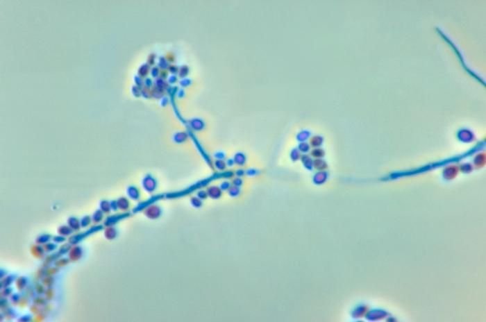
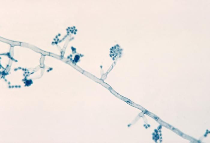
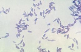
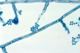
Magnified 500X, this photomicrograph revealed the presence of Sporothrix sp. fungal organisms that were isolated from a peat moss specimen.Created: 1971
-
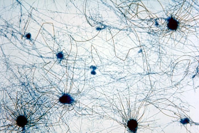
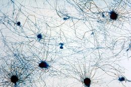
This photomicrograph reveals multiple perithecia, or fruiting bodies, of a Chaetomium spp. fungus.Created: 1955
-
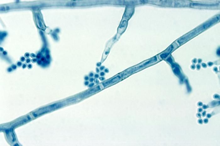
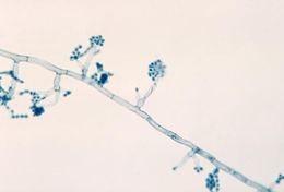
This photomicrograph reveals the conidiophores and conidia of the fungus Sporothrix schenckii.Created: 1972
-
This photomicrograph reveals multiple perithecia, or fruiting bodies, of a Chaetomium spp. fungus.Created: 1955
-
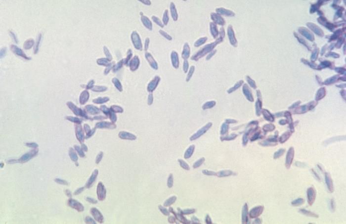
Shown here is a photomicrograph of the fungus Sporothrix schenckii during yeast phase.Created: 1964
-
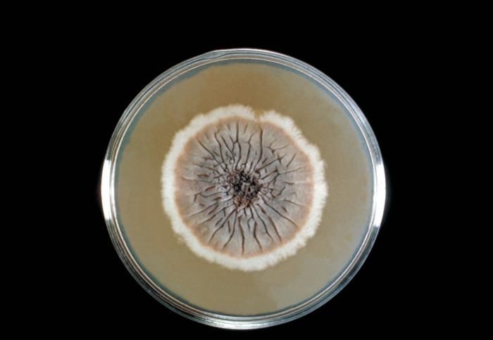
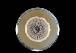
This Sabouraud's dextrose agar plate culture is growing the fungus Sporothrix schenckii.Created: 1964
-
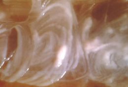
Shown here is a close-up of a Sporothrix schenckii culture during yeast phase.Created: 1964
-
This photomicrograph shows the presence of Sporothrix schenckii in a smear obtained from a rat.Created: 1964
-
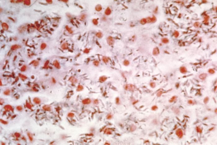
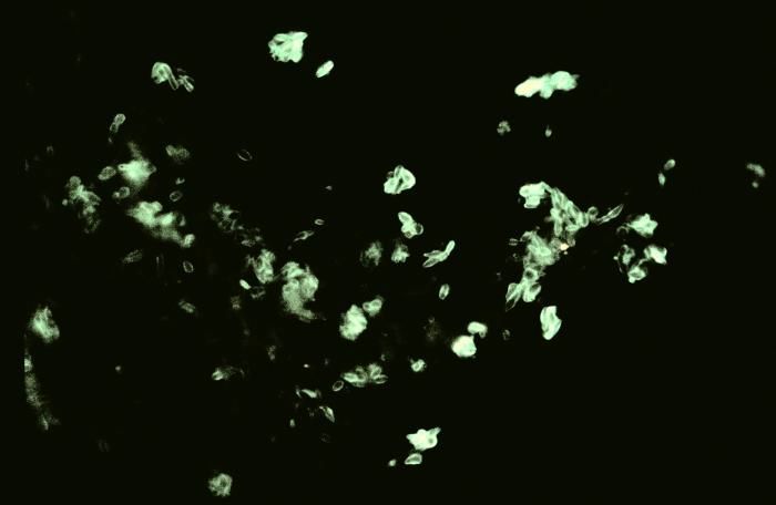
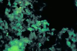
Using a direct FA stain, this slide demonstrates the histopathology of sporotrichosis due to Sporothrix schenckii.Created: 1977
-
Using a direct FA stain, this slide demonstrates the histopathology of sporotrichosis due to Sporothrix schenckii.Created: 1977
-
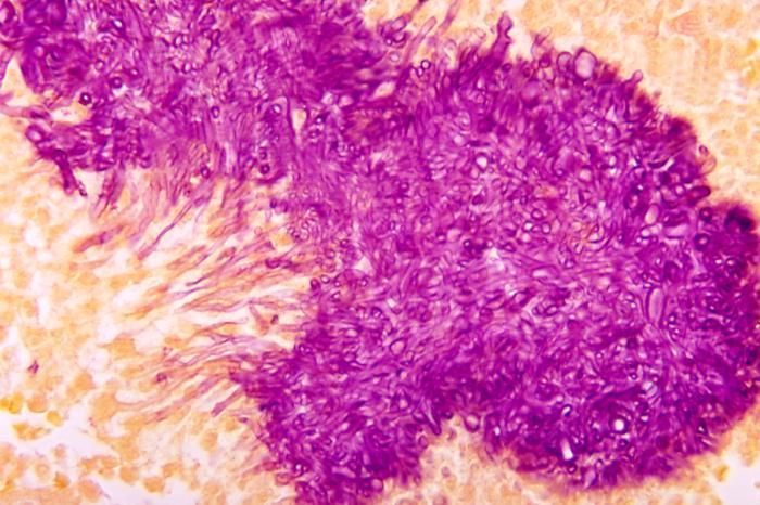
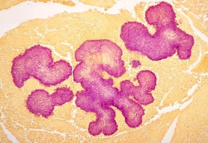
Note the histopathologic appearance of black grain mycetoma due to Madurella mycetomatis using a Gridley stain.Created: 1972
-
Note the histopathologic appearance of black grain mycetoma due to Madurella mycetomatis using a Gridley stain.Created: 1972
-
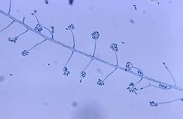
This photomicrograph shows phialides with terminal conidia of the Madurella mycetomatis fungus.Created: 1961
-
This photomicrograph shows phialides with terminal conidia of the Madurella mycetomatis fungus.Created: 1961