-
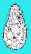
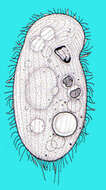
Tetrahymena (tet-ra-high-men-a) is an oligohymenophoran ciliate. There are cilia in about 20 kineties (rows) over the body and which are used for cell locomotion. There is also a group of three membranelles and an undulating membrane around the cytostome (upper right), and these are the buccal or oral cilia and are used in food capture. In nature often associated with damaged animals or dead tissue, may eat bacteria. Widely used in laboratory studies, and axenic (bacteria-free) cultures are maintained within high protein medium. Cells are pear-shaped. Differential interference contrast.
-
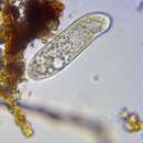
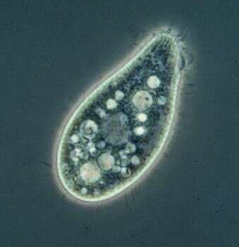
Tetrahymena (tet-ra-high-men-a) is an oligohymenophoran ciliate. There are cilia in about 20 kineties (rows) over the body and which are used for cell locomotion. There is also a group of three membranelles and an undulating membrane around the cytostome (upper left), and these are the buccal or oral cilia and are used in food capture. In nature often associated with damaged animals or dead tissue, may eat bacteria. Widely used in laboratory studies, and axenic (bacteria-free) cultures are maintained within high protein medium. Cells pear-shaped. Phase contrast.
-
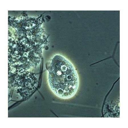
Tetrahymena (tet-ra-high-men-a) is an oligohymenophoran ciliate. Widely used in laboratory studies, and axenic (bacteria-free) cultures are maintained within high protein medium. In nature often associated with damaged animals or dead tissue. This occurrence within the cytoskeleton where they may feed on residual tissue (pieces left behind if the cytoskeleton has been shed) or on the body tissue (if the crustacea was damaged) is a good example of where this genus might be found in nature. Dark ground.
-
-
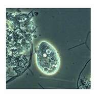
Tetrahymena: One of the most studied protozoans. This image was taken by Krishnakumar B. in a sample from an anaerobic bioreactor for organic rich wastewater treatment in Regional Research Laboratory-Trivandrum (CSIR-India).
-
Portrait (ventral view) of the hymenostome ciliate,Tetrahymena pyriformis complex.
-
Right ventrolateral view of the silverline system (argyrome) of a ciliate from the Tetrahymena pyriformis complex.The oral aperture is to the viewer's upper right. Stained by the Klein-Foissner dry silver nitrate technic (see Foissner, W. Europ. J. Protistol., 27:313-330;1991). Brightfield, black and white.
-
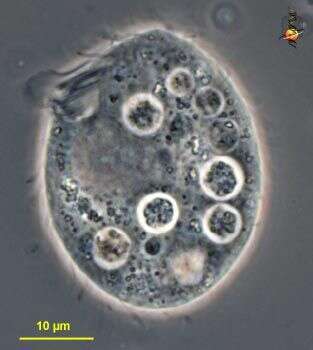
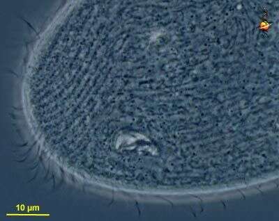
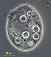
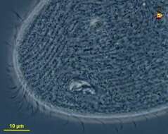
Cell from strain W, oral cilia are near the front of the cell (top of image), large clear area is the macronucleus, there is no micronucleus in this strain. Phase contrast image.
-
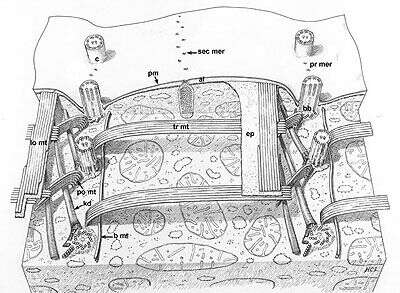
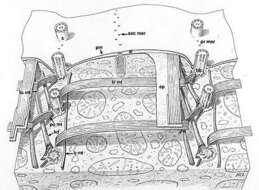
A classic drawing of the organization of this model ciliate. Segments of two kineties or rows of basal body (bb)/cilium (c) complexes. Kinetodesmal fibers (kd) arise from the left side of the proximal end of each basal body and angle up to the cell pellicle. Transverse microtubules (tr mt) extend to the right and postciliary microtubules (po mt) angle posteriorly and to the left of the kinety. One or two basal microtubules (b mt) extend along the right of the kinety at the proximal end of the basal bodies. A band of longitudinal microtubules (lo mt) lies between the epiplasm (ep) and the alveoli (al) to the left of the kinety. Mucocysts (mu) approach the plasma membrane (pm) through the septa separating alveoli along the primary (pr mer) and secondary meridians (sec mer). Drawn by H. C. Lyman.
This image is available in Richard Allen's collection.
-
The protargol stains the bases of the cilia and the nucleus. The image shows the kineties, the three membranelles and the single curved undulating membrane that make up the mouth ciliature, and the macronucleus (this strain lacks a micronucleus).
-
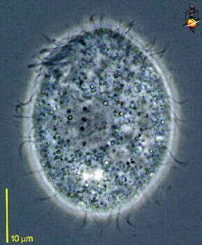
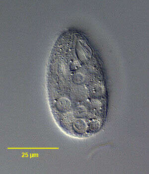
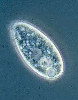
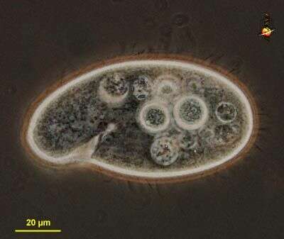
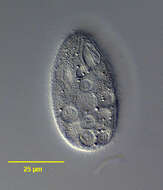
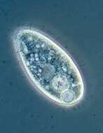
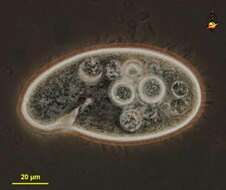
Phase contrast image of living cell. The mouth and some of the associated ciliature can be seen upper left, the surface is coated with somatic cilia that are used for cell locomotion, there is a contractile vacuole near the posterior end of the cell, and a light-coloured macronucleus near the centre of the cell. the other vacuoles are food vacuoles (phagosomes)
-
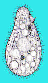
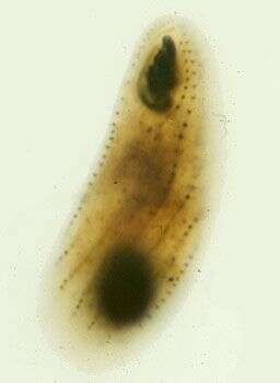
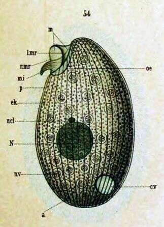
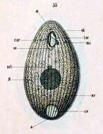
Originally described as Glaucoma pyriformis (Ehrenberg) Key to Schewiakoff's abbreviations: a -- Anus cv -- Contractile vacuole ek -- Ectoplasm l. mr -- Left membrane edge m -- Undulating membrane mi -- Inner undulating membrane N -- Macronucleus ncl -- Micronucleus oe -- Throat r.mr --Right membrane edge p -- Pellicle
-
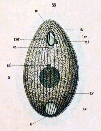
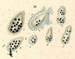
(Originally described under the name Glaucoma pyriformis) Ventral view. Key to Schewiakoff's abbreviations: a -- Anus cv -- Contractile vacuole el -- Ectoplasm l.or -- Left edge of mouth m -- Undulating membrane mi -- Inner Undulating membrane N -- Macronucleus ncl -- Micronucleus nv -- Food vacuole oe -- Throat r.or -- Right edge of mouth
-
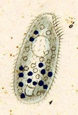
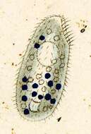
Originally described by Ehrenberg under the name Leucophrys pyriformis.
-
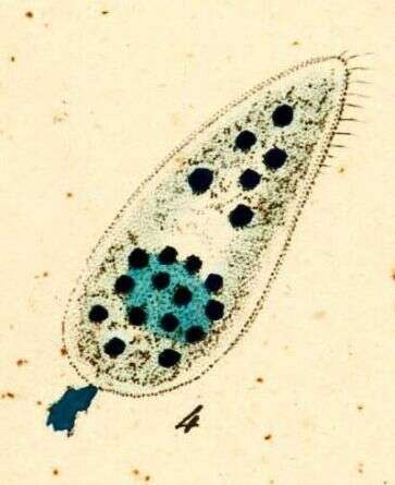
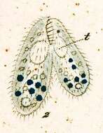
Originally described by Ehrenberg under the name Leucophrys pyriformis.
-
Originally described by Ehrenberg under the name Leucophrys pyriformis.
-
Originally described by Ehrenberg under the name Leucophrys pyriformis.
-
Colpidium (coll-pid-ee-um) is an oligohymenophoran ciliate, very closely related to Tetrahymena. The mouth is located near the front end, it is recessed, and the body is slightly twisted in front of the mouth. Eats bacteria and often found in organically enriched sites with little available oxygen. Phase contrast.
-
Colpidium (coll-pid-ee-um) is an oligohymenophoran ciliate, very closely related to Tetrahymena. The mouth is located near the front end, it is recessed, and the body is slightly twisted in front of the mouth. This detail shows the kineties at an angle anterior to the mouth. Eats bacteria and often found in organically enriched sites with little available oxygen. Phase contrast.
-
-
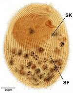
An early stage of stomatogenesis in Colpidium colpoda (LOSANA,1829) STEIN,1860.Stomatogenesis is of the monoparakinetal type. The stomatogenic field (SF) is seen to the left of the midportion of K1, the stomatogenic kinety (SK).From a putrefying raw culture from a freshwater pond near Boise, Idaho.October 2007. Stained by the silver carbonate technique (see Foissner, W. Europ. J. Protistol., 27:313-330;1991).Brightfield.
-
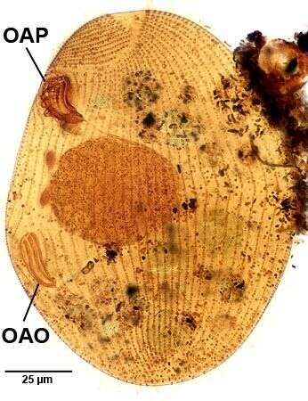
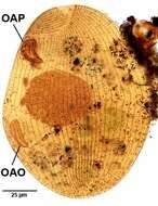
Late stage of stomatogenesis in Colpidium colpoda (LOSANA,1829) STEIN,1860.Stomatogenesis is of the monoparakinetal type. The adoral organelles and paraoral membranelles of the opisthe (OAO)have developed from a stomatogenic field adjacent to the stomatogenic kinety in the mid-ventral portion of the cell.OAP=oral apparatus of the proter or parental cell.From a putrefying raw culture from a freshwater pond near Boise, Idaho.October 2007. Stained by the silver carbonate technique (see Foissner, W. Europ. J. Protistol., 27:313-330;1991).Brightfield.
-
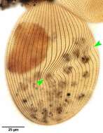
Dorsal infraciliature of Colpidium colpoda (LOSANA,1829) STEIN,1860.The green arrowheads mark the oblique furrow that extends from the oral aperture accross the right side of the cell to the center of the dorsal surface.The somatic kineties are more closely spaced and bend strongly to the left in this depression.In the living cell this area appears as a more densely ciliated region on the right. From a putrefying raw culture from a freshwater pond near Boise, Idaho.October 2007. Stained by the silver carbonate technique (see Foissner, W. Europ. J. Protistol., 27:313-330;1991).Brightfield.