-
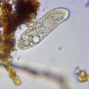
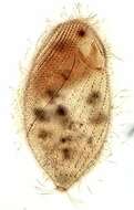
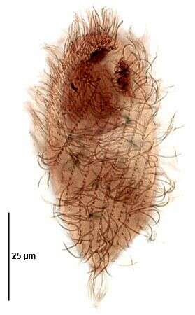
Phase contrast micrograph of the tetrahymenine ciliate. The anterior end of the cell is slightly twisted, the mouth being located at the base of this anterior region.
-
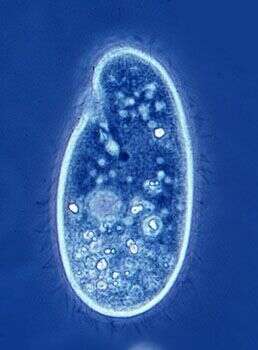
Anterior is to the bottom of the image, there are two mouth structures - the original near the anterior end and the mouth of the daughter cell developing behind where the division furrow will form.
-
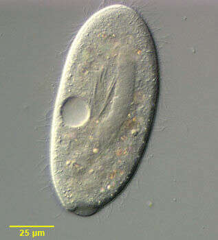
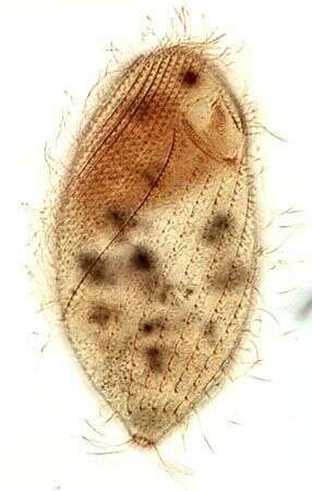
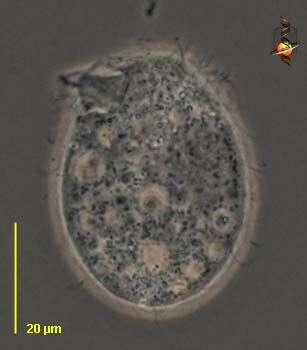
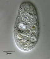
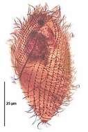
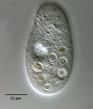
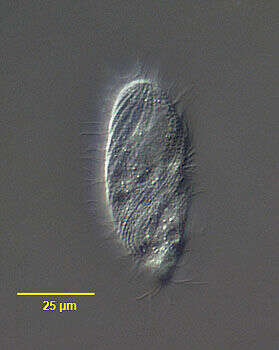
Portrait of Clathrostoma viminale (Penard, 1922) a frontoniine ciliate. Similar in overall appearance to but smaller than Frontonia leucas. The cytostome is located in a slight depression in the anterior half of the body. There is a cytopharyngeal basket (seen well here) composed of fibrils (as found in Loxodes) rather than true trichites. The somatic ciliation is uniform with a postoral suture. The sausage-shaped macronucleus is seen here with an adjacent cluster of several (1-4) micronuclei at its posterior end. A single contractile vacuole is located in the posterior 1/3. Collected from a freshwater agricultural irrigation ditch near McCall, Idaho in September 2003. DIC optics.
-
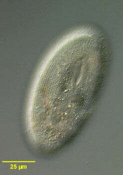
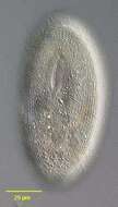
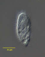
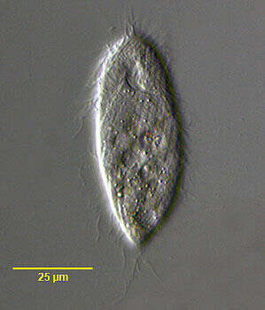
Portrait of Clathrostoma viminale (Penard, 1922) a frontoniine ciliate (surface view). Similar in overall appearance to but smaller than Frontonia leucas. The cytostome is located in a slight depression in the anterior half of the body (seen well in this image). There is a cytopharyngeal basket composed of fibrils (as found in Loxodes) rather than true trichites. The somatic ciliation is uniform with a postoral suture (seen in this image). The sausage-shaped macronucleus (not seen in this image) has an adjacent cluster of several (1-4) micronuclei at its posterior end. A single contractile vacuole is located in the posterior 1/3. Collected from a freshwater agricultural irrigation ditch near McCall, Idaho in September 2003. DIC optics.
-
Portrait of Clathrostoma viminale (Penard, 1922) a frontoniine ciliate. Similar in overall appearance to but smaller than Frontonia leucas. The cytostome is located in a slight depression in the anterior half of the body. There is a cytopharyngeal basket composed of fibrils (as found in Loxodes) rather than true trichites. The somatic ciliation is uniform with a postoral suture. The sausage-shaped macronucleus is seen here with an adjacent cluster of several (1-4) micronuclei at its posterior end. A single contractile vacuole is located in the posterior 1/3. Collected from a freshwater agricultural irrigation ditch near McCall, Idaho in September 2003. DIC optics.
-
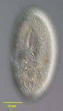
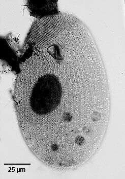
Portrait (left anterolateral view) of the hymenostome ciliate Colpidium kleini (Foissner, 1969). Very similar in overall appearance to C. colpoda although usually more slender and with fewer somatic kineties. The cytostome is in the anterior 1/4 of the cell. There is a curved paraoral membrane along the convex right margin of the cytostome. The left margin is slightly concave. There are three adoral membranelles. There are 32 to 44 somatic kineties. The kineties to the right and left of the oral aperture meet at a curved preoral suture. There is an anterior apical area bare of cilia. There are rows of inconspicuous mucocysts between the somatic kineties. The ellipsoid macronucleus and adjacent micronucleus are centrally located. The single contractile vacuole is located in the midbody with a single excretory pore on the right surface. The feature most clearly distinguishing Colpidium kleini from C. coploda is the silverline system (as demonstrated by silver nitrate staining). Collected from an organically enriched freshwater pond near Boise, Idaho. DIC.
-
Ventral infraciliature of the hymenostome ciliate Colpidium kleini (Foissner, 1969). C. kleini is very similar in overall appearance to C. colpoda although usually more slender and with fewer somatic kineties. The cytostome is in the anterior 1/4 of the cell. There is a curved paraoral membrane along the convex right margin of the cytostome. The left margin is slightly concave. There are three adoral membranelles. There are 32 to 44 somatic kineties. The kineties to the right and left of the oral aperture meet at a curved preoral suture. The right somatic kineties bend leftward at the level of the cytostome.There is an anterior apical area bare of cilia. There are rows of inconspicuous mucocysts between the somatic kineties. The ellipsoid macronucleus and adjacent micronucleus are centrally located. The single contractile vacuole is located in the midbody with a single excretory pore on the right surface. The feature most clearly distinguishing Colpidium kleini from C. coploda is the silverline system (as demonstrated by silver nitrate staining).Stained by the silver carbonate technic (see Foissner, W.Europ. J. Protistol.27,313-330;1991). Collected from an organically enriched freshwater pond near Boise, Idaho. Brightfield.
-

Right lateral infraciliature of the hymenostome ciliate Colpidium kleini (Foissner, 1969). C. kleini is very similar in overall appearance to C. colpoda although usually more slender and with fewer somatic kineties. The cytostome is in the anterior 1/4 of the cell. There is a curved paraoral membrane along the convex right margin of the cytostome. The left margin is slightly concave. There are three adoral membranelles. There are 32 to 44 somatic kineties. The kineties to the right and left of the oral aperture meet at a curved preoral suture. The right somatic kineties bend leftward at the level of the cytostome. There is an anterior apical area bare of cilia. There are rows of inconspicuous mucocysts between the somatic kineties. The ellipsoid macronucleus and adjacent micronucleus are centrally located. The single contractile vacuole is located in the midbody with a single excretory pore on the right surface. The feature most clearly distinguishing Colpidium kleini from C. coploda is the silverline system (as demonstrated by silver nitrate staining).Stained by the silver carbonate technic (see Foissner, W.Europ. J. Protistol.27,313-330;1991). Collected from an organically enriched freshwater pond near Boise, Idaho.Brightfield.
-
Right lateral view of the silverline system of the hymenostome ciliate Colpidium kleini (Foissner, 1969). C. kleini is very similar in overall appearance to C. colpoda although usually more slender and with fewer somatic kineties. The cytostome is in the anterior 1/4 of the cell. There is a curved paraoral membrane along the convex right margin of the cytostome. The left margin is slightly concave. There are three adoral membranelles. There are 32 to 44 somatic kineties. The kineties to the right and left of the oral aperture meet at a curved preoral suture.The right somatic kineties bend leftward at the level of the cytostome. There is an anterior apical area bare of cilia. There are rows of inconspicuous mucocysts between the somatic kineties. The ellipsoid macronucleus and adjacent micronucleus are centrally located. The single contractile vacuole is located in the midbody with a single excretory pore on the right surface. The feature most clearly distinguishing Colpidium kleini from C. coploda is the silverline system. In C. kleini there is only one secondary meridian (silverline) between two primary meridians (primary meridians correspond to somatic kineties). In some cases short segments of the secondary meridians may be duplicated. Short transverse L or T-shaped branches arise from both primary and secondary meridians at irregular intervals.Stained by the dry silver nitrate technic (see Foissner, W.Europ. J. Protistol.27,313-330;1991). Collected from an organically enriched freshwater pond near Boise, Idaho. Brightfield. Black and white.
-
Left lateral view of the silverline system of the hymenostome ciliate Colpidium kleini (Foissner, 1969). C. kleini is very similar in overall appearance to C. colpoda although usually more slender and with fewer somatic kineties. The cytostome is in the anterior 1/4 of the cell. There is a curved paraoral membrane along the convex right margin of the cytostome. The left margin is slightly concave. There are three adoral membranelles. There are 32 to 44 somatic kineties. The kineties to the right and left of the oral aperture meet at a curved preoral suture. There is an anterior apical area bare of cilia. There are rows of inconspicuous mucocysts between the somatic kineties. The ellipsoid macronucleus and adjacent micronucleus are centrally located. The single contractile vacuole is located in the midbody with a single excretory pore on the right surface. The feature most clearly distinguishing Colpidium kleini from C. coploda is the silverline system. In C. kleini there is only one secondary meridian (silverline) between two primary meridians (primary meridians correspond to somatic kineties seen here as the wavier lines). Short transverse L or T-shaped branches arise from both primary and secondary meridians at irregular intervals. In some cases short segments of the secondary meridians may be duplicated. Stained by the dry silver nitrate technic (see Foissner, W.Europ. J. Protistol.27,313-330;1991). Collected from an organically enriched freshwater pond near Boise, Idaho. Brightfield. Black and white.
-
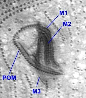
Oral infraciliature of Colpidium kleini (FOISSNER, 1969).There are three adoral membranelles (M1-3) and a right paraoral membrane (POM).Stained by the silver carbonate technique (see Foissner, W. Europ. J. Protistol., 27:313-330;1991).Brightfield.
-
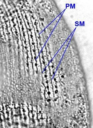
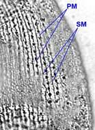
Silverline system of Colpidium kleini (FOISSNER, 1969).There is a single secondary meridian (SM) between each pair of primary meridians (PM).This feature distinguishes C. kleini from the larger C. colpoda whose silverline system shows two secondary meridians between pairs of primary meridians.Stained by the dry silver nitrate technique (see Foissner, W. Europ. J. Protistol., 27:313-330;1991).Brightfield.
-

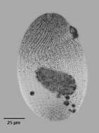
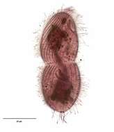
Infraciliature (ventral view) of the trichostomatid ciliate, Spirozona caudata (Kahl,1926)in middle division. The cell is elongate and rounded anteriorly and tapers posteriorly to a narrow truncate cone. The cell is ellipsoid in cross section. The cytostome is located in the anterior ¼. Its right margin is curved and the left relatively straight. There are several oral polykinetids on the left and an undulating membrane on the right. The somatic ciliature is distinctive with a wide swath of closely spaced kineties spiraling from the right anterior to the posterior midline. A single spiral kinety of more densely packed kinetosomes bearing longer cilia originates to the right of the cytostome and spirals around the right side to the dorsum of the cell. There are 3 postoral kineties and one shorta paraoral kinetid on the left margin of the cytostome. More widely spaced kineties with less densely packed kinetids originate to the left of the cytostome and follow a less spiral course to the posterior end ventrally. The narrow truncate cone of the posterior end bears a circular row of kinetids. The spherical macronucleus and micronucleus are located in the anterior half. The single contractile vacuole is located at the posterior end. Collected from sapropelic bottom sediments from standing freshwater near Boise, Idaho November, 2004. Stained by the silver carbonate technique (see Foissner, W.Europ. J. Protistol.27,313-330;1991). Brightfield optics.
-
Infraciliature (dorsal view) of the trichostomatid ciliate, Spirozona caudata (Kahl,1926). The cell is elongate and rounded anteriorly and tapers posteriorly to a narrow truncate cone. The cell is ellipsoid in cross section. The cytostome is located in the anterior 1/4. Its right margin is curved and the left relatively straight. There are several oral polykinetids on the left and an undulating membrane on the right. The somatic ciliature is distinctive with a wide swath of closely spaced kineties spiraling from the right anterior to the posterior midline. A single spiral kinety of more densely packed kinetosomes bearing longer cilia originates to the right of the cytostome and spirals around the right side to the dorsum of the cell. There are 3 postoral kineties and one short paraoral kinetid on the left margin of the cytostome. More widely spaced kineties with less densely packed kinetids originate to the left of the cytostome and follow a less spiral course to the posterior end ventrally. The narrow truncate cone of the posterior end bears a circular row of kinetids.There is a small unciliated anterior apical area or "frontal plate" (seen here). The spherical macronucleus and micronucleus are located in the anterior half. The single contractile vacuole is located at the posterior end. Collected from sapropelic bottom sediments from standing freshwater near Boise, Idaho November, 2004. Stained by the silver carbonate technique (see Foissner, W.Europ. J. Protistol.27,313-330;1991). Brightfield optics.
-
Infraciliature (ventral view) of the trichostomatid ciliate, Spirozona caudata (Kahl,1926). The cell is elongate and rounded anteriorly and tapers posteriorly to a narrow truncate cone. The cell is ellipsoid in cross section. The cytostome is located in the anterior �. Its right margin is curved and the left relatively straight. There are several oral polykinetids on the left and an undulating membrane on the right. The somatic ciliature is distinctive with a wide swath of closely spaced kineties spiraling from the right anterior to the posterior midline. A single spiral kinety of more densely packed kinetosomes bearing longer cilia originates to the right of the cytostome and spirals around the right side to the dorsum of the cell. There are 3 postoral kineties and one shorta paraoral kinetid on the left margin of the cytostome. More widely spaced kineties with less densely packed kinetids originate to the left of the cytostome and follow a less spiral course to the posterior end ventrally. The narrow truncate cone of the posterior end bears a circular row of kinetids. There is a small unciliated anterior apical area or "frontal plate". The spherical macronucleus and micronucleus are located in the anterior half. The single contractile vacuole is located at the posterior end. Collected from sapropelic bottom sediments from standing freshwater near Boise, Idaho November, 2004. Stained by the silver carbonate technique (see Foissner, W.Europ. J. Protistol.27,313-330;1991). Brightfield optics.
-
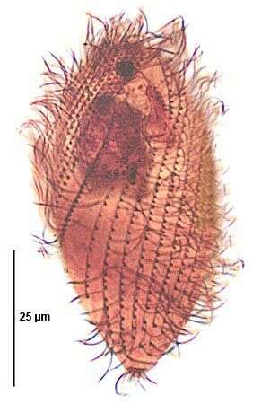
Infraciliature (ventral view) of the trichostomatid ciliate, Spirozona caudata (Kahl,1926). The cell is elongate and rounded anteriorly and tapers posteriorly to a narrow truncate cone. The cell is ellipsoid in cross section. The cytostome is located in the anterior 1/4. Its right margin is curved and the left relatively straight. There are several oral polykinetids on the left and an undulating membrane on the right. The somatic ciliature is distinctive with a wide swath of closely spaced kineties spiraling from the right anterior to the posterior midline. A single spiral kinety of more densely packed kinetosomes bearing longer cilia originates to the right of the cytostome and spirals around the right side to the dorsum of the cell. There are 3 postoral kineties and one shorta paraoral kinetid on the left margin of the cytostome. More widely spaced kineties with less densely packed kinetids originate to the left of the cytostome and follow a less spiral course to the posterior end ventrally. The narrow truncate cone of the posterior end bears a circular row of kinetids. There is a small unciliated anterior apical area or "frontal plate". The spherical macronucleus and micronucleus are located in the anterior half. The single contractile vacuole is located at the posterior end. Collected from sapropelic bottom sediments from standing freshwater near Boise, Idaho November, 2004. Stained by the silver carbonate technique (see Foissner, W.Europ. J. Protistol.27,313-330;1991). Brightfield optics.
-
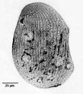
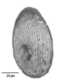
Portrait (dorsal view) of the trichostomatid ciliate, Spirozona caudata (Kahl,1926). The cell is elongate and rounded anteriorly and tapers posteriorly to a narrow truncate cone. The cell is ellipsoid in cross section. The cytostome is located in the anterior ¼. Its right margin is curved and the left relatively straight. There are several oral polykinetids on the left and an undulating membrane on the right. The somatic ciliature is distinctive with a wide swath of closely spaced kineties spiraling from the right anterior to the posterior midline. A single spiral kinety of more densely packed kinetosomes bearing longer cilia originates to the right of the cytostome and spirals around the right side to the dorsum of the cell. There are 3 postoral kineties and one short paraoral kinetid on the left margin of the cytostome. More widely spaced kineties with less densely packed kinetids originate to the left of the cytostome and follow a less spiral course to the posterior end ventrally. The narrow truncate cone of the posterior end bears a circular row of kinetids. There is a small unciliated anterior apical area or "frontal plate". The spherical macronucleus and micronucleus are located in the anterior half. The single contractile vacuole is located at the posterior end. Collected from sapropelic bottom sediments from standing freshwater near Boise, Idaho November, 2004. DIC.
-
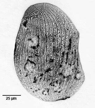
Portrait (ventral view) of the trichostomatid ciliate, Spirozona caudata (Kahl,1926). The cell is elongate and rounded anteriorly and tapers posteriorly to a narrow truncate cone. The cell is ellipsoid in cross section. The cytostome is located in the anterior ¼. Its right margin is curved and the left relatively straight. There are several oral polykinetids on the left and an undulating membrane on the right. The somatic ciliature is distinctive with a wide swath of closely spaced kineties spiraling from the right anterior to the posterior midline. A single spiral kinety of more densely packed kinetosomes bearing longer cilia originates to the right of the cytostome and spirals around the right side to the dorsum of the cell. There are 3 postoral kineties and one shorta paraoral kinetid on the left margin of the cytostome. More widely spaced kineties with less densely packed kinetids originate to the left of the cytostome and follow a less spiral course to the posterior end ventrally. The narrow truncate cone of the posterior end bears a circular row of kinetids. There is a small unciliated anterior apical area or "frontal plate". The spherical macronucleus and micronucleus are located in the anterior half. The single contractile vacuole is located at the posterior end. Collected from sapropelic bottom sediments from standing freshwater near Boise, Idaho November, 2004. DIC.
-
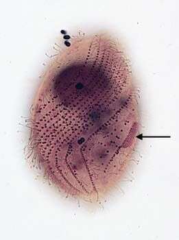
Dorsolateral view of the infraciliature of Spirozona caudata (Kahl,1926) in early division. The stomatogenic field of the opisthe is seen as a patch of kinetosomes adjacent to the first somatic kinety (k1) (black arrow).Stained by the silver carbonate technique (see Foissner, W. Europ. J. Protistol., 27:313-330;1991).Brightfield.
-
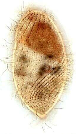
Infraciliature (dorsal view) of the trichostomatid ciliate, Spirozona caudata (Kahl,1926). The cell is elongate and rounded anteriorly and tapers posteriorly to a narrow truncate cone. The cell is ellipsoid in cross section. The cytostome is located in the anterior 1/4. Its right margin is curved and the left relatively straight. There are several oral polykinetids on the left and an undulating membrane on the right. The somatic ciliature is distinctive with a wide swath of closely spaced kineties spiraling from the right anterior to the posterior midline. A single spiral kinety of more densely packed kinetosomes bearing longer cilia originates to the right of the cytostome and spirals around the right side to the dorsum of the cell. There are 3 postoral kineties and one short paraoral kinetid on the left margin of the cytostome. More widely spaced kineties with less densely packed kinetids originate to the left of the cytostome and follow a less spiral course to the posterior end ventrally. The narrow truncate cone of the posterior end bears a circular row of kinetids.There is a small unciliated anterior apical area or "frontal plate". The spherical macronucleus and micronucleus are located in the anterior half. The single contractile vacuole is located at the posterior end. Collected from sapropelic bottom sediments from slow flowing freshwater near Boise, Idaho March,2007. Stained by the silver carbonate technique (see Foissner, W.Europ. J. Protistol.27,313-330;1991). Brightfield optics.
-
Infraciliature (ventral view) of the trichostomatid ciliate, Spirozona caudata (Kahl,1926).
-
Tetrahymena (tet-ra-high-men-a), only one thread on the entire slide and it has to land on me.
-
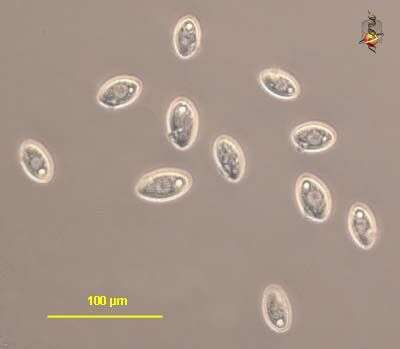
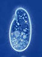
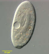
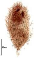
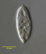
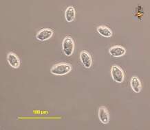
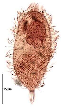
Tetrahymena (tet-ra-high-men-a) is an oligohymenophoran ciliate. There are cilia in about 20 kineties (rows) over the body and which are used for cell locomotion. There is also a group of three membranelles and an undulating membrane around the cytostome (upper left), and these are the buccal or oral cilia and are used in food capture. In nature often associated with damaged animals or dead tissue, may eat bacteria. Widely used in laboratory studies, and axenic (bacteria-free) cultures are maintained within high protein medium. This cell is slightly compressed. Phase contrast.
-
Tetrahymena (tet-ra-high-men-a) is an oligohymenophoran ciliate. There are cilia in about 20 kineties (rows) over the body and which are used for cell locomotion. There is also a group of three membranelles and an undulating membrane around the cytostome, and these are the buccal or oral cilia and are used in food capture. In nature often associated with damaged animals or dead tissue, may eat bacteria. Widely used in laboratory studies, and axenic (bacteria-free) cultures are maintained within high protein medium. cells pear-shaped. Phase contrast.