cortex schematic
Açıklama:
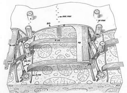
A classic drawing of the organization of this model ciliate. Segments of two kineties or rows of basal body (bb)/cilium (c) complexes. Kinetodesmal fibers (kd) arise from the left side of the proximal end of each basal body and angle up to the cell pellicle. Transverse microtubules (tr mt) extend to the right and postciliary microtubules (po mt) angle posteriorly and to the left of the kinety. One or two basal microtubules (b mt) extend along the right of the kinety at the proximal end of the basal bodies. A band of longitudinal microtubules (lo mt) lies between the epiplasm (ep) and the alveoli (al) to the left of the kinety. Mucocysts (mu) approach the plasma membrane (pm) through the septa separating alveoli along the primary (pr mer) and secondary meridians (sec mer). Drawn by H. C. Lyman. This image is available in Richard Allen's collection.
Aşağıdaki Sayfalarda Bulunmaktadır:
- Life
- Cellular
- Eukaryota (Ökaryot)
- SAR (Stramenopiles, Alveolates, Rhizaria)
- Alveolata
- Ciliophora (Silliler)
- Intramacronucleata
- Oligohymenophorea
- Hymenostomatida
- Tetrahymenina
- Tetrahymenidae
- Tetrahymena
- Tetrahymena pyriformis
Bu resim hiçbir koleksiyonda yer almıyor.
Kaynak Bilgileri
- lisans
- cc-by-nc
- yazar
- R. D. Allen
- sağlayıcı
- micro*scope
- orijinal
- orijinal medya dosyası
- kaynağı ziyaret et
- ortak site
- micro*scope
- ID