-
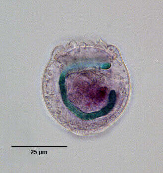
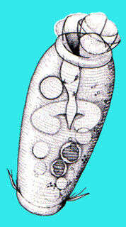
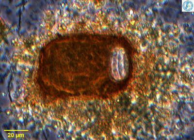
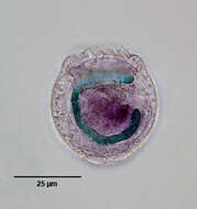
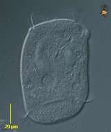
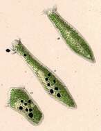
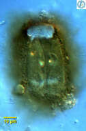
Macronucleus (stained green in this image) of the free-swimming peritrich ciliate, Astylozoon faurei (complex). Since A. faurei and A. enriquesi differ only by the macronuclear morphology (reniform in the former and C-shaped in the latter) and since overlap in this feature between individuals, Foissner has grouped these two species into "Astylozoon faurei complex" (See Foissner, W., Berger, H. and Schaumburg, J.: Identification of Limnetic Plankton Ciliates. Bavarian State Office for Water Management. Munich, 1999; pp. 455-459). This cell has been moderately squashed, dislodging the macronucleus from its usual transverse orientation. Stained by the methyl green-pyronin Y technic (see Foissner, W. Europ. J. Protistol. 27:313-330;1991). Collected from a temporary rainwater pool containing dead grass near Boise, Idaho March 2005. DIC.
-
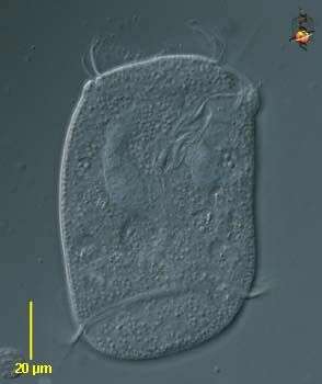
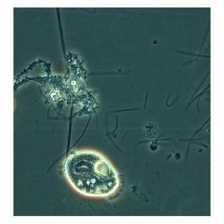
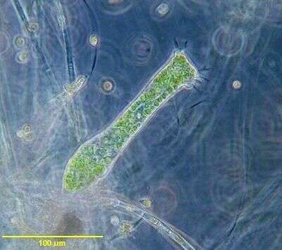
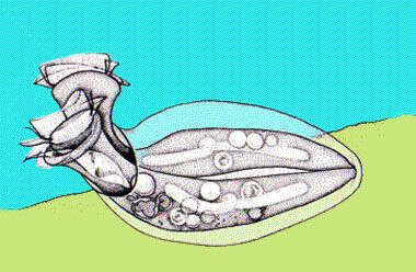
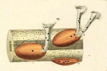
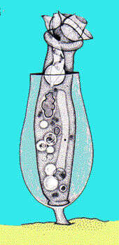
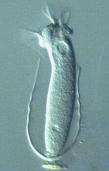
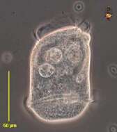
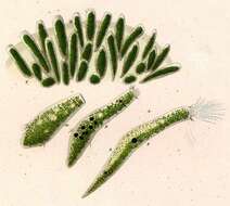
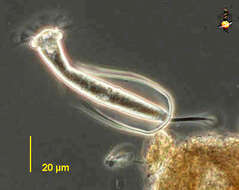
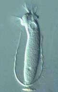
Opisthonecta (owe-pissed-though-neck-ta) - one of the peritrich ciliate, closely related to the sessile forms. However, this one is not sessile, but swims around. At the anterior end (upper) are the oral cilia (membranelles and undulating membrane) which form a spiral wreath which then enters into a narrowing channel in the cell to end at the cytostome. This is where food is packaged into food vacuoles, and several large food vacuoles are evident in this picture. Posteriorly, there is another wreath of cilia which help to propel the cell. Large curving macronucleus seen in the upper part of the cell. Differential interference contrast.
-
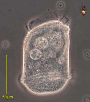
Opisthonecta (owe-pissed-though-neck-ta) - one of the peritrich ciliate, closely related to the sessile forms. However, this one is not sessile, but swims around. At the anterior end (upper) are the oral cilia (membranelles and undulating membrane) which form a spiral wreath which then enters into a narrowing channel in the cell to end at the cytostome. This is where food is packaged into food vacuoles, and several large food vacuoles are evident in this picture. Posteriorly, there is another wreath of cilia which help to propel the cell. Large curving macronucleus seen in the upper part of the cell. Phase contrast.
-
-
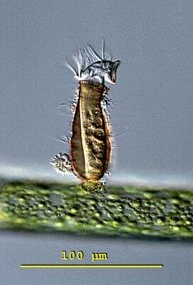
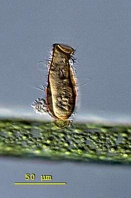
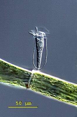
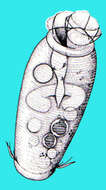
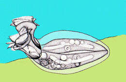
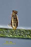
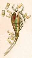
Telotrochidium attached to stalk. The free swimming form of this peritrich is barrel shaped without oral bristle. This image was taken by Krishnakumar B. in a sample from an anaerobic bioreactor for organic rich wastewater treatment in Regional Research Laboratory-Trivandrum (CSIR-India).
-
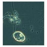
This is a phase contrast picture taken using 40x/N.A. 0.75 objective.
-
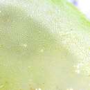
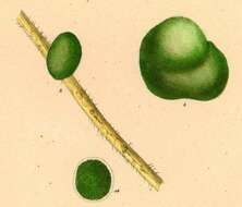
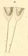
This is a brightfield picture of a pair of Ophrydium versatile taken using 25x/N.A. 0.60 objective.
-
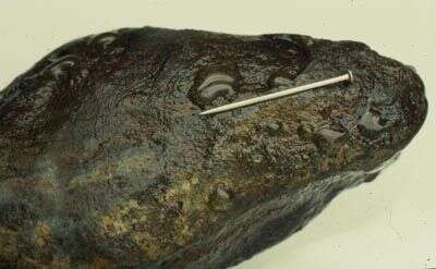
A dozen or so gelatinous colonies of this peritrich ciliate on the underside of a rock from a stream near Bristol, England. It's a pin.
-
-
-
Mucilaginous colonies of Ophrydium versatile.
-
-
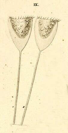
Portrait of Platycola. This peritrich ciliate resides in a simple non-valved lorica with a curved neck. The lorica adheres to the substrate along its length. Most often two individuals per lorica. From freshwater pond with abundant filamentous algae near Boise, Idaho. Oblique illumination.
-
Platycola, a loricate peritrich. This one forms a flattened lorica with apical apertures through which the cells extend while feeding. The cell cannot be seen clearly. The lorica appears transparent when first formed but gets darker with age. From Lake Donghu, China. Phase contrast micrograph.
-
Platycola, a loricate peritrich. This one forms a flattened lorica with apical apertures through which the cells extend while feeding. Two contracted cells lie inside the lorica. The lorica appears transparent when first formed but gets darker with age. From Lake Donghu, China. Differential interference contrast micrograph.
-
Originally described by Ehrenberg under the name Vaginicola decumbens.
-
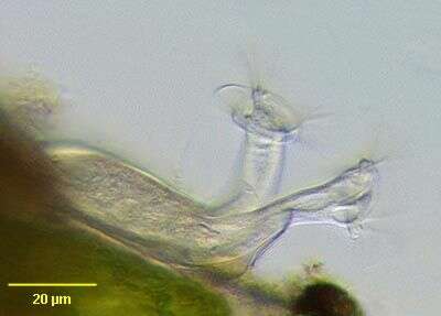
Pyxicola (pig-sick-cola) carteri with opened cap of the lorica. The cap is part of the cell and not of the lorica. This specimen was collected in freshwater ponds near Konstanz, Germany. Differential interference contrast.
-
Pyxicola (pig-sick-cola) carteri with opened cap of the lorica. The cap is part of the cell and not of the lorica. This cell has a closed lorica. The individual can pull down and tighten the cap by contraction of the body. This specimen was collected in freshwater ponds near Konstanz, Germany. Differential interference contrast.
-
Cothurnia (co-thur-knee-a) is a sessile peritrich ciliate. The cells live within a lorica which is itself stalked. Cells attach to the base of the lorica by the posterior ends of the cell. Cells can contract into the lorica. The oral cilia form a wreath around the anterior end of the cell. One of the peritrich ciliates, distinguished by having a wreath of cilia around the anterior of the cell. Normally feeding on bacteria. No cilia on the body. Phase contrast.
-
-
Differential interference contrast image showing living cell and lorica with a short stalk.
-
-
Cothurnia (coe-thurr-knee-ah) annulata built up a colourless transparent lorica. The lorica is fixed by a stalk on the ground. Cothurnia annulata has a distinctive ringshape bulge in the middle of the cell. Differential interference contrast.
-