-
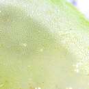
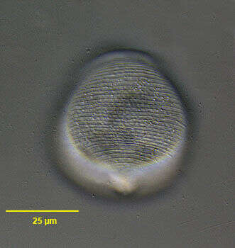
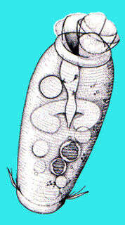
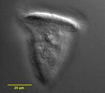
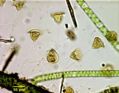
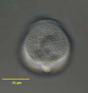
Surface detail of the peritrich ciliate, Pseudovorticella chlamydophora (Penard, 1922) Jankowski, 1976. Pseudovorticella is distinguished from Vorticella by silver staining which reveals a lattice-like silver line system in the former and circumferential lines without vertical connections in the latter. Pseudovorticella also has two contractile vacuoles. P. chlamydophora is distinguished by a distinct hyaline layer consisting of large cuboid pellicular blebs. The lattice-like pattern of these blebs is visible here. Feeds on bacteria. From freshwater pond near Boise, Idaho. DIC.
-
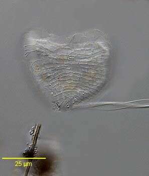
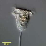
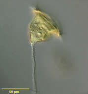
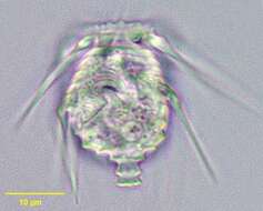
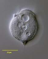
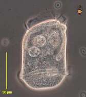
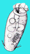
Portrait of the peritrich ciliate, Pseudovorticella chlamydophora (Penard,1922) Jankowski, 1976. This genus is distinguished from the genus Vorticella by its grid-like silver line system. The transverse components of the silverline system of Vorticella species have no vertical connections. P. Chlamydophora has a thick clear pellicular layer composed of cuboid units, which give the cell surface a distinctive quilted appearance. The extended cell is an inverted bell shape connected at the aboral scopula to a contractile stalk. The cell is spherical when contracted. The stalk contracts as a coil rather than a zigzag (e.g. Haplocaulus). The peristomal disc is almost flush. The ciliature is reduced to two rows of peristomal cilia, which beat counterclockwise toward the funnel-shaped buccal cavity (seen here to the viewers left anteriorly). The roughly C-shaped macronucleus is oriented in the long axis (to the viewers left of midline here). A single contractile vacuole is seen adjacent to the buccal cavity. The otherwise identical P. vestita has two contractile vacuoles. Multiple yellowish food vacuoles are seen here. P. chlamydophora may be gregarious but does not form true colonies. Collected from a freshwater pond near Boise, Idaho May 2004. DIC optics.
-
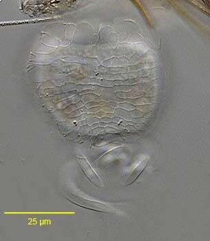
Portrait of the peritrich ciliate, Pseudovorticella chlamydophora (Penard,1922) Jankowski, 1976. This genus is distinguished from the genus Vorticella by its grid-like silver line system. The transverse components of the silverline system of Vorticella species have no vertical connections. P. Chlamydophora has a thick clear pellicular layer composed of cuboid units, which give the cell surface a distinctive quilted appearance (seen en face here). The extended cell is an inverted bell shape connected at the aboral scopula to a contractile stalk. The cell is spherical when contracted. The stalk contracts as a coil rather than a zigzag (e.g. Haplocaulus). The otherwise identical P. vestita has two contractile vacuoles. P. chlamydophora may be gregarious but does not form true colonies. Collected from a freshwater pond near Boise, Idaho.June 2005. DIC.
-
Surface detail of the peritrich ciliate, Pseudovorticella chlamydophora (Penard,1922) Jankowski, 1976. This genus is distinguished from the genus Vorticella by its grid-like silver line system. The transverse components of the silverline system of Vorticella species have no vertical connections. P. Chlamydophora has a thick clear pellicular layer composed of cuboid units, which give the cell surface a distinctive quilted appearance (seen en face here). The extended cell is an inverted bell shape connected at the aboral scopula to a contractile stalk. The cell is spherical when contracted. The stalk contracts as a coil rather than a zigzag (e.g. Haplocaulus). The otherwise identical P. vestita has two contractile vacuoles. P. chlamydophora may be gregarious but does not form true colonies. Collected from a freshwater pond near Boise, Idaho.June 2005. DIC.
-
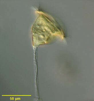
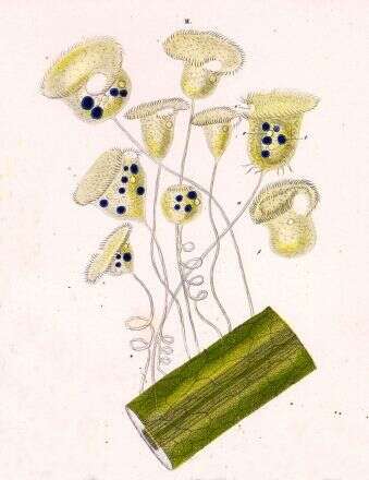
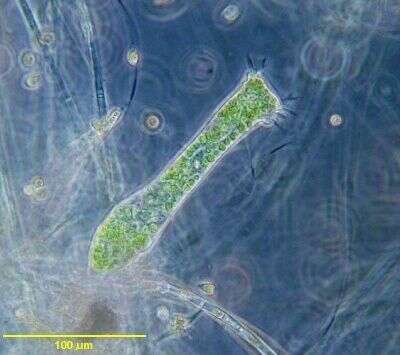
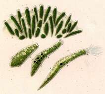
Group portrait of Vorticella citrina (Muller 1786) a sessiline peritrich ciliate. Part of the Vorticella convallaria complex.This species is lemon yellow to light green in color. The body has typical inverted bell shape. There is a peristomal lip. Peristomal cilia wind counterclockwise to the cytostome. There are fine annular striations on the cell body. At the aboral pole is a scopula, the organelle that secretes the contractile stalk. The stalk is a contractile myonemes enclosed in by a sheath, which is ovoid in cross section. The stalk contracts in corkscrew fashion unlike the zigzag contraction of the stalk in the similar genus, Haplocaulus. The nucleus is short and horseshoe shaped. There is a single contractile vacuole. Vorticella is not colonial but may be gregarious. Primarily bactiverous. Collected from freshwater pond near Boise, Idaho October 2003. Brightfield optics.
-
Individual portrait of Vorticella citrina (Muller 1786) a sessiline peritrich ciliate. Part of the Vorticella convallaria complex. This species is lemon yellow to light green in color. The body has typical inverted bell shape. There is a peristomal lip. Peristomal cilia wind counterclockwise to the cytostome. There are fine annular striations on the cell body (seen here). At the aboral pole is a scopula, the organelle that secretes the contractile stalk. The stalk is a contractile myonemes enclosed in by a sheath, which is ovoid in cross section. The stalk contracts in corkscrew fashion unlike the zigzag contraction of the stalk in the similar genus, Haplocaulus. The nucleus is short and horseshoe shaped. There is a single contractile vacuole. Vorticella is not colonial but may be gregarious. Primarily bactiverous. Collected from freshwater pond near Boise, Idaho October 2003. DIC optics.
-
-
-
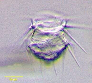
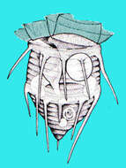
Hastatella. Portrait of uncommon colorless peritrich ciliate H. radians. It has two girdles of spines which distinguishes it from H. aesculacantha, which has four. Swimming interrupted by intermittent jumping movements. Bacterivorous. Pellicle has annular ridges. Wreath of cilia around anterior peristomal disc. These individuals have a blunt posterior scopula but sometimes this may appear more spinous. Macronucleus is roughly equatorial and C-shaped. horough description and illustrations of H. radians can be found in: Foissner,W.,Berger,H. and Schaumberg,J:Identification and Ecology of Limnetic Plankton Ciliates. Bavarian State Office for Water Management; Munich,1999.
-
Hastatella. Portrait of uncommon colorless peritrich ciliate H. radians. It has two girdles of spines which distinguishes it from H. aesculacantha, which has four. Swimming interrupted by intermittent jumping movements. Bacterivorous. Pellicle has annular ridges. Wreath of cilia around anterior peristomal disc. These individuals have a blunt posterior scopula but sometimes this may appear more spinous. Macronucleus is roughly equatorial and C-shaped. From freshwater aquaculture pond near Boise, Idaho.Thorough description and illustrations of H. radians can be found in: Foissner,W.,Berger,H. and Schaumberg,J:Identification and Ecology of Limnetic Plankton Ciliates. Bavarian State Office for Water Management; Munich,1999.
-
-
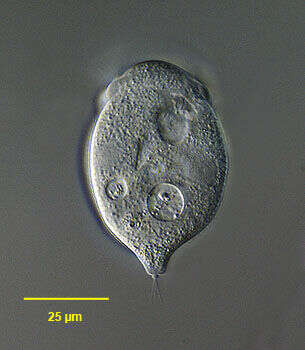
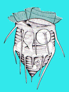
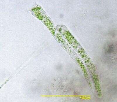
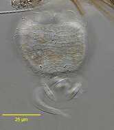
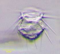
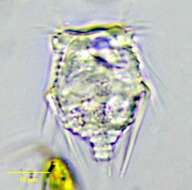
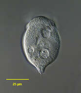
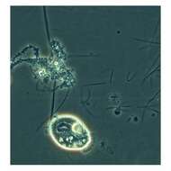
Portrait of the free-swimming peritrich ciliate, Astylozoon faurei (complex). Since A. faurei and A. enriquesi differ only by the macronuclear morphology (reniform in the former and C-shaped in the latter) and since overlap in this feature between individuals, Foissner has grouped these two species into "Astylozoon faurei complex" (See Foissner, W., Berger, H. and Schaumburg, J.: Identification of Limnetic Plankton Ciliates. Bavarian State Office for Water Management. Munich, 1999; pp. 455-459). The cell has a globular inverted teardrop shape. Contracted cells are spherical. The oral aperture is anterior. There is a peristomial collar and the central peristomial disc is dome-shaped. The adoral ciliary field winds counterclockwise to the cytostome. There is a short posterior conical process bearing one or more short bristles. The single contractile vacuole lies dorsal to and empties into the vestibulum. The pellicle is marked by conspicuous transverse striae. In this population the transversely oriented C-shaped macronucleus is equatorial. Collected from a temporary rainwater pool containing dead grass near Boise, Idaho March 2005. DIC.
-
Pellicular detail of the free-swimming peritrich ciliate, Astylozoon faurei (complex). Since A. faurei and A. enriquesi differ only by the macronuclear morphology (reniform in the former and C-shaped in the latter) and since overlap in this feature between individuals, Foissner has grouped these two species into "Astylozoon faurei complex" (See Foissner, W., Berger, H. and Schaumburg, J.: Identification of Limnetic Plankton Ciliates. Bavarian State Office for Water Management. Munich, 1999; pp. 455-459). The pellicle is marked by conspicuous transverse striae (seen here). In this population the transversely oriented C-shaped macronucleus is equatorial. Collected from a temporary rainwater pool containing dead grass near Boise, Idaho March 2005. DIC.
-
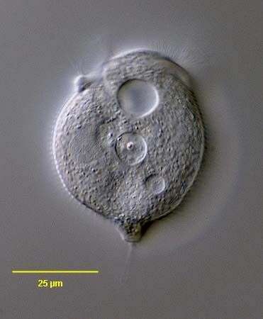
Portrait of the free-swimming peritrich ciliate, Astylozoon faurei (complex). Since A. faurei and A. enriquesi differ only by the macronuclear morphology (reniform in the former and C-shaped in the latter) and since overlap in this feature between individuals, Foissner has grouped these two species into "Astylozoon faurei complex" (See Foissner, W., Berger, H. and Schaumburg, J.: Identification of Limnetic Plankton Ciliates. Bavarian State Office for Water Management. Munich, 1999; pp. 455-459). The cell has a globular inverted teardrop shape. Contracted cells are spherical. The oral aperture is anterior. There is a peristomial collar and the central peristomial disc is dome-shaped. The adoral ciliary field winds counterclockwise to the cytostome. There is a short posterior conical process bearing one or more short bristles. The single contractile vacuole lies dorsal to and empties into the vestibulum. The pellicle is marked by conspicuous transverse striae. In this population the transversely oriented C-shaped macronucleus is equatorial. Collected from a temporary rainwater pool containing dead grass near Boise, Idaho March 2005. DIC.
-
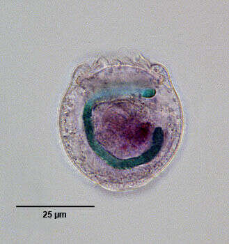
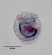
Macronucleus (stained green in this image) of the free-swimming peritrich ciliate, Astylozoon faurei (complex). Since A. faurei and A. enriquesi differ only by the macronuclear morphology (reniform in the former and C-shaped in the latter) and since overlap in this feature between individuals, Foissner has grouped these two species into "Astylozoon faurei complex" (See Foissner, W., Berger, H. and Schaumburg, J.: Identification of Limnetic Plankton Ciliates. Bavarian State Office for Water Management. Munich, 1999; pp. 455-459). This cell has been moderately squashed, dislodging the macronucleus from its usual transverse orientation. Stained by the methyl green-pyronin Y technic (see Foissner, W. Europ. J. Protistol. 27:313-330;1991). Collected from a temporary rainwater pool containing dead grass near Boise, Idaho March 2005. DIC.
-
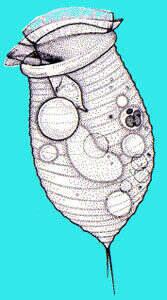
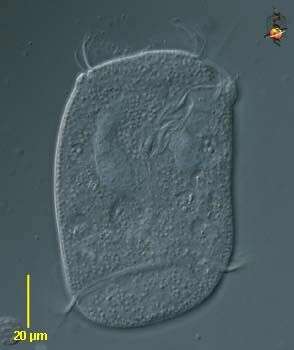
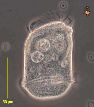
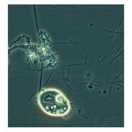
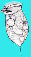
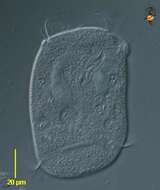
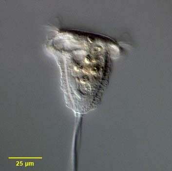
Opisthonecta (owe-pissed-though-neck-ta) - one of the peritrich ciliate, closely related to the sessile forms. However, this one is not sessile, but swims around. At the anterior end (upper) are the oral cilia (membranelles and undulating membrane) which form a spiral wreath which then enters into a narrowing channel in the cell to end at the cytostome. This is where food is packaged into food vacuoles, and several large food vacuoles are evident in this picture. Posteriorly, there is another wreath of cilia which help to propel the cell. Large curving macronucleus seen in the upper part of the cell. Differential interference contrast.
-
Opisthonecta (owe-pissed-though-neck-ta) - one of the peritrich ciliate, closely related to the sessile forms. However, this one is not sessile, but swims around. At the anterior end (upper) are the oral cilia (membranelles and undulating membrane) which form a spiral wreath which then enters into a narrowing channel in the cell to end at the cytostome. This is where food is packaged into food vacuoles, and several large food vacuoles are evident in this picture. Posteriorly, there is another wreath of cilia which help to propel the cell. Large curving macronucleus seen in the upper part of the cell. Phase contrast.
-
-
Telotrochidium attached to stalk. The free swimming form of this peritrich is barrel shaped without oral bristle. This image was taken by Krishnakumar B. in a sample from an anaerobic bioreactor for organic rich wastewater treatment in Regional Research Laboratory-Trivandrum (CSIR-India).
-
This is a phase contrast picture taken using 40x/N.A. 0.75 objective.
-
This is a brightfield picture of a pair of Ophrydium versatile taken using 25x/N.A. 0.60 objective.
-
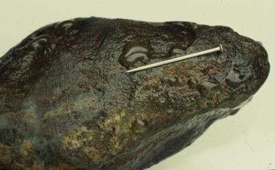
A dozen or so gelatinous colonies of this peritrich ciliate on the underside of a rock from a stream near Bristol, England. It's a pin.
-