-
-
-
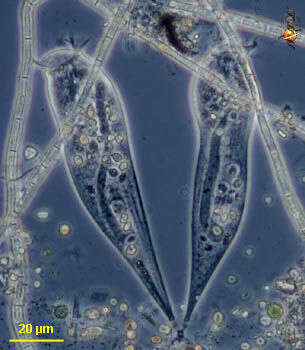
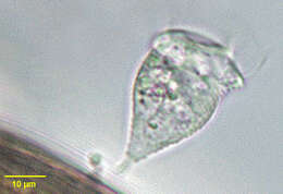
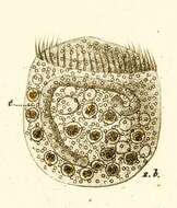
Rhabdostyla (rab-doe-style-a) a peritrich ciliate, attached to the substrate by the posterior end of the cell, but not with a differentiated stalk. Feeding cilia located as a wreath at the anterior end. The long dark bodies within these cells are their macronuclei. Phase contrast.
-
-
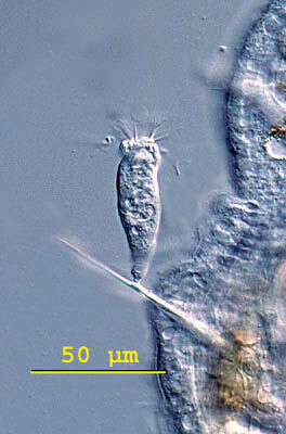
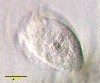
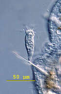
Rhabdostyla (rad-doe-stile-a) are solitary peritrich ciliates with a very short noncontractile stalk. The body is mostly inverted bell-shaped. The peristome shows a definite lip. Some species of the genus live epizooic on freshwater invertebrates, particularly crustacea, insects and worms. This genus can be confused with Apiosoma which is restricted to being epizooic on vertebrates. Rhabdostyla inclinans is an epizooic species and this cell was attached by a short stalk to the bristle of a worm. Differential interference contrast.
-
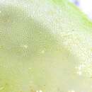
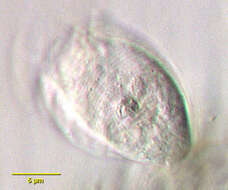
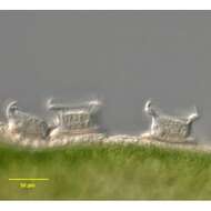
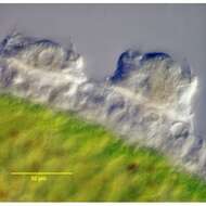
Rhabdostyla, peritrich ciliate attached to substrate by non-contractile stalk. This image shows a detail of the surface. Differential interference contrast optics.
-
Rhabdostyla, peritrich ciliate attached to substrate by non-contractile stalk. The oral ciliate forms a wreath around the anterior end, the inner wreath being an extended membranelle, the outer one being the undulating membrane.
-
Rhabdostyla, peritrich ciliate attached to substrate by non-contractile stalk. The oral ciliate forms a wreath around the anterior end, the inner wreath being an extended membranelle, the outer one being the undulating membrane. The surface of the body is ridged.
-
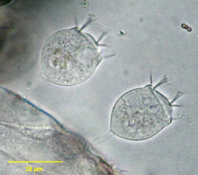
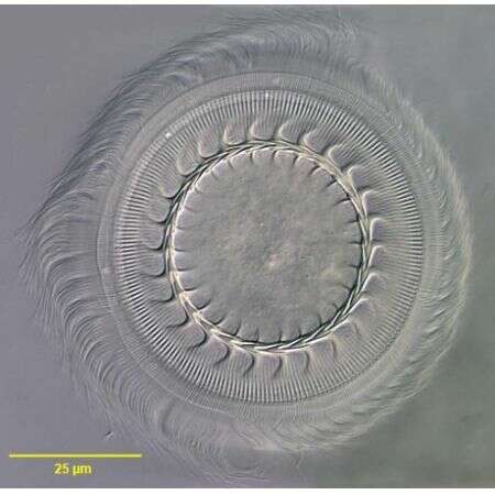
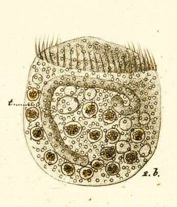
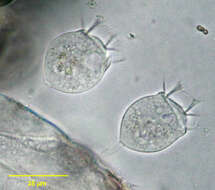
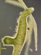
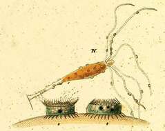
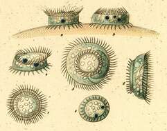
Description: Lateral view of the motile peritrich ciliate. Specimens were found on Eudiaptomus vulgaris, a common copepod. Compared with the bobbin-shaped Trichodina pediculus the lateral view of these cells is round, more dome-shaped, the size varying from 42 to 52 µm. The macronucleus is found in the transverse axis of the cell, almost circular. The large micronucleus lies at the outside of the c-shaped macronucleus. The conspicuous adhesive disk is located at the aboral side of the cell and, functioning as a holdfast organelle, attaches the ciliate to the surface of itâs host. The anatomy of the adhesive disk differs between species of the genus. The picture in the upper right angle shows an adhesive disk separated from itâs cell, counting 20 denticles.
-
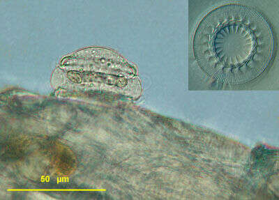
Lateral view of the mobiline peritrich ciliate, Trichodina pediculus (Ehrenberg,1851). These individuals are ectocommensals on Chlorohydra viridissimus. The bobbin-shaped cell body attaches to the epidermis of the host by an aboral adhesive disc or holdfast organelle that has a complex skeletal ring of denticles. When contracted the cells appear as flattened discs in lateral view. There is a ring of cilia around the aboral end. The peristomal ciliary field makes 1 1/4 turns around the anterior end. The pellicle between the anterior end and aboral end is bare of cilia. The C-shaped macronucleus lies in the transverse plane. There is one contractile vacuole.The cells scurry allong the surface of the host by means of the aboral cilia and adhesive disc. T. pediculus can be an important harmful parasite of freshwater fish. Collected from slow-flowing freshwater stream near Boise, Idaho February 2005. DIC.
-
Lateral view of the mobiline peritrich ciliate, Trichodina pediculus (Ehrenberg,1851). These individuals are ectocommensals on Chlorohydra viridissimus. The bobbin-shaped cell body attaches to the epidermis of the host by an aboral adhesive disc or holdfast organelle that has a complex skeletal ring of denticles .When contracted the cells appear as flattened discs in lateral view (e.g. individual to viewer's right). There is a ring of cilia around the aboral end. The peristomal ciliary field makes 1 1/4 turns around the anterior end. The pellicle between the anterior end and aboral end is bare of cilia. The C-shaped macronucleus lies in the transverse plane. There is one contractile vacuole.The cells scurry allong the surface of the host by means of the aboral cilia and adhesive disc. T. pediculus can be an important harmful parasite of freshwater fish. Collected from slow-flowing freshwater stream near Boise, Idaho February 2005. DIC.
-
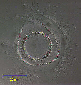
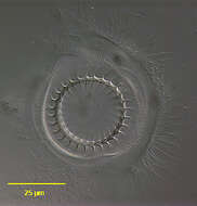
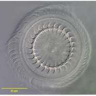
Aboral view of the mobiline peritrich ciliate, Trichodina pediculus (Ehrenberg,1851). T. pediculus is ectocommensal on the coelenterate, Chlorohydra viridissimus.This individual has been dislodged from its host. The bobbin-shaped cell body attaches to the epidermis of the host by an aboral adhesive disc or holdfast organelle that has a complex skeletal ring of denticles (seen here).When contracted the cells appear as flattened discs in lateral view. There is a ring of cilia around the aboral end. The peristomal ciliary field makes 1 1/4 turns around the anterior end. The pellicle between the anterior end and aboral end is bare of cilia. The C-shaped macronucleus lies in the transverse plane. There is one contractile vacuole.The cells scurry allong the surface of the host by means of the aboral cilia and adhesive disc. T. pediculus can be an important harmful parasite of freshwater fish. Collected from slow-flowing freshwater stream near Boise, Idaho February 2005. DIC.
-
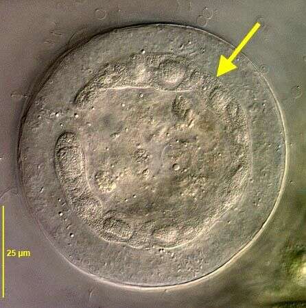
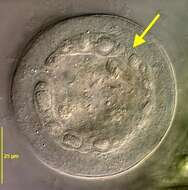
Optical section from anterior view showing C-shaped macronucleus (yellow arrow) of Trichodina pediculus (EHRENBERG,1851). DIC.
-
Aboral view of the mobiline peritrich ciliate, Trichodina pediculus (EHRENBERG,1851). T. pediculus is ectocommensal on the coelenterate, Chlorohydra viridissimus.This individual has been dislodged from its host. The bobbin-shaped cell body attaches to the epidermis of the host by an aboral adhesive disc or holdfast organelle that has a complex skeletal ring of denticles (seen here).When contracted the cells appear as flattened discs in lateral view. There is a ring of cilia around the aboral end. The peristomal ciliary field makes 1 1/4 turns around the anterior end. The pellicle between the anterior end and aboral end is bare of cilia. The C-shaped macronucleus lies in the transverse plane. There is one contractile vacuole.The cells scurry allong the surface of the host by means of the aboral cilia and adhesive disc. T. pediculus can be an important harmful parasite of freshwater fish. Collected on Hydra from bottom sediments of a freshwater pond near Boise, Idaho February 2005. DIC.
-
Lateral view of the mobiline peritrich ciliate, Trichodina pediculus (EHRENBERG,1851). These individuals are ectocommensals on Chlorohydra viridissimus. The bobbin-shaped cell body attaches to the epidermis of the host by an aboral adhesive disc or holdfast organelle that has a complex skeletal ring of denticles .When contracted the cells appear as flattened discs in lateral view (e.g. individual to viewer's right). There is a ring of cilia around the aboral end. The peristomal ciliary field makes 1 1/4 turns around the anterior end. The pellicle between the anterior end and aboral end is bare of cilia. The C-shaped macronucleus lies in the transverse plane. There is one contractile vacuole.The cells scurry allong the surface of the host by means of the aboral cilia and adhesive disc. T. pediculus can be an important harmful parasite of freshwater fish. Collected from slow-flowing freshwater stream near Boise, Idaho February 2005. DIC.
-
-
-
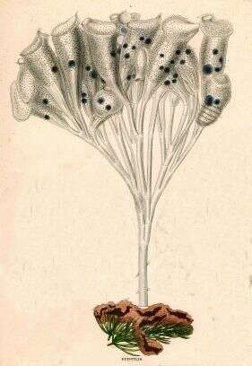
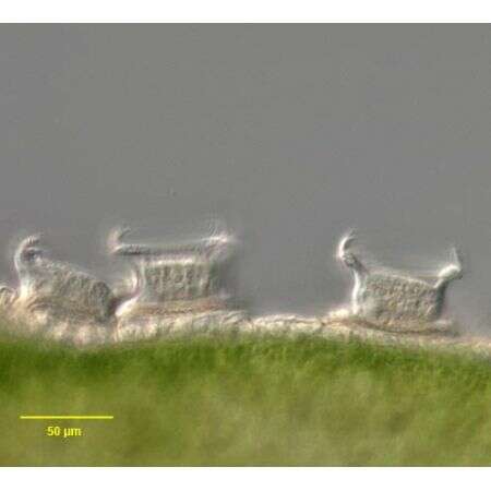
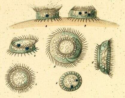
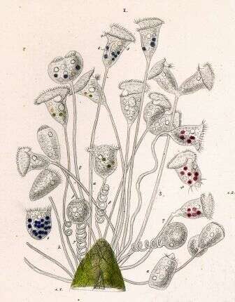
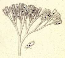
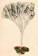
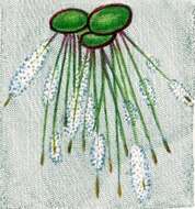
Described by Ehrenberg under the name Vorticella nebulifera. The species was moved to the genus Pseudovorticella by Jankowski, in 1976.
-
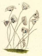
Described by Ehrenberg under the name Vorticella nebulifera. The species was moved to the genus Pseudovorticella by Jankowski, in 1976.
-
-