-
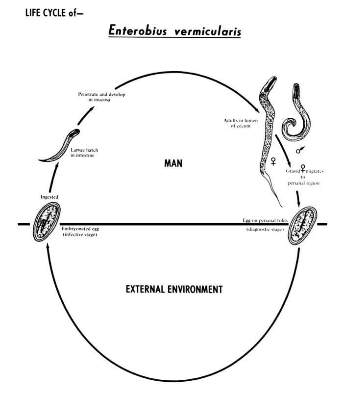
This diagram depicts the various stages in the life cycle of the human pinworm nematode Enterobius vermicularis.Created: 1982
-
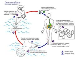
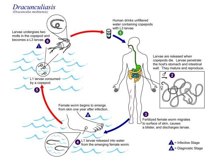
This is an illustration of the life cycle of Dracunculus medinensis, the causal agent of Dracunculiasis.Created: 2002
-
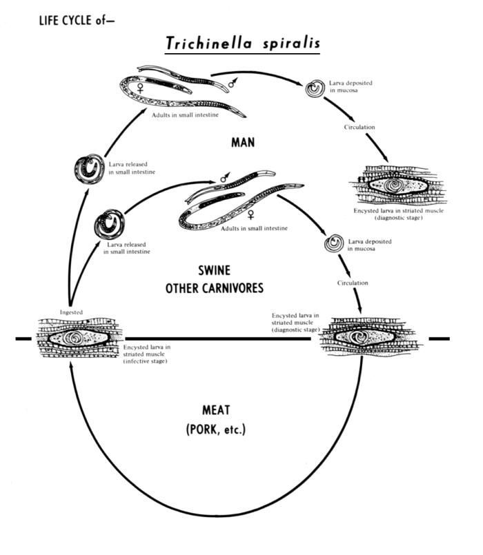
This diagram depicts the various stages in the life cycle of the nematode Trichinella spiralis.Created: 1982
-
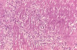
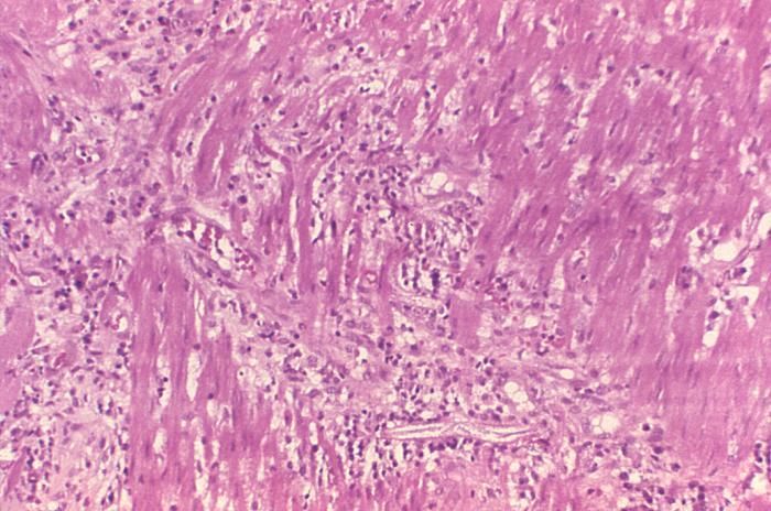
This photomicrograph reveals histopathologic changes indicative of the presence of the intestinal parasitic nematode (roundworm), Strongyloides stercoralis.Clinical Features:Frequently asymptomatic. Gastrointestinal symptoms include abdominal pain and diarrhea. Pulmonary symptoms (including Loefflers syndrome) can occur during pulmonary migration of the filariform larvae. Dermatologic manifestations include urticarial rashes in the buttocks and waist areas. Disseminated strongyloidiasis occurs in immunosuppressed patients, can present with abdominal pain, distension, shock, pulmonary and neurologic complications and septicemia, and is potentially fatal. Blood eosinophilia is generally present during the acute and chronic stages, but may be absent with dissemination.Created: 1972
-
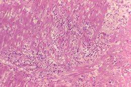
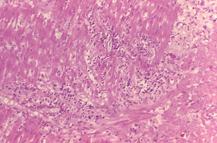
This photomicrograph reveals histopathologic changes indicative of the presence of the intestinal parasitic nematode (roundworm), Strongyloides stercoralis.Clinical Features:Frequently asymptomatic. Gastrointestinal symptoms include abdominal pain and diarrhea. Pulmonary symptoms (including Loefflers syndrome) can occur during pulmonary migration of the filariform larvae. Dermatologic manifestations include urticarial rashes in the buttocks and waist areas. Disseminated strongyloidiasis occurs in immunosuppressed patients, can present with abdominal pain, distension, shock, pulmonary and neurologic complications and septicemia, and is potentially fatal. Blood eosinophilia is generally present during the acute and chronic stages, but may be absent with dissemination.Created: 1972
-
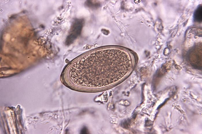
Magnified 240X, this photomicrograph revealed the presence of a Heterodera sp. nematode egg.Created: 1974
-
Magnified 240X, this photomicrograph revealed the presence of a Heterodera sp. nematode egg.Created: 1974
-
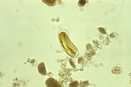
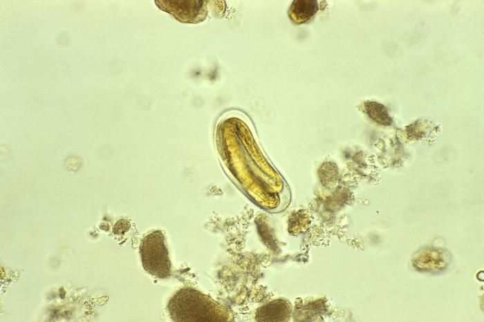
Magnified 500X, this photomicrograph of an unstained, formalin-preserved stool specimen mount, revealed the presence of a Trichuris vulpis nematode egg. T. vulpis is a whipworm that is common to dogs.Adult worms are 30 50nm in length, and as parasites, reside in the ceceum, a region of the large intestine where it is joined by the small intestine or ileum. This specie of whipworm, though common to canines, has been found in the human digestive tract.Created: 1973
-
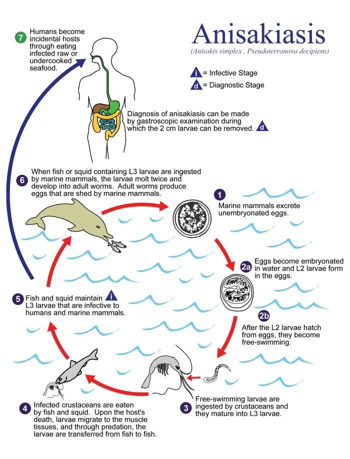
This illustration depicts the life cycle of A. simplex and P. decipiens, the causal agents of Anisakiasis.Created: 2002
-
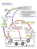
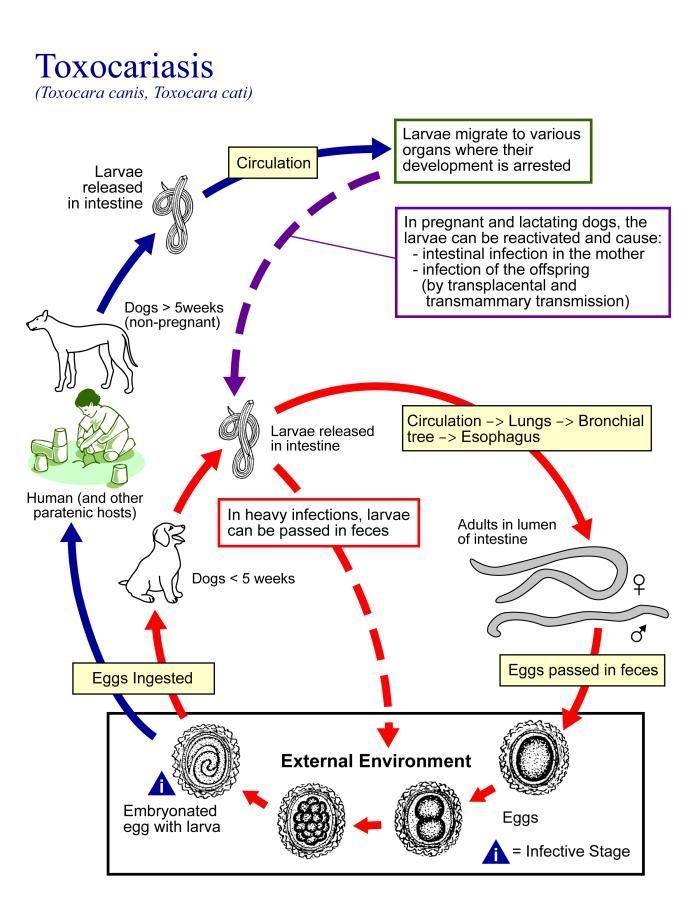
This is an illustration of the life cycle of Toxocara canis and Toxocara cati, the causal agents of Toxocariasis.Created: 2002
-
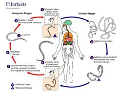
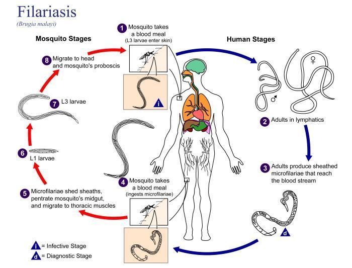
This illustration depicts the life cycle of Brugia malayi, the causal agent of Filariasis.Created: 2002
-
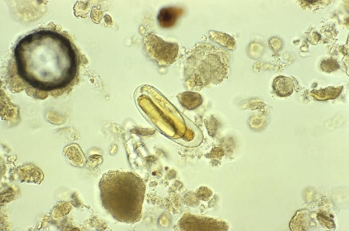
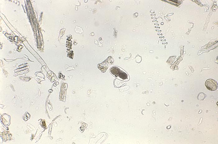
Magnified 128X, this photomicrograph depicted an egg from the parasitic nematode, Trichostrongylus.Created: 1979
-
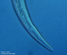
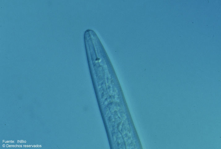
Instituto Nacional de Biodiversidad - INBio, Costa Rica.
INBio
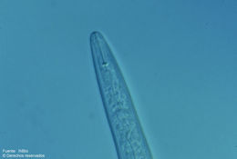
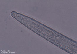
Fig. 1. Fotomicrografía de la región anterior de un juvenil (J2) de Meloidogyne incognita. Foto: A. Esquivel
-
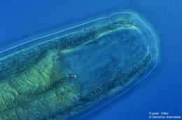
Instituto Nacional de Biodiversidad - INBio, Costa Rica.
INBio
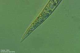
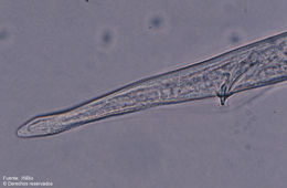
Fig. 2. Fotomicrografía de la región posterior de un juvenil (J2) de Meloidogyne incognita. Foto: A. Esquivel
-
Instituto Nacional de Biodiversidad - INBio, Costa Rica.
INBio
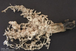
Fig.4. Nódulos radicales en Cucurbita moschata causados por M.incognita
-
Instituto Nacional de Biodiversidad - INBio, Costa Rica.
INBio
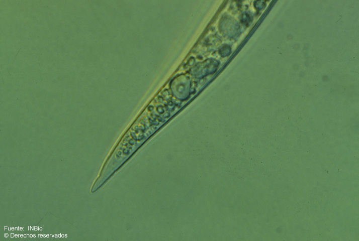
Fig. 1. Región anterior de la hembra. Foto: A. Esquivel
-
Instituto Nacional de Biodiversidad - INBio, Costa Rica.
INBio
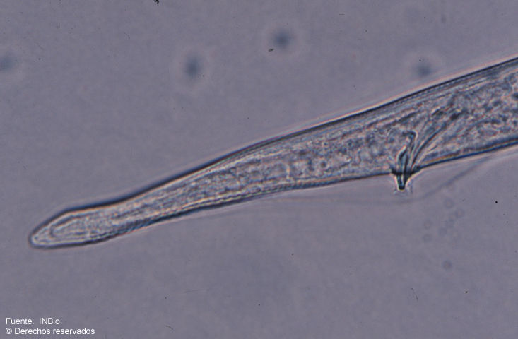
Fig. 2. Región anterior del macho. Foto: A. Esquivel
-
Instituto Nacional de Biodiversidad - INBio, Costa Rica.
INBio
Fig. 3. Región posterior del macho. Foto: A. Esquivel
-
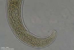
Instituto Nacional de Biodiversidad - INBio, Costa Rica.
INBio
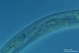
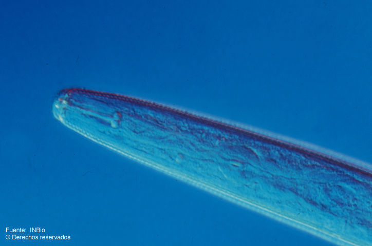
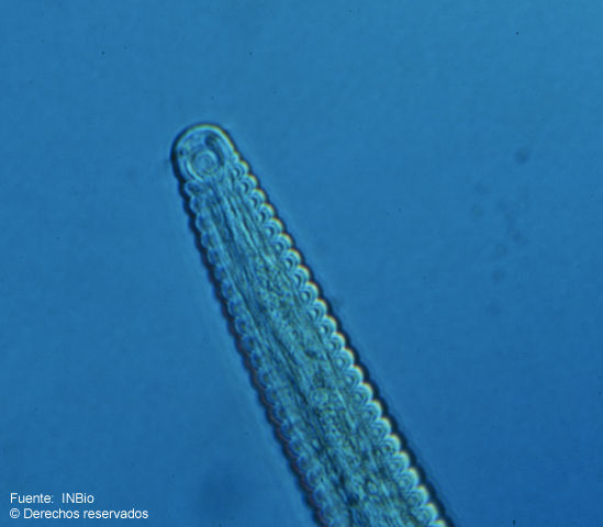
Fig.1. Fotomicrografía de la región anterior de la hembra. Anfidios grandes y cutícula fuertemente anulada. Foto: A. Esquivel
-
Instituto Nacional de Biodiversidad - INBio, Costa Rica.
INBio
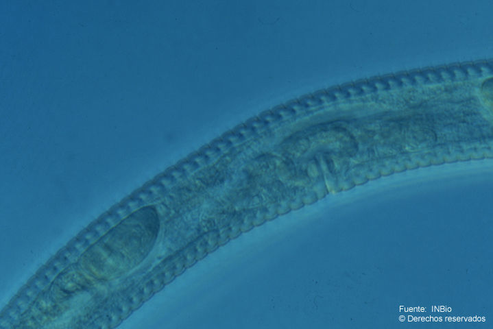
Fig.2. Región genital de la hembra Nótese el ovario flexionado y la abertura vulvar. Foto: A. Esquivel
-
Instituto Nacional de Biodiversidad - INBio, Costa Rica.
INBio
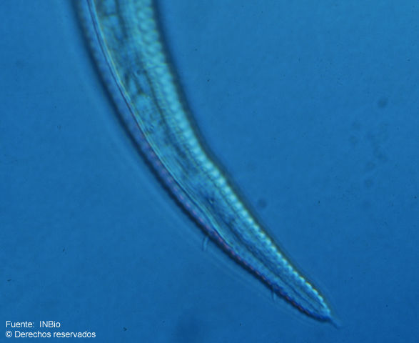
Fig.3. Región posterior del macho. Se aprecia la cuticula anulada, el campo lateral, setas y espinerete. Foto: A. Esquivel
-
Instituto Nacional de Biodiversidad - INBio, Costa Rica.
INBio
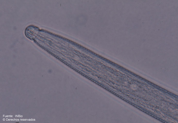
Fig. 1. Fotomicrografía de la región anterior de la hembra. Foto: A. Esquivel
-
Instituto Nacional de Biodiversidad - INBio, Costa Rica.
INBio
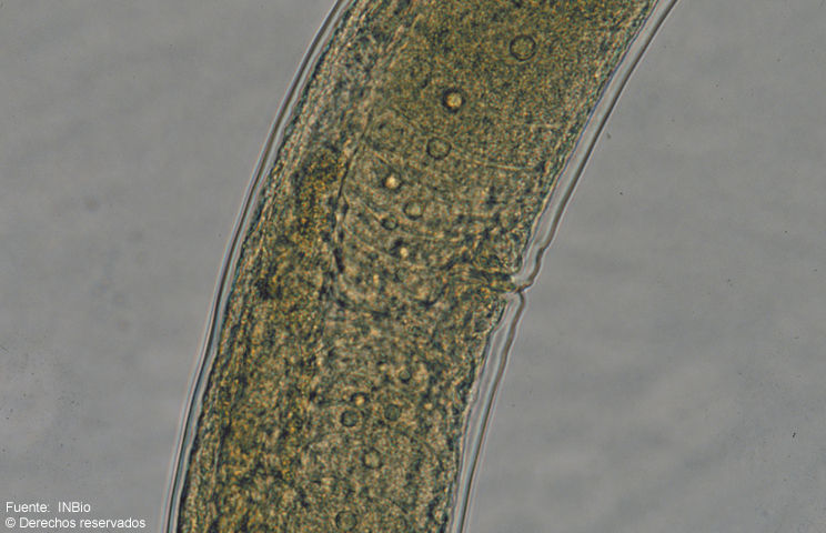
Fig. 2. Fotomicrografía de la región genital de la hembra. Foto: A. Esquivel
-
Instituto Nacional de Biodiversidad - INBio, Costa Rica.
INBio
Fig. 3. Fotomicrografía de la región posterior de la hembra. Foto: A. Esquivel