-
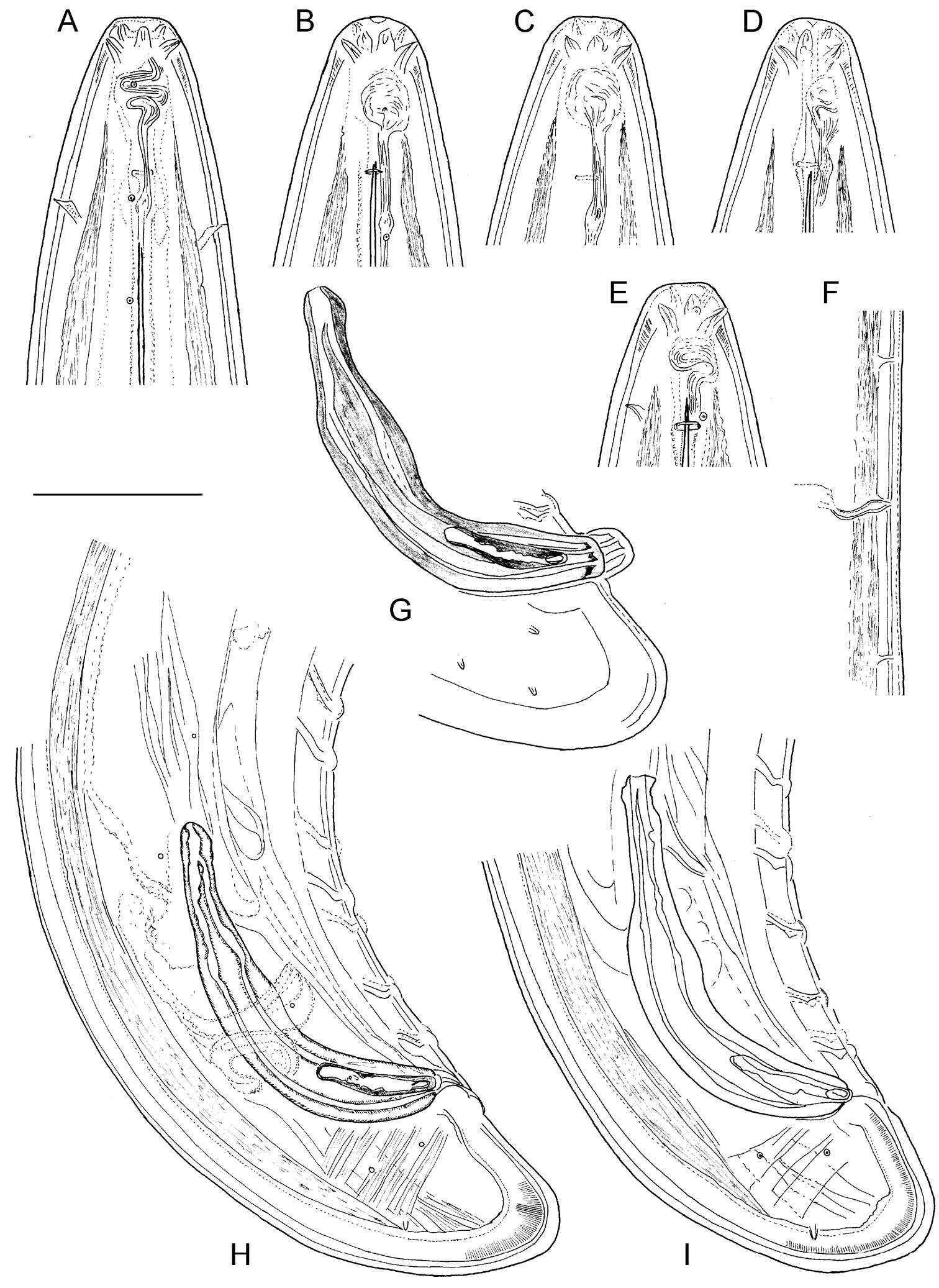
Sevdan Nedelchev, Milka Elshishka, Stela Lazarova, Georgi Radoslavov, Peter Hristov, Vlada Peneva
Zookeys
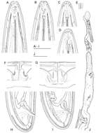
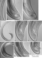
Figure 4.Calcaridorylaimus castaneae sp. n. Female: A Anterior end C Amphid F–I Tail shapes Male B Anterior end D Posterior end with extruded spicules, arrow indicating the spur E Posterior end. Scale bars: A, B, D–I – 20 μm; C – 6 μm.
-
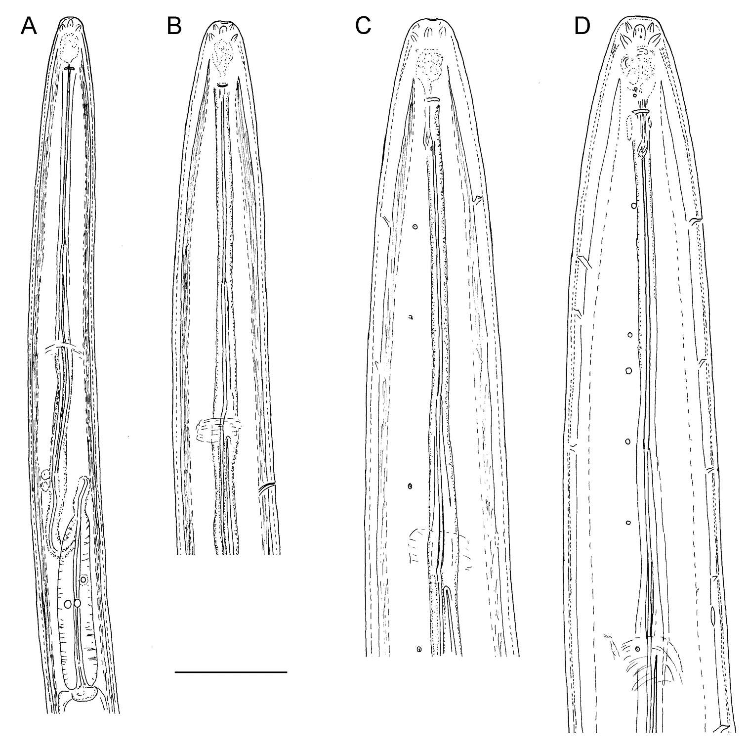
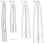
Figure 5. Longior longior Morffe & García sp. n. (female). A Cephalic end B Egg. Longior longior Morffe & García sp. n. (male) C Cephalic end D Tail, lateral view E Pre-cloacal median mammiform papilla, lateral view F Post-cloacal dorso-lateral papilla. Longior similis Morffe, García & Ventosa, 2009 (male) G Cephalic end H Tail, lateral view. Scale bars: A, B, C, D, G, H. 0.05 mm. E, F. 0.020 mm.
-
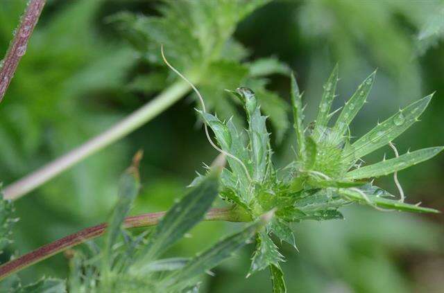
Hobro, Jylland, Danmark
-
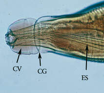
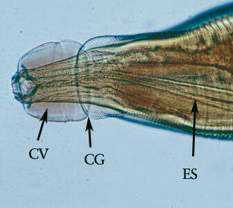
Öesophagostomum adultMagnification of the anterior end. Note the presence of the cephalic vesicle (CV), cephalic groove (CG) and esophagus (ES).From
CDC DPDx website
-
Saša Širca, Gregor Urek, Stela Lazarova, Milka Elshishka, Vlada Peneva
Zookeys
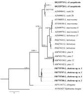
Figure 12.Phylogenetic tree of rDNA D2/D3 expansion region sequences of Longidorus carniolensis sp. n. from Slovenia (square mark) and sequences of closely related Longidorus species (NCBI GenBank). Sequences were analysed using Neighbour Joining Method. Bootstrap support values higher than 50% are presented.
-
Vlada K. Peneva, Stela S. Lazarova, Francesca De Luca, Derek J. F. Brown
Zookeys
Figure 11.Phylogenetic relationships of Longidorus cholevae sp. n. and its closest species for the partial 18S-ITS1 rDNA regions. Bayesian Inference strict consensus tree acquired under K2+G model. Numbers at the nodes indicating posterior probabilities higher that 0.8 and bootstrap values more that 70% for ML and NJ are presented.
-
Sevdan Nedelchev, Milka Elshishka, Stela Lazarova, Georgi Radoslavov, Peter Hristov, Vlada Peneva
Zookeys
Figure 5.Calcaridorylaimus castaneae sp. n. Female: A Entire body B Lip region D Prerectum, arrow pointing tongue-like valve E Pharyngeal bulb F Vulval region with posterior uterus G Vulval region with egg in posterior uterus H, I Vulval region J Cardia L Lateral field. Male: A Entire body C Lip region K Cardia M Supplements. Scale bars: A – 200 μm; B, C, M – 6 μm; E–L – 20 μm.
-
Hobro, Jylland, Danmark
-
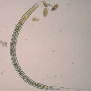
OesophagostomumL3 larva of Oesophagostomum sp., obtained via coproculture from the feces of a baboon (Papio ursinus) in South Africa. Note the long, thin, pointed tail. Image courtesy of the UTC Baboon Research Unit, University of Cape Town, South Africa.The definitive hosts of Oesophagostomum spp. become infected from the ingestion of infective L3 larvae, which develop in the environment. L3 larvae are longer than the L3 larvae of hookworms, measuring 710-950 µm in length. The intestine is made-up of alternating triangular-shaped cells (zigzag gut cells). The tail end has a long, thin, tapered sheath, with a considerable gap between the end of the tail and the end of the sheath.From
CDC-DPDx
-
Saša Širca, Gregor Urek, Stela Lazarova, Milka Elshishka, Vlada Peneva
Zookeys
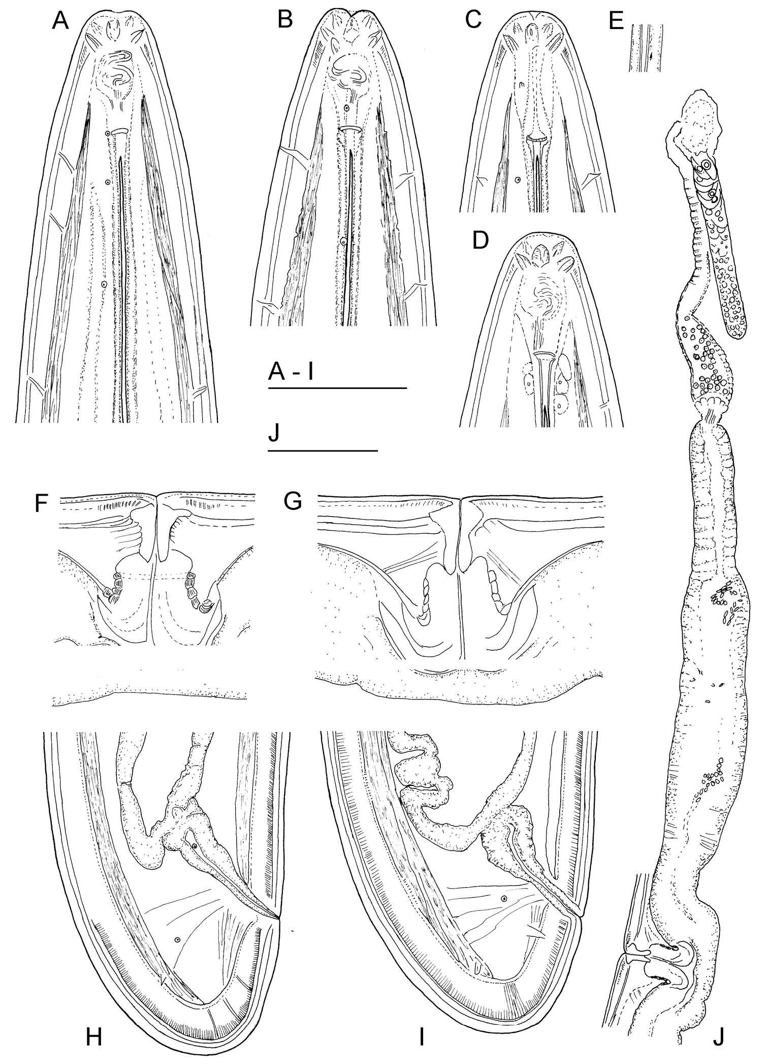
Figure 2.Longidorus carniolensis sp. n. Female: A–D Anterior ends E Vestigium in the walls of the slender part of pharynx F, G Vulval region G Anterior genital branch. Scale bars: A–I 50 μm, J 100 μm.
-
Sevdan Nedelchev, Milka Elshishka, Stela Lazarova, Georgi Radoslavov, Peter Hristov, Vlada Peneva
Zookeys
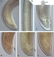
Figure 6.Calcaridorylaimus castaneae sp. n. Male: A Posterior end B Sperm cells in testis C–F Spicular region C Lateral piece of spicules D, E Extruded spicules, arrows pointing the spur F Spicules in the body. Scale bars: A, B – 20 μm; C–F – 18 μm.
-
Hobro, Jylland, Danmark
-
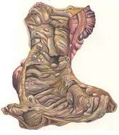
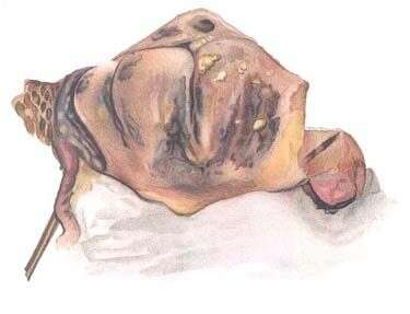
ÖesophagostomumPost-Mortem Pathology Plate borrowed from Brumpti and Thomas. "The Pathological Report of Oesophagostomiasis in Man."Colon and small intestine tissue samples from patient infected with Oesophagostomum. Important to note are the nodules lining the lumen of the colon (right).From:
"Introduction to the parasite family Oesophagostomum" (Stanford University Human Biology 103 class)
-
Saša Širca, Gregor Urek, Stela Lazarova, Milka Elshishka, Vlada Peneva
Zookeys
Figure 3.Longidorus carniolensis sp. n. Female: A Anterior region B–D Amphidial fovea E Vestigium F–H Vulval region I Vulval region, uterus and egg J Pharyngeal bulb, dorsal and subventral glands K, L Tail – different optical sections M Sphincter N Prerectum O–Q Variation in tail shape. Scale bars: I, N 200 μm; A–G, H–M, O–Q 50 μm.
-
ÖesophagostomumPost-Mortem Pathology Plate borrowed from Brumpti and Thomas. "The Pathological Report of Oesophagostomiasis in Man." Worm protruding from nodule on the lining of the colon. From:
"Introduction to the parasite family Oesophagostomum" (Stanford University Human Biology 103 class)
-
Saša Širca, Gregor Urek, Stela Lazarova, Milka Elshishka, Vlada Peneva
Zookeys
Figure 4.Longidorus carniolensis sp. n. Male: A–E Anterior end B, D, E in sublateral view F Excretory pore and ventral pores G Partly protracted spicules H–I Tail end. Scale bar: 50 μm.
-
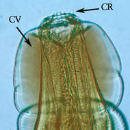
Öesophagostomum adultHigher magnification of the anterior end. Note the presence of the cephalic vesicle (CV) and corona radiata (CR).
-
Saša Širca, Gregor Urek, Stela Lazarova, Milka Elshishka, Vlada Peneva
Zookeys
Figure 5.Longidorus carniolensis sp. n. Male: A, B Variation in tail shape. Scale bar: 50 μm.
-
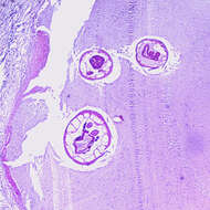
Öesophagostomum sp. adult in host tissueCross-section of an adult of Oesophagostomum sp. in a colon biopsy specimen from a patient from Africa, stained with hematoxylin and eosin (H&E). Image taken at 40x magnification.From
CDC DPDx
-
Saša Širca, Gregor Urek, Stela Lazarova, Milka Elshishka, Vlada Peneva
Zookeys
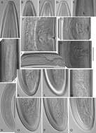
Figure 6.Longidorus carniolensis sp. n. Male: A Anterior region B, C Head region D–G Amphidial fovea H Vestigium (white arrow), excretory pore (thick arrow) and ventral pores (slender arrows) I Ejaculatory glands (marked by arrows) J Lateral field K, L Pharyngeal bulb with glandular bodies (marked by arrows) M, N Sperm cells at different stage of development. Scale bars: A 200 μm; B–N 50 μm.
-
ÖesophagostomumPost-Mortem Pathology Plate borrowed from Brumpti and Thomas. "The Pathological Report of Oesophagostomiasis in Man." Outside covering of encysted worm. From:
"Introduction to the parasite family Oesophagostomum" (Stanford University Human Biology 103 class)
-
Saša Širca, Gregor Urek, Stela Lazarova, Milka Elshishka, Vlada Peneva
Zookeys
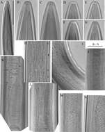
Figure 7.Longidorus carniolensis sp. n. Male: A Posterior genital branch B, C, E, F Tail and copulatory apparatus – different optical sections D, G Posterior end H Rectum (marked by arrow), spicules and lateral piece I Partly protracted spicules. Scale bars: A, D, G – 200 μm; B, C, E–F, H, I – 50 μm.
-
Centers for Disease Control/Division of Parasitic Diseases and Malaria
EOL staff
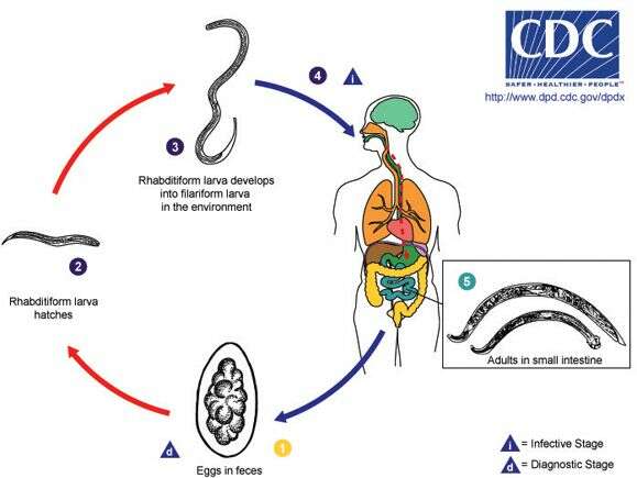
Life cycle of Trichostrongylus nematodes infecting humans Trichostrongylus eggs are passed in the stool of the definitive host (usually an herbivorous mammal) (1) and under favorable conditions (moisture, warmth, shade) larvae hatch within several days. The released rhabditiform larvae grow in the soil or on vegetation (2) and after 5 to 10 days (and two molts) they become filariform (third-stage) larvae that are infective (3). Infection of the human host occurs upon ingestion of these filariform larvae (4). The larvae reach the small intestine, where they reside and mature into adults. Adult worms inhabit the digestive tract of the definitive host and may occur as incidental infections in humans (5). (From
Centers for Disease Control Parasites and Health website)
-
Saša Širca, Gregor Urek, Stela Lazarova, Milka Elshishka, Vlada Peneva
Zookeys
Figure 8.Longidorus carniolensis sp. n. Juveniles: A Neck region of first stage B–D Spear region of second, third and fourth stage. Scale bar: 50 μm.