Anna Halász, Catherine S. McFadden, Dafna Aharonovich, Robert Toonen, Yehuda Benayahu
Zookeys
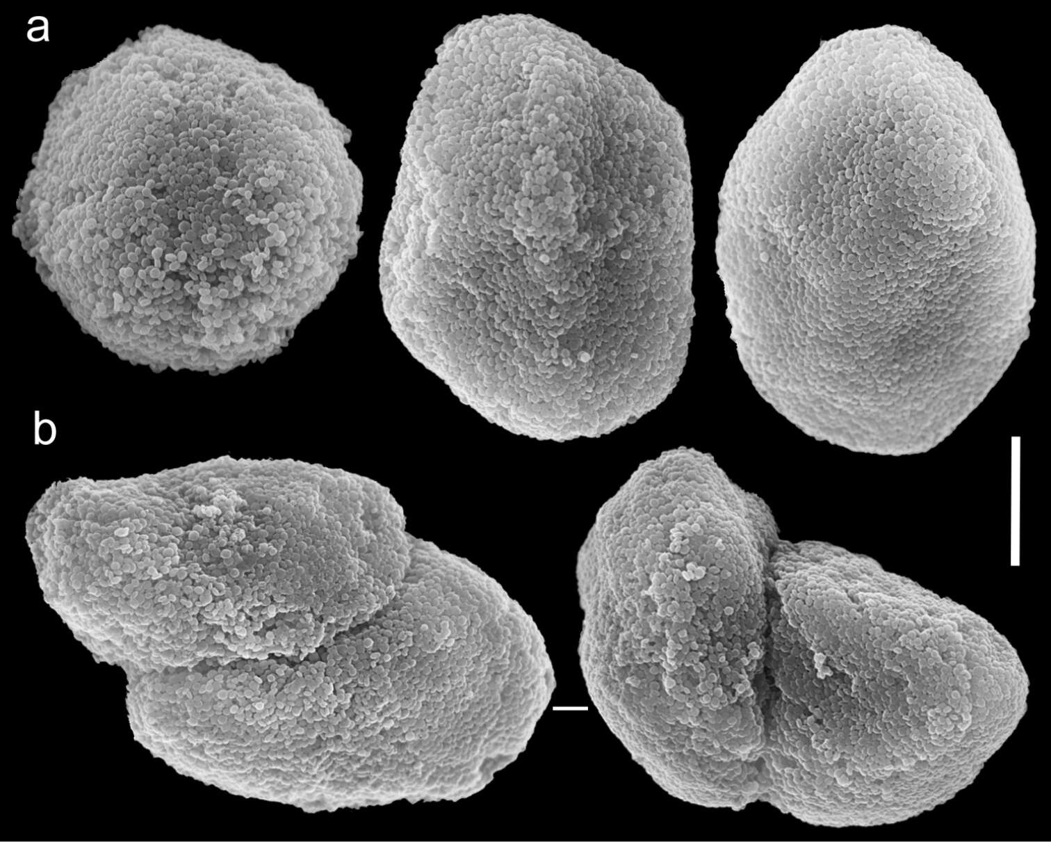
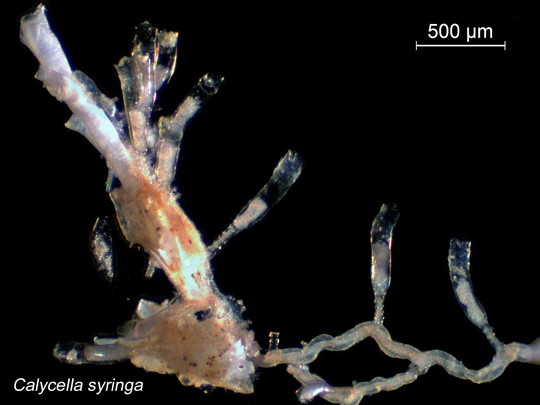
Figure 14.Scanning electron micrographs of polyp sclerites of Ovabunda impulsatilla (Verseveldt & Cohen, 1971) holotype (HUJ I Co. 84). a Regular sclerites b Fused sclerites. Scale bar 10 µm.
Tullia I. Terraneo, Michael L. Berumen, Roberto Arrigoni, Zarinah Waheed, Jessica Bouwmeester, Annalisa Caragnano, Fabrizio Stefani, Francesca Benzoni
Zookeys
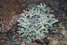
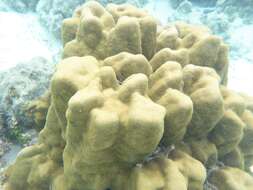
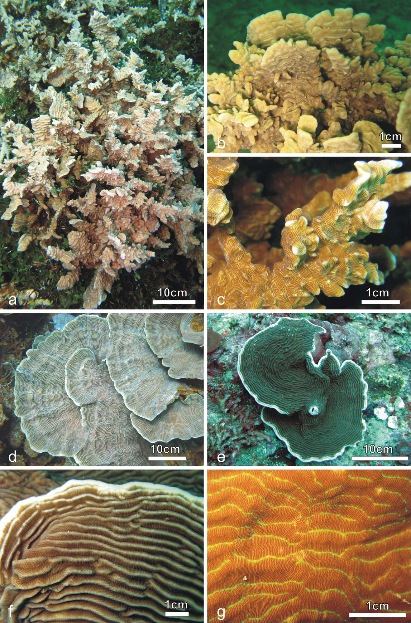
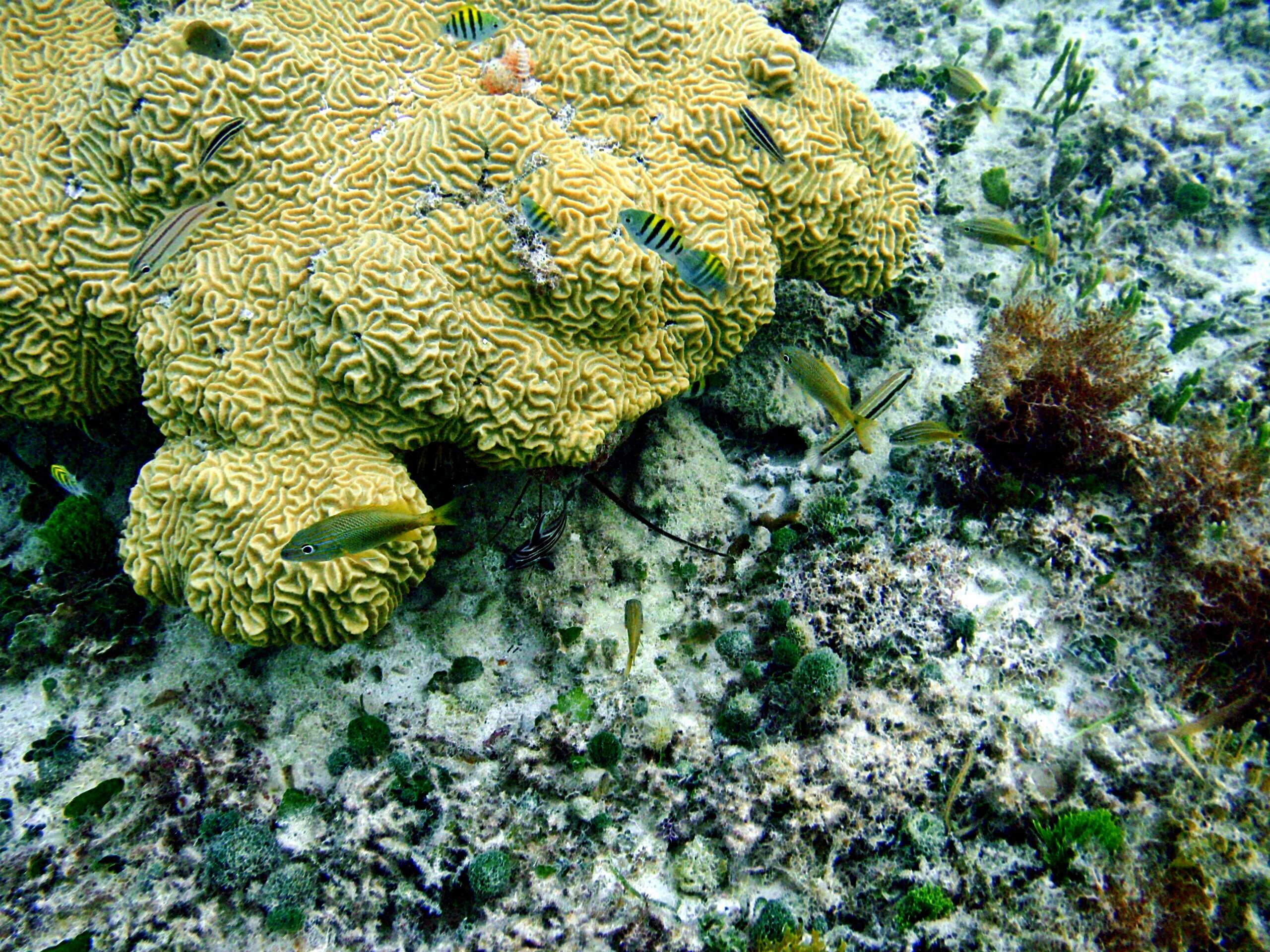
Figure 1.Colonies of Pachyseris rugosa (a–c) and Pachyseris speciosa (d–g) in situ. a Image of the whole colony of specimen IRD HS2893, Prony Bay, New Caledonia b Lateral view of the fronds of specimen IRD HS2594, Prony Bay, New Caledonia c Fronds of specimen IRD HS2856 viewed from above, Prony Bay, New Caledonia d Tiers of foliose projections of a colony from New Caledonia e Image of specimen UNIMIB SO040, Socotra Island f Part of specimen KAUST SA714, Saudi Arabia g Detail of a colony with reduced carinae and brightly colored polyp mouths, growing in very turbid environment, Banc des Japonais, New Caledonia.
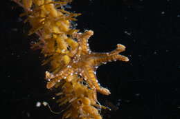
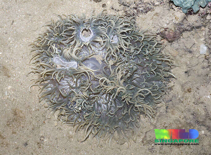
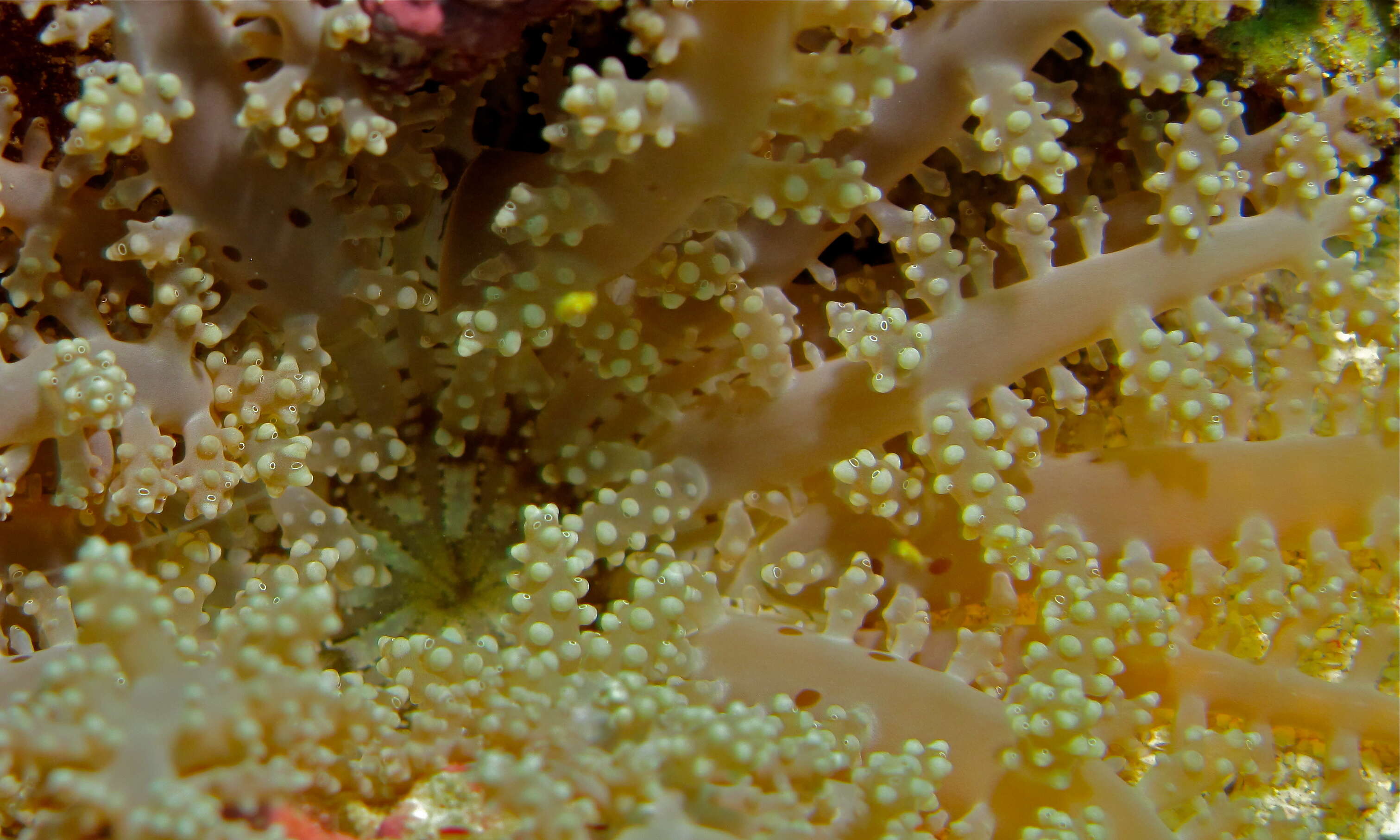
Sasakiella cruciformis, photographed in lab with the equipment of Yakko Hirano, Hokkaido, Japan, 2006, Collected as part of the Cnidarian Tree of Life expedition.
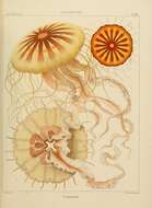
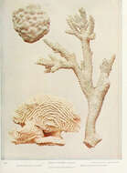
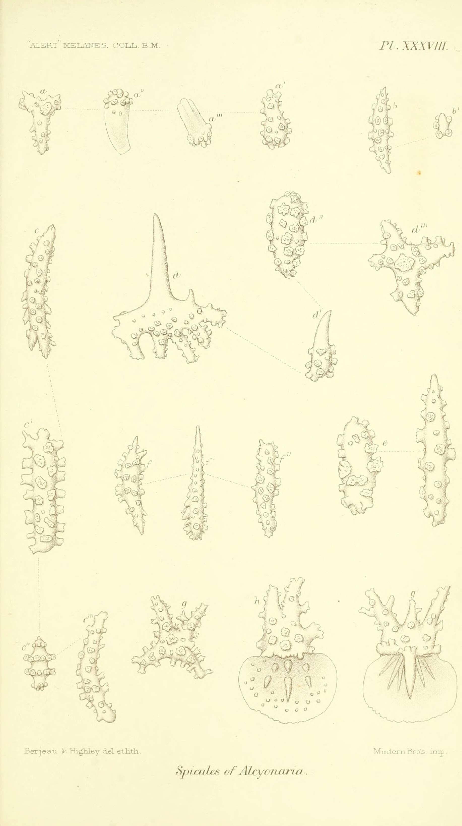
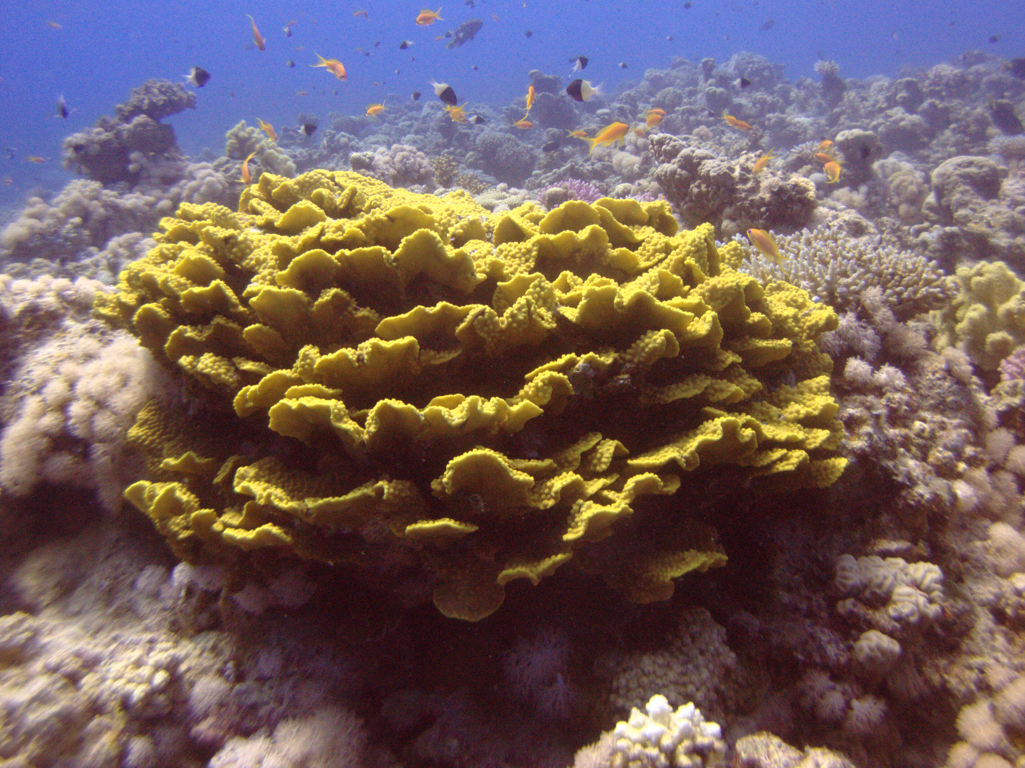
Report on the zoological collections made in the Indo-Pacific Ocean during the voyage of H.M.S. 'Alert' 1881-2.London :Printed by order of the Trustees,1884. biodiversitylibrary.org/page/12067762
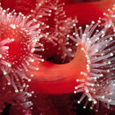
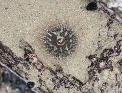
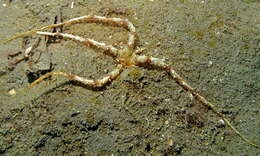
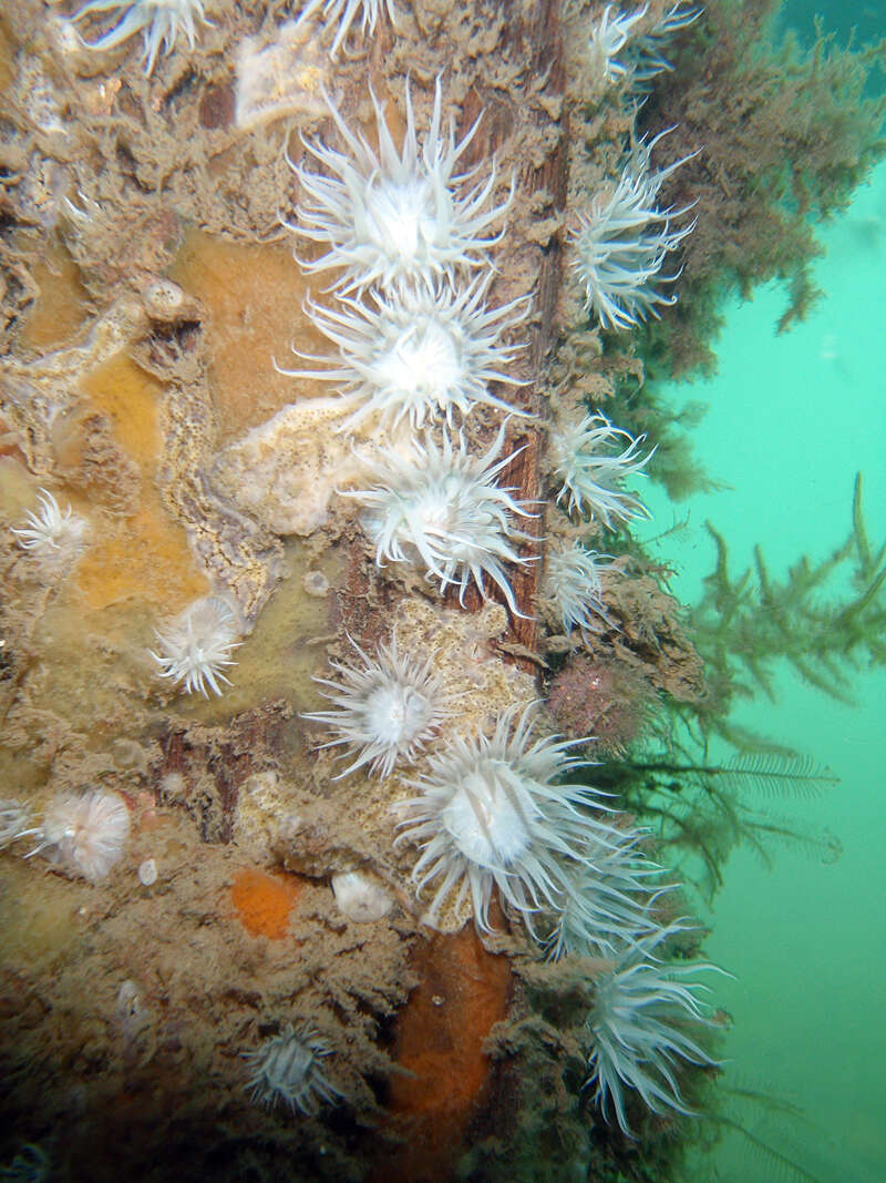
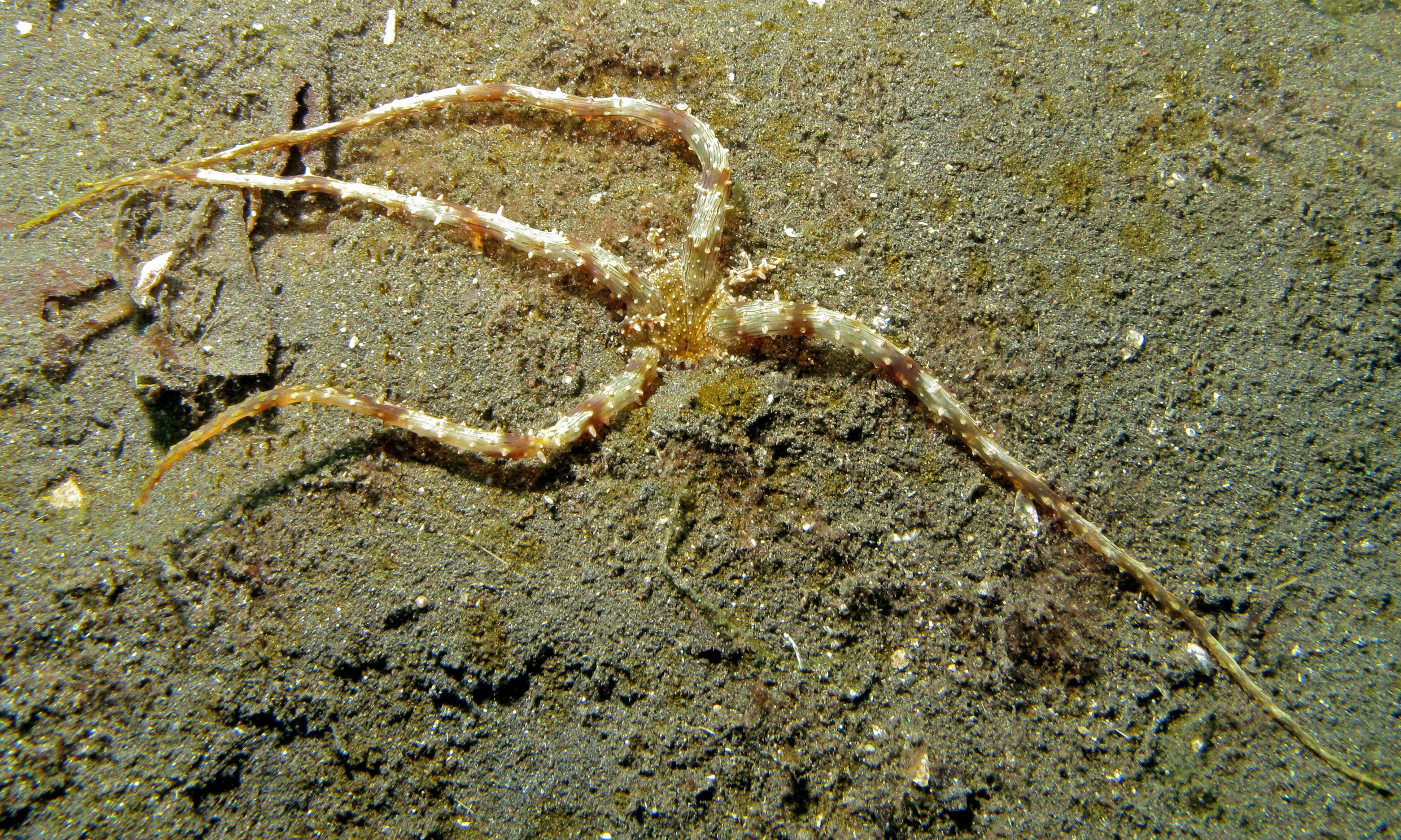
Eyes Under Puget SoundThis species image was collected from Puget Sound sediments and photographed by the Washington State Department of Ecologys Marine Sediment Monitoring Team. For more information about this teams work visit: www.ecy.wa.gov/programs/eap/psamp/index.htm.Cant get enough benthos? Check out our Eyes Under Puget Sound Critter of the Month species profile blogs at bit.ly/critterofthemonth