-
All Biocode files are based on field identifications to the best of the researcher’s ability at the time.
-
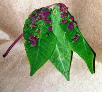
Resmo Alvar, Öland, Sverige
-
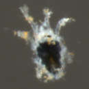
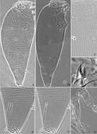
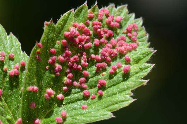
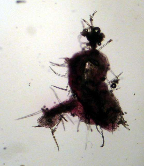
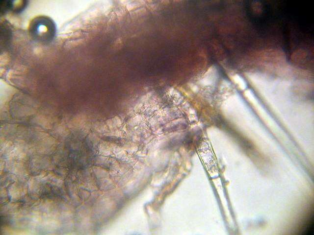
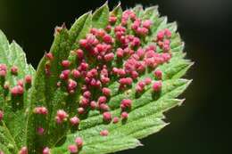
Mushroom Observer Image 103174: Aculops
-
Xiao-Feng Xue, Hussein Sadeghi, Xiao-Yue Hong, Samira Sinaie
Zookeys
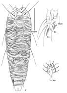
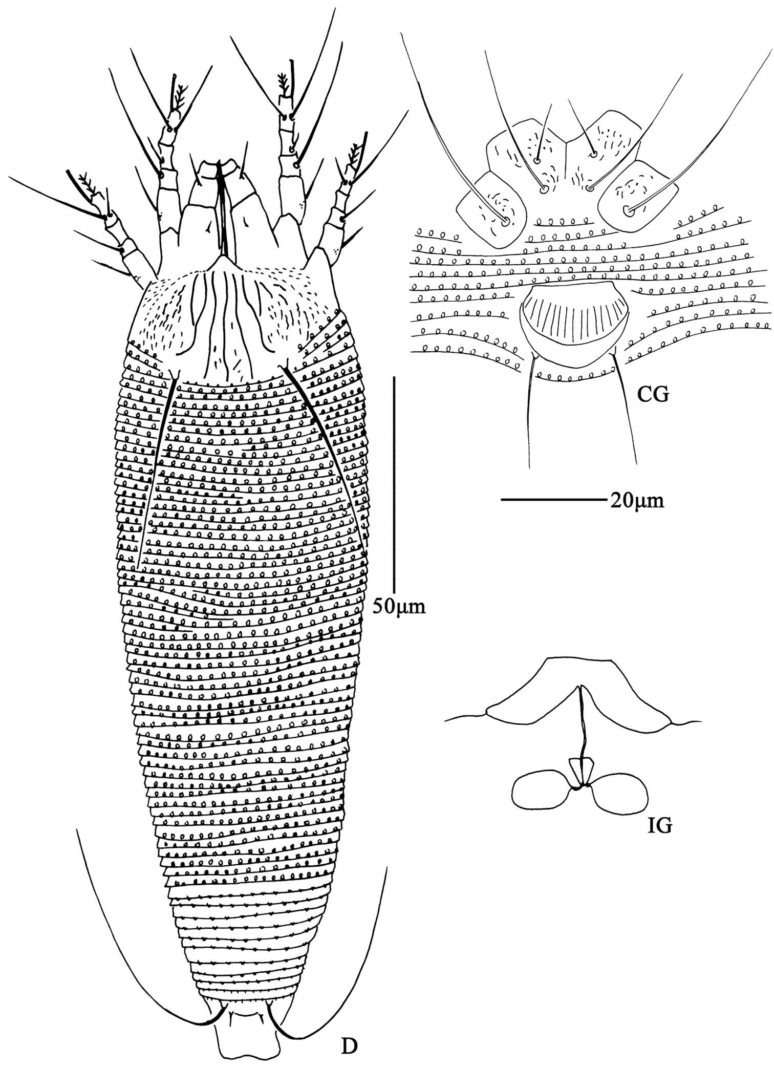
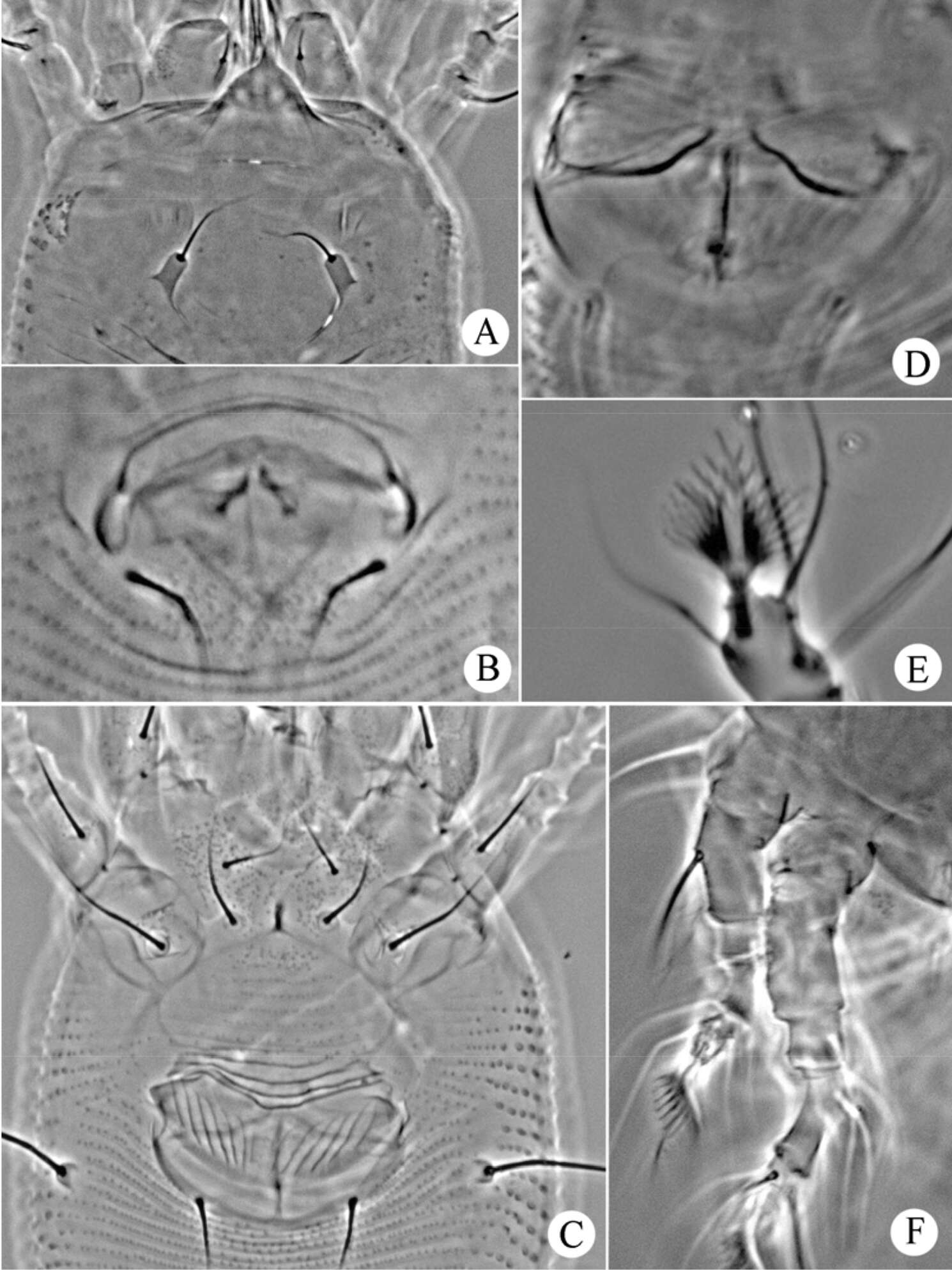
Figure 6. Aceria pulicaris sp. n. D dorsal view of female CG coxae and female genitalia IG female internal genitalia.
-
Hao-Sen Li, Xiao-Feng Xue, Xiao-Yue Hong
Zookeys
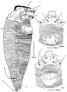
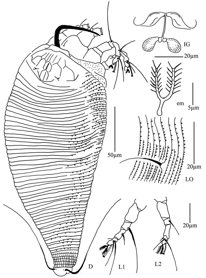
Figure 27.Diptacus berberinus sp. n.: D dorsal view of female IG female internal genitalia LO lateral microtubercles L1 leg I L2 leg II em empodium.
-
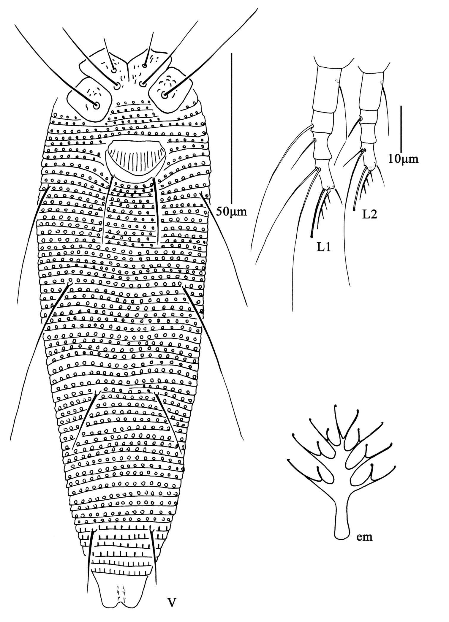
Qiong Wang, Xiao Han, Xiao-Feng Xue, Xiao-Yue Hong
Zookeys
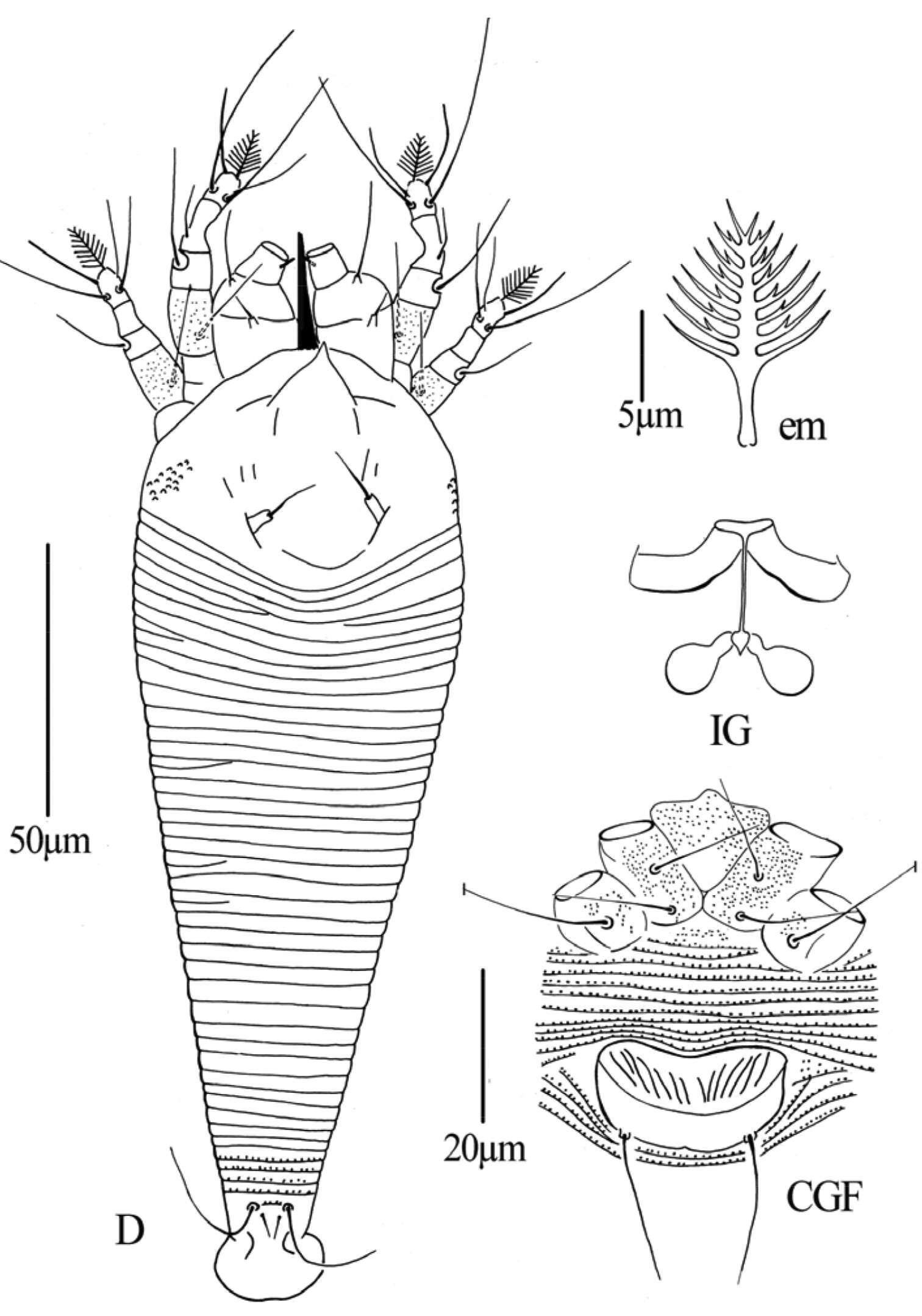
Figure 5.Phyllocoptes setalsolenidion sp. n.: D dorsal view of female em empodium IG female internal genitalia CGF female coxae and genitalia.
-
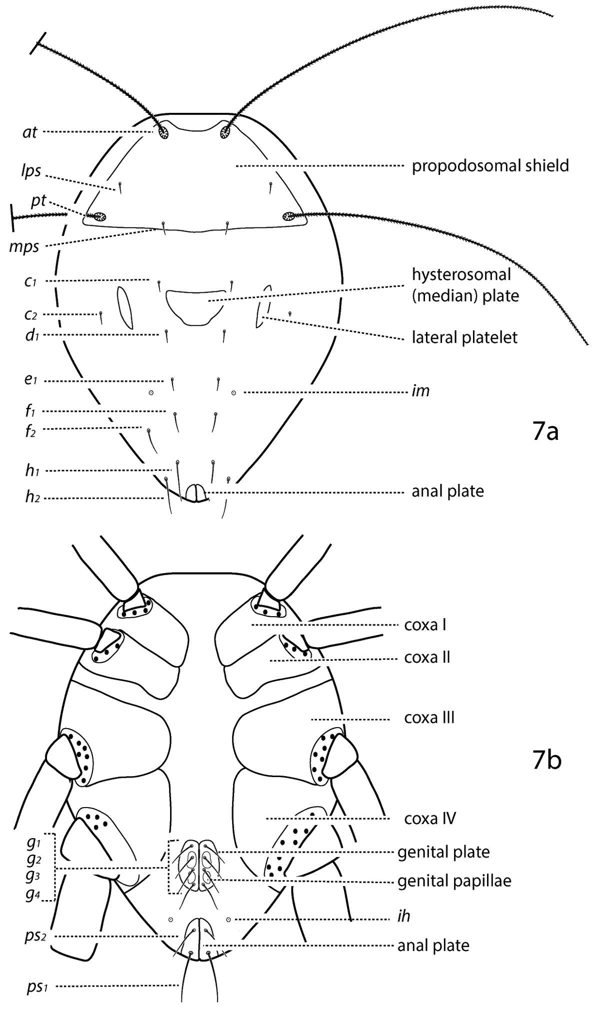
Michael J. Skvarla, J. Ray Fisher, Ashley P. G. Dowling
Zookeys
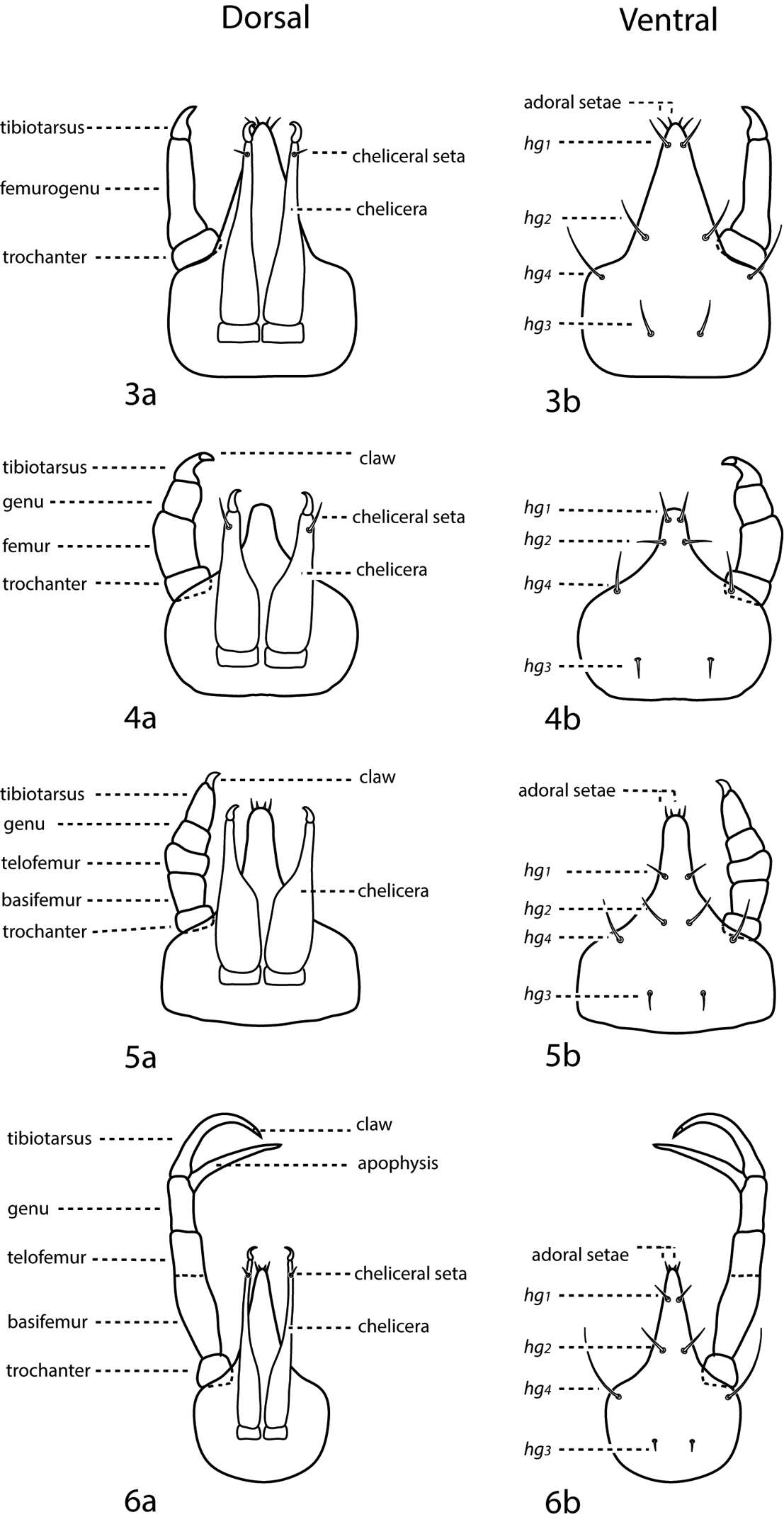
Figures 3–6.a. dorsal. b. ventral. 3 3-segmented pedipalp (Cunaxoidinae) 4 4-segmented pedipalp (Scirulinae) 5 5-segmented pedipalp that does not extend beyond the subcapitulum by more than the distal half of the genua (Bonziinae, Coleoscirinae, and Orangescirulinae) 6 5-segmented pedipalp that reaches beyond the subcapitulum by at least the distal half of the genua (Cunaxinae).
-
Resmo Alvar, Öland, Sverige
-
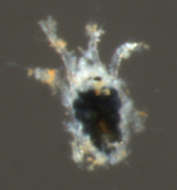
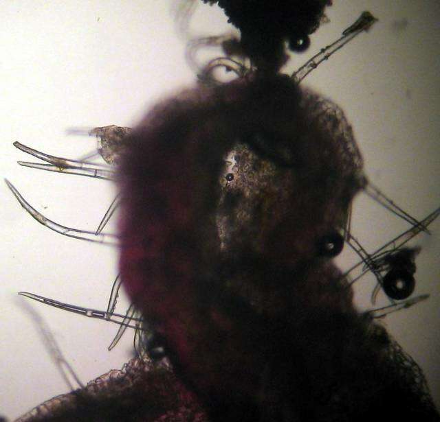
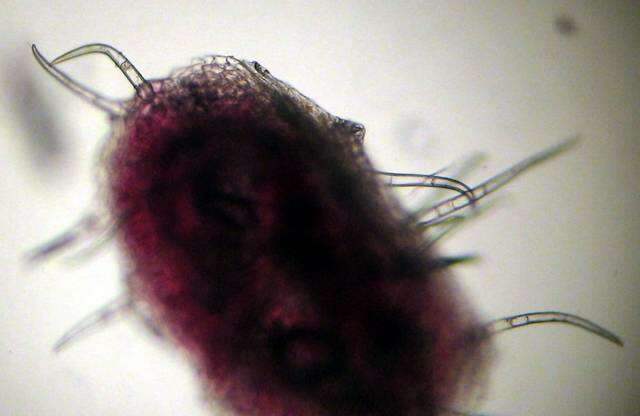
Mushroom Observer Image 103175: Aculops
-
Xiao-Feng Xue, Hussein Sadeghi, Xiao-Yue Hong, Samira Sinaie
Zookeys
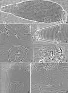
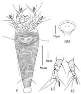
Figure 7. Aceria pulicaris sp. n. V ventral view of female em empodium L1 leg І L2 leg ІІ.
-
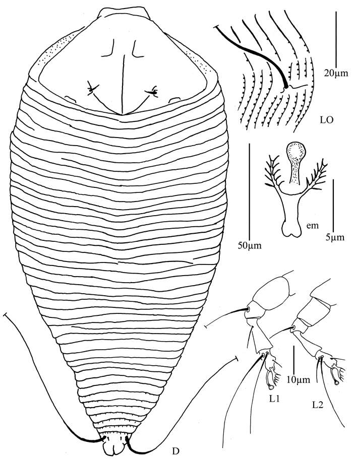
Hao-Sen Li, Xiao-Feng Xue, Xiao-Yue Hong
Zookeys
Figure 28.Diptacus berberinus sp. n.: L lateral view of female CMG coxae and male genitalia CG coxae and female genitalia.
-
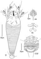
Qiong Wang, Xiao Han, Xiao-Feng Xue, Xiao-Yue Hong
Zookeys
Figure 6.Phyllocoptes setalsolenidion sp. n.: V ventral view of female GM male genital region L1 leg I L2 leg II.
-
Michael J. Skvarla, J. Ray Fisher, Ashley P. G. Dowling
Zookeys
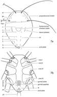
Figure 7.Generalized schematic of cunaxid idiosomal morphology. 7a Dorsal. 7b Ventral.
-
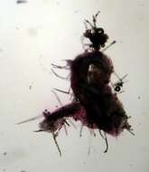
Mushroom Observer Image 103176: Aculops
-
Xiao-Feng Xue, Hussein Sadeghi, Xiao-Yue Hong, Samira Sinaie
Zookeys
Figure 8. Aceria pulicaris sp. n. A dorsal view of female B ventral view of female C prodorsal shield D coxae and female genitalia.
-
Hao-Sen Li, Xiao-Feng Xue, Xiao-Yue Hong
Zookeys
Figure 29.Diptacus berberinus sp. n.: A dorsal view of female B ventral view of female C lateral microtubercles D empodium E dorsal view of female posterior part F ventral view of female posterior part G leg I and leg II.
-
Qiong Wang, Xiao Han, Xiao-Feng Xue, Xiao-Yue Hong
Zookeys
Figure 7.Phyllocoptes setalsolenidion sp. n.: A prodorsal shield B male genitalia C coxae and female genitalia D female internal genitalia E empodium F leg I and leg II.
-
Michael J. Skvarla, J. Ray Fisher, Ashley P. G. Dowling
Zookeys
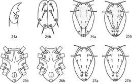
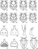
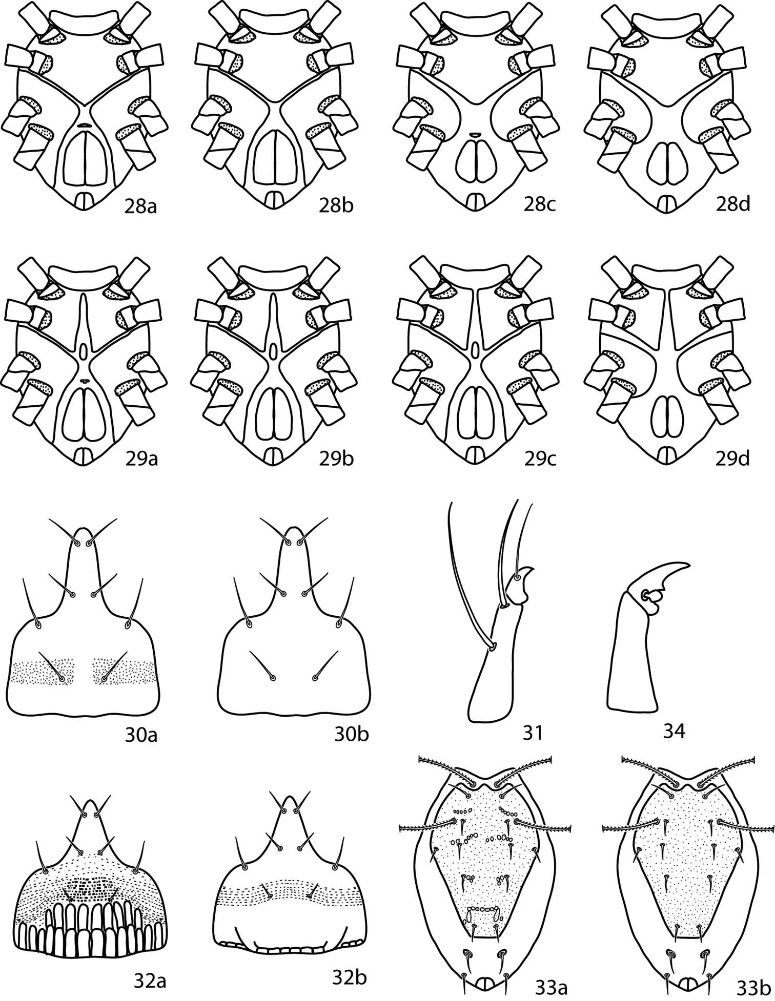
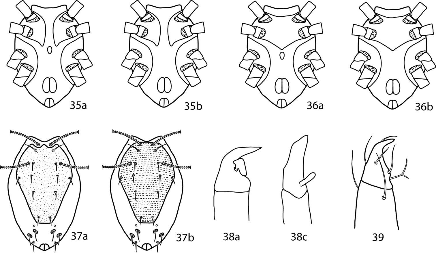
Figures 24–27.Lupaeus illustrations. 24a Pedipalp tibiotarsus 24b Genital setae not in a row, g3 out of line 25–27 Lupaeus key illustrations. Setae and cupules removed from figures 25a, b to increase clairity 25a Lupaeus longisetus, dorsal 25b Lupaeus polilloensis, dorsal 26a Ventral, small platelet present 26b Ventral, small platelet absent 27a Setae f1, f2 born on small platelets 27b Setae f1, f2 born on integument.
-
Mushroom Observer Image 103177: Aculops
-
Hao-Sen Li, Xiao-Feng Xue, Xiao-Yue Hong
Zookeys
Figure 30.Diptacus berberinus sp. n.: H lateral view of female I coxae and female genitalia J lateral view of female posterior part K female internal genitalia L coxae and male genitalia M prodorsal shield.
-
Michael J. Skvarla, J. Ray Fisher, Ashley P. G. Dowling
Zookeys
Figures 28–34.Neocunaxoides key illustrations. See key for explanations of figures.
-
Mushroom Observer Image 103178: Aculops
-
Hao-Sen Li, Xiao-Feng Xue, Xiao-Yue Hong
Zookeys
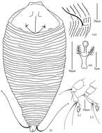
Figure 31.Diptacus mengdaensis sp. n.: D dorsal view of female LO lateral microtubercles em empodium L1 leg I L2 leg II.
-
Michael J. Skvarla, J. Ray Fisher, Ashley P. G. Dowling
Zookeys
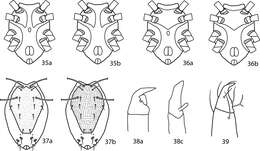
Figures 35–39.Pulaeus illustrations. 35 Genital setae in a row 36–39 Pulaeus key illustrations 36, 37 Venter, setae removed for clairity 36a Coxae I–II not coalesced medially, median platelet present 36b Coxae I–II not coalesced medially, median platelet absent 37a Coxae I–II coalesced medially, median platelet present 37b Coxae I–II coalesced medially, median platelet absent 38a Dorsal shield with punctures 38b Dorsal shield with broken striae 39a–c Pedipalp tibiotarsus 39a Tibiotarsus with elongate apophysis 39b Tibiotarsus with flat apophysis 39c Tibiotarsus with flange-like apophysis.