Chlamydomonas reinhardtii és una alga verda unicel·lular que es fa servir d'organisme model. Mesura uns 10 micròmetres de diàmetre amb dos flagels. La seva paret cel·lular està feta de glucoproteïnes riques en hidroxiprolina, un gran cloroplast en forma de copa, un gran pirenoide, i una taca que detecta la llum (fotosensible). Aquesta alga té una distribució cosmopolita en sòls i aigua dolça. També és d'interès com biocombustible per la producció d'hidrogen. Aquesta espècie d'alga no només fa la fotosíntesi sinó que pot capturar energia d'altres plantes (digerint la cel·lulosa), essent la primera vegada que s'ha observat això en un vegetal i això pot tenir un gran impacte en el futur de la bioenergia.[1][2]
Es va començar a estudiar en laboratori a partir de 1945 .[3][4]
Les Chlamydomonas es fan servir per contestar les preguntes de la biologia cel·lular i la biologia molecular referides al moviment, resposta a la llum, reconeixement, regulació del proteoma per controlar els flagels, resposta als canvis en nutrició mineral. Es fan servir molts mutants d'aquesta espècie per estudiar per exemple la fotosíntesi, la síntesi de proteïnes o l'evolució dels organismes.
Les cèl·lules vegetatives de C. reinhardtii són haploides amb 17 cromosomes petits. Sota privació de nitrogen es desenvolupen gàmetes haploides. Els progenitors són d'aparença idèntica (s'anomenen mt(+) i mt(-)), que es fusionen per donar un zigot diploide. Aquest zigot no està flagellat i és inactiu al sòl. Amb la llum el zigot experimenta la meiosi i allibera quatre cèl·lules flagelades haploides. Sota condicions ideals les cèl·lules poden de vegades tenir dos o tres cicles de mitosi i així es pot tenir de 4 a 8 cèl·lules filles per cada cèl·lula materna. El seu cicle cel·lular es pot sincronitzar alternant períodes de llum i foscor.[5]
Chlamydomonas reinhardtii és una alga verda unicel·lular que es fa servir d'organisme model. Mesura uns 10 micròmetres de diàmetre amb dos flagels. La seva paret cel·lular està feta de glucoproteïnes riques en hidroxiprolina, un gran cloroplast en forma de copa, un gran pirenoide, i una taca que detecta la llum (fotosensible). Aquesta alga té una distribució cosmopolita en sòls i aigua dolça. També és d'interès com biocombustible per la producció d'hidrogen. Aquesta espècie d'alga no només fa la fotosíntesi sinó que pot capturar energia d'altres plantes (digerint la cel·lulosa), essent la primera vegada que s'ha observat això en un vegetal i això pot tenir un gran impacte en el futur de la bioenergia.
Es va començar a estudiar en laboratori a partir de 1945 .
Chlamydomonas reinhardtii ist eine einzellige Grünalgenart aus der Gattung Chlamydomonas. Sie wurde von der Sektion Phykologie der Deutschen Botanischen Gesellschaft als Alge des Jahres 2014 ausgewählt.[1]
Chlamydomonas reinhardtii ist ein 3 bis 10 Mikrometer großer Hüllenflagellat mit eiförmiger Gestalt. Die beiden gleich langen (isokonten) Geißeln dienen der Fortbewegung. Die Schläge der Geißeln erfolgen nacheinander, und zwar mit einer kleinen Verzögerung. Dadurch entsteht eine Überlagerung ihrer Vorwärtsbewegung durch eine Rotation um die Längsachse der Alge mit einer Frequenz von etwa 1 Hertz. Die Fortbewegung der Zelle ist lichtgesteuert.
Die Zellmembran ist an der Vorderseite nicht zu einer Papille verdickt. Der Chloroplast besitzt ein großes Pyrenoid.
Es ist ein Augenfleck (Stigma) von etwa 1 Mikrometer Durchmesser vorhanden. Er ist durch eingelagerte Carotinoide orange gefärbt und gut erkennbar. Der Augenfleck liegt mit einem Abstand von etwa 10 Mikrometer von den Geißeln leicht oberhalb des Zelläquators. Als Photorezeptor sind Rhodopsin-ähnliche Proteine, sogenannte Channelrhodopsine, darin eingelagert.
Die Art lebt im Süßwasser, man findet sie vor allem in nährstoffreichen Kleingewässern. Sie ist weltweit verbreitet.
Die Alge kann sich sowohl vegetativ als auch geschlechtlich durch Gameten vermehren.[2] Die geschlechtliche Fortpflanzung wird durch Nährstoffmangel (insbesondere Stickstoffmangel) induziert. Dabei ist die Zygote das einzige diploide Stadium, das widrige Umstände überdauert und beim Keimen nach einer Meiose wieder haploide Nachkommen hervorbringt.
Chlamydomonas reinhardtii besitzt mit dem Augenfleck einen lichtsensitiven Apparat zur Phototaxis. Die Alge kann die Richtung und die Intensität von einfallendem Licht ermitteln. Bei niedriger Beleuchtungsstärke schwimmt sie auf die Lichtquelle zu, bei hoher Intensität von dieser weg. Dadurch optimiert sie ihre Photosynthese und damit die Zellernährung.[3] Die Alge kann sowohl phototroph (unter Ausnutzung des Lichtes) als auch heterotroph (auf Nährmedium) leben.
Die Geißeln von C. reinhardtii sind ebenso aufgebaut wie die menschlicher Spermien und vieler anderer Lebewesen. Diese Alge wurde als Modellorganismus für das Studium der Struktur und Funktion der Geißeln gewählt, weil sie sich sehr leicht kultivieren lässt und weil sie etwa durch einen pH-Schock zum Abwerfen der Geißeln veranlasst werden kann.[4]
Das Genom von C. reinhardtii ist vollständig sequenziert.[5] Zu Forschungszwecken wurden viele unterschiedliche Mutanten des Wildtyps gezüchtet, die zum Beispiel Zellwanddefekte aufweisen oder „blind“ sind.[6]
Ein Forschungsschwerpunkt ist die genetische Modifikation von C.reinhardtii hin zu Stämmen, die in industriell nutzbarem Maße Wasserstoff erzeugen können. Parallel dazu gibt es eine Reihe von wissenschaftlichen Projekten zur Entwicklung von leistungsfähigen Bioreaktoren zur Wasserstoffproduktion.[7]
Interessant erscheint auch die Fähigkeit, Cellulose zu spalten und zur Energiegewinnung zu nutzen.[8] Die Alge könnte somit als Biokatalysator für die Herstellung von Cellulose-Biokraftstoffen (z. B. Cellulose-Ethanol) dienen.
In einer 2021 veröffentlichten Studie konnte gezeigt werden, dass infolge Selektionsdruck durch Fressfeinde innerhalb von nur 500 Generationen Mutationen entstanden, die C. reinhardtii zur Bildung von Kolonien befähigten.[9]
Chlamydomonas reinhardtii ist eine einzellige Grünalgenart aus der Gattung Chlamydomonas. Sie wurde von der Sektion Phykologie der Deutschen Botanischen Gesellschaft als Alge des Jahres 2014 ausgewählt.
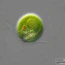
Chlamydomonas reinhardtii is a single-cell green alga about 10 micrometres in diameter that swims with two flagella. It has a cell wall made of hydroxyproline-rich glycoproteins, a large cup-shaped chloroplast, a large pyrenoid, and an eyespot that senses light.
Chlamydomonas species are widely distributed worldwide in soil and fresh water. Chlamydomonas reinhardtii is an especially well studied biological model organism, partly due to its ease of culturing and the ability to manipulate its genetics. When illuminated, C. reinhardtii can grow photoautotrophically, but it can also grow in the dark if supplied with organic carbon. Commercially, C. reinhardtii is of interest for producing biopharmaceuticals and biofuel, as well being a valuable research tool in making hydrogen.
The C. reinhardtii wild-type laboratory strain c137 (mt+) originates from an isolate collected near Amherst, Massachusetts, in 1945 by Gilbert M. Smith.[1][2]
The species' name has been spelled several different ways because of different transliterations of the name from Russian: reinhardi, reinhardii, and reinhardtii all refer to the same species, C. reinhardtii Dangeard.[3]
Chlamydomonas is used as a model organism for research on fundamental questions in cell and molecular biology such as:
There are many known mutants of C. reinhardtii. These mutants are useful tools for studying a variety of biological processes, including flagellar motility, photosynthesis, and protein synthesis. Since Chlamydomonas species are normally haploid, the effects of mutations are seen immediately without further crosses.
In 2007, the complete nuclear genome sequence of C. reinhardtii was published.[4]
Channelrhodopsin-1 and Channelrhodopsin-2, proteins that function as light-gated cation channels, were originally isolated from C. reinhardtii.[5][6] These proteins and others like them are increasingly widely used in the field of optogenetics.[7]
The genome of C. reinhardtii is significant for mitochondrial study as it is one species where the genes for 6 of the 13 proteins encoded for the mitochondria are found in the nucleus of the cell, leaving 7 in the mitochondria. In all other species these genes are present only in the mitochondria and are unable to be allotopically expressed. This is significant for the testing and development of therapies for genetic mitochondrial diseases.
Vegetative cells of reinhardtii species are haploid with 17 small chromosomes. Under nitrogen starvation, vegetative cells differentiate into haploid gametes.[8] There are two mating types, identical in appearance, thus isogamous, and known as mt(+) and mt(-), which can fuse to form a diploid zygote. The zygote is not flagellated, and it serves as a dormant form of the species in the soil. In the light, the zygote undergoes meiosis and releases four flagellated haploid cells that resume the vegetative lifecycle.
Under ideal growth conditions, cells may sometimes undergo two or three rounds of mitosis before the daughter cells are released from the old cell wall into the medium. Thus, a single growth step may result in 4 or 8 daughter cells per mother cell.
The cell cycle of this unicellular green algae can be synchronized by alternating periods of light and dark. The growth phase is dependent on light, whereas, after a point designated as the transition or commitment point, processes are light-independent.[9]
C. reinhardtii has an eyespot similar to that of dinoflagellates.[10] The eyespot is located near the cell equator. It is composed of a carotenoid-rich granule layer in the chloroplast which act like a light reflector.[11] The main function of the eyespot is the phototaxis, which consist of the movement (with the flagella) related to a light stimulus.[12] The phototaxis is crucial for the alga and allows for localization of the environnement with optimal light conditions for photosynthesis.[13] Phototaxis can be positive or negative depending on the light intensity.[10] The phototactic pathway consists of four steps leading to a change in the beating balance between the two flagella (the cis-flagellum which is the one closest to the eyespot, and the trans-flagellum which is the one farthest from the eyespot).[12]
The attractiveness of the algae as a model organism has recently increased with the release of several genomic resources to the public domain. The Chlre3 draft of the Chlamydomonas nuclear genome sequence prepared by Joint Genome Institute of the U.S. Dept of Energy comprises 1557 scaffolds totaling 120 Mb. Roughly half of the genome is contained in 24 scaffolds all at least 1.6 Mb in length. The current assembly of the nuclear genome is available online.[14]
The ~15.8 Kb mitochondrial genome (database accession: NC_001638) is available online at the NCBI database.[15] The complete ~203.8 Kb chloroplast genome (database accession: NC_005353) is available online.[16][17]
In addition to genomic sequence data, there is a large supply of expression sequence data available as cDNA libraries and expressed sequence tags (ESTs). Seven cDNA libraries are available online.[18] A BAC library can be purchased from the Clemson University Genomics Institute.[19] There are also two databases of>50 000[20] and>160 000[21] ESTs available online.
A genome-wide collection of mutants with mapped insertion sites covering most nuclear genes[22][23] is available: https://www.chlamylibrary.org/.
The genome of C. reinhardtii has been shown to contain N6-Methyldeoxyadenosine (6mA), a mark common in prokaryotes but much rarer in eukaryotes.[24] Some research has indicated that 6mA in Chlamydomonas may be involved in nucleosome positioning, as it is present in the linker regions between nucleosomes as well as near the transcription start sites of actively transcribed genes.[25]
C. reinhardtii appears to be capable of several DNA repair processes.[26] These include recombinational repair, strand break repair and excision repair.
Chlamydomonas has been used to study different aspects of evolutionary biology and ecology. It is an organism of choice for many selection experiments because (1) it has a short generation time, (2) it is both an autotroph and a facultative heterotroph, (3) it can reproduce both sexually and asexually, and (4) there is a wealth of genetic information already available.
Some examples (nonexhaustive) of evolutionary work done with Chlamydomonas include the evolution of sexual reproduction,[27] the fitness effect of mutations,[28] and the effect of adaptation to different levels of CO2.[29]
According to one frequently cited theoretical hypothesis,[30] sexual reproduction (in contrast to asexual reproduction) is adaptively maintained in benign environments because it reduces mutational load by combining deleterious mutations from different lines of descent and increases mean fitness. However, in a long-term experimental study of C. reinhardtii, evidence was obtained that contradicted this hypothesis. In sexual populations, mutation clearance was not found to occur and fitness was not found to increase.[31]
C. reinhardtii swims thanks to its two flagella,[32] in a movement analogous to human breaststroke. Repeating this elementary movement 50 times per second the algae have a mean velocity of 70 µm/s;[33] the genetic diversity of the different strains results in a huge range of values for this quantity. After few seconds of run, an asynchronous beating of the two flagella leads to a random change of direction, a movement called "run and tumble".[32] At a larger time and space scale, the random movement of the alga can be described as an active diffusion phenomenon.[34]
Gene transformation occurs mainly by homologous recombination in the chloroplast and heterologous recombination in the nucleus. The C. reinhardtii chloroplast genome can be transformed using microprojectile particle bombardment or glass bead agitation, however this last method is far less efficient. The nuclear genome has been transformed with both glass bead agitation and electroporation. The biolistic procedure appears to be the most efficient way of introducing DNA into the chloroplast genome. This is probably because the chloroplast occupies over half of the volume of the cell providing the microprojectile with a large target. Electroporation has been shown to be the most efficient way of introducing DNA into the nuclear genome with maximum transformation frequencies two orders of magnitude higher than obtained using glass bead method.
Genetically engineered C. reinhardtii has been used to produce a mammalian serum amyloid protein (needs citation), a human antibody protein (needs citation), human Vascular endothelial growth factor, a potential therapeutic Human Papillomavirus 16 vaccine,[35] a potential malaria vaccine (an edible algae vaccine),[36] and a complex designer drug that could be used to treat cancer.[37]
C. reinhardtii is in production as a new algae-based nutritional source. Compared to Chlorella and Spirulina, C. reinhardtii was found to have more Alpha-linolenic acid, and a lower quantity of heavy metals while also containing all the essential amino acids and similar protein content.[38] Triton Algae Innovations is developing a commercial alternative protein product made from C reinhardtii.
In 1939, the German researcher Hans Gaffron (1902–1979), who was at that time attached to the University of Chicago, discovered the hydrogen metabolism of unicellular green algae. C reinhardtii and some other green algae can, under specified circumstances, stop producing oxygen and convert instead to the production of hydrogen. This reaction by hydrogenase, an enzyme active only in the absence of oxygen, is short-lived. Over the next thirty years, Gaffron and his team worked out the basic mechanics of this photosynthetic hydrogen production by algae.[39]
To increase the production of hydrogen, several tracks are being followed by the researchers.
{{cite journal}}: Cite journal requires |journal= (help) {{cite journal}}: Cite journal requires |journal= (help) Chlamydomonas reinhardtii is a single-cell green alga about 10 micrometres in diameter that swims with two flagella. It has a cell wall made of hydroxyproline-rich glycoproteins, a large cup-shaped chloroplast, a large pyrenoid, and an eyespot that senses light.
Chlamydomonas species are widely distributed worldwide in soil and fresh water. Chlamydomonas reinhardtii is an especially well studied biological model organism, partly due to its ease of culturing and the ability to manipulate its genetics. When illuminated, C. reinhardtii can grow photoautotrophically, but it can also grow in the dark if supplied with organic carbon. Commercially, C. reinhardtii is of interest for producing biopharmaceuticals and biofuel, as well being a valuable research tool in making hydrogen.
Chlamydomonas reinhardtii estas verdalga specio el genro Chlamydomonas.
Chlamydomonas reinhardtii estas 14 ĝis 22 mikrometrojn granda verdalgo kun preskaŭ kugla formo kaj kun du flageloj. La ĉelmembrano dikiĝis ĉe la antaŭa flanko, la unuĉelulo havas grandan okulmakulon. La kloroplasto havas grandan pirenoidon. La specio larĝe disvastiĝis kaj troviĝas ĉefe en etaj akvoj.
La Chlamydomonas reinhardtii es un alga unicelular de 10 micrómetros de diámetro que nada con dos flagelos.
C. reinhardtii contiene un cloroplasto en forma de copa, que ocupa el 40% del volumen de toda la célula. Los depósitos de almidón típicamente rodean el pirenoide y también se acumulan entre las membranas de tilacoides. Las mitocondrias aparecen como orgánulos alargados que forman una red interconectada. El núcleo tiene 2-4 µm de diámetro. El aparato de Golgi se localiza en las cercanías del núcleo y el retículo ER. [1]
En este género la célula vegetativa haplonte sufre cambios hasta funcionar como un gametangio. Se divide y se forman 4, 8, 16 a 32 gametos flagelados, que son liberadas al agua. Allí se unen dos gametos formando un huevo o cigoto. Este cigoto luego se divide meióticamente dando cuatro coosporas flageladas, las que aumentan de volumen dando cada una célula vegetativa.
Es de la División de las clorofitas, flagelados en alguna fase, poseen clorofila a y c y carotenoides, sus reservas de energía es el almidón.
La Chlamydomonas reinhardtii es un alga unicelular de 10 micrómetros de diámetro que nada con dos flagelos.
C. reinhardtii contiene un cloroplasto en forma de copa, que ocupa el 40% del volumen de toda la célula. Los depósitos de almidón típicamente rodean el pirenoide y también se acumulan entre las membranas de tilacoides. Las mitocondrias aparecen como orgánulos alargados que forman una red interconectada. El núcleo tiene 2-4 µm de diámetro. El aparato de Golgi se localiza en las cercanías del núcleo y el retículo ER.
En este género la célula vegetativa haplonte sufre cambios hasta funcionar como un gametangio. Se divide y se forman 4, 8, 16 a 32 gametos flagelados, que son liberadas al agua. Allí se unen dos gametos formando un huevo o cigoto. Este cigoto luego se divide meióticamente dando cuatro coosporas flageladas, las que aumentan de volumen dando cada una célula vegetativa.
Video de la reconstrucción 3D.Es de la División de las clorofitas, flagelados en alguna fase, poseen clorofila a y c y carotenoides, sus reservas de energía es el almidón.
Chlamydomonas reinhardtii est une espèce d'algues vertes, utilisée comme modèle en biologie végétale. Ses caractéristiques génétiques, structurale (unicellulaire) et de croissance (culture sur boîte de Petri ou en milieu liquide) lui valent parfois le surnom de « levure verte », par analogie avec la levure Saccharomyces cerevisiae, organisme modèle des eucaryotes unicellulaires non photosynthétiques.
Cette algue mobile a été utilisée comme modèle depuis les années 1960 pour, notamment, étudier le fonctionnement des flagelles végétaux, la photosynthèse et certaines particularités génomiques qu’elle partage avec des animaux. C'est chez cet organisme que la biogenèse des flagelles a été étudiée et que la présence d'ADN dans les chloroplastes a été découverte.
Le décryptage de son génome a commencé en 2001 et a été publié six ans plus tard par la revue Science (oct 2007). 120 millions de paires de bases forment environ 15 000 gènes. Ceux-ci comprennent un mélange de gènes animaux et végétaux : 35 % de ces gènes sont communs à cette espèce, aux plantes à fleurs et à l'humain, 10 % sont typiquement animaux, dont ceux codant des enzymes (nucléotides cyclases ou ceux synthétisant des sélénoprotéines chez certaines lignées animales dont des vertébrés)[2], ce qui est beaucoup pour une algue unicellulaire. Le décryptage complet a associé une centaine de chercheurs mobilisés par le Joint Genome Institute (JGI) du département américain de l'énergie (D0E). Olivier Vallon (CNRS) a coordonné l’annotation du génome (description fonctionnelle des gènes), ce qui pourrait être utile pour mieux comprendre son fonctionnement, ou l’exploiter pour le Génie génétique.
Les télomères sont en général constitués de répétitions d'une séquence TTTTAGGG. Ils sont précédés de sous-télomères (en), constitués (sur 31 des 34 extrémités de chromosomes) non pas de transposons mais de séquences répétitives spécifiques appelées « Sultan » (SUbtelomeric Long TANdem repeat), longues d'environ 850 nucléotides, se répétant de 2 à 46 fois par extrémité[3].
En 2012, l’équipe d’Olga Blifernez-Klassen[4], de l'université allemande de Bielefeld, en Rhénanie-du-Nord-Westphalie, a observé que placée dans un environnement extrême pauvre en dioxyde de carbone, élément nécessaire au processus de photosynthèse, la micro-algue verte sécrète une enzyme qui lui permet de digérer la cellulose des autres végétaux. La digestion a lieu en dehors de la cellule, et les sucres sont ensuite transportés dans la cellule. C'est la première fois qu'on observe chez un organisme chlorophyllien unicellulaire la capacité de produire de la matière organique par digestion, donc autrement que par photosynthèse.
Depuis, on sait que les microorganismes unicellulaires mixotrophes, comme mesodinium chamaeleon sont capables de digérer des microalgues vertes, ou de conserver en leur sein des microalgues rouges et leurs pigments photosynthetiques afin de se nourrir à la fois des nutriments digérés et de profiter de la photosynthese.
Dans des conditions favorables, avec des nutriments comme de l’azote, Chlamydomonas reinhardtii croît et se multiplie de façon asexuée, qu'elle soit éclairée ou non. Mais lors d’une carence en azote, et avec de la lumière, elle se différencie en gamètes en se divisant de manière mitotique. La lumière semble agir de façon indirecte, par la photosynthèse pour avoir l’énergie de se différencier, et n’est pas nécessaire pour la fusion des gamètes par la suite. Cependant, l’ajout d’azote à des gamètes cause la dédifférenciation des gamètes en cellules végétatives. Les deux types de gamètes sont identiques en apparence, et sont appelés mt(+) et mt(-). Leur fusion forme un zygote diploïde stable non flagellé pouvant rester dormant en attendant des conditions plus favorables, ayant une taille cellulaire et nucléaire supérieure ainsi que plus d’ADN par noyau et un plus grand nombre de chromosomes. Il possède aussi une paroi plus épaisse. On parle de zygote mature. Lorsque le milieu est plus favorable au développement de l’algue, le zygote subit une méiose et relâche 4 ou 8 cellules haploïdes[5],[6].
En 2013, l'équipe du professeur Tran[7] à San Diego a réussi à transfecter des cellules de Chlamydomonas reinhardtii avec un vecteur contenant une immunotoxine anticancéreuse. Ce vecteur est composé de :
Cette immunotoxine se fixe spécifiquement sur les antigènes tumoraux CD22 et inhibe la prolifération des cellules cancéreuses par action des domaines de l'exotoxine.
Chlamydomonas reinhardtii est un modèle émergent pour la production de biocarburants. En effet, son génome est modifiable facilement (miRNA, CrispR-Cas9, transfection) ce qui permet d'améliorer les souches de culture sur plusieurs aspects :
Il est donc possible de modifier génétiquement les souches de Chlamydomonas reinhardtii et/ou de les produire dans divers milieux de culture en fonction de ce que l'on cherche à obtenir. [8]
Chlamydomonas reinhardtii est une espèce d'algues vertes, utilisée comme modèle en biologie végétale. Ses caractéristiques génétiques, structurale (unicellulaire) et de croissance (culture sur boîte de Petri ou en milieu liquide) lui valent parfois le surnom de « levure verte », par analogie avec la levure Saccharomyces cerevisiae, organisme modèle des eucaryotes unicellulaires non photosynthétiques.
Chlamydomonas reinhardtii é unha especie de alga verde unicelular de aproximadamente 10 micrómetros de diámetro que nada impulsándose con dous flaxelos e vive en augas doces e no solo. Ten unha parede celular feita de glicoproteínas ricas en hidroxiprolina, un cloroplasto grande con forma de copa, un grande pirenoide, e unha "mancha ocular" que percibe a luz.
Está amplamente distribuída por todo o mundo en augas doces e no solo, e utilízase principalmente como organismo modelo en estudos biolóxicos de diversos campos. Cando recibe luz, C. reinhardtii pode crecer en medios carentes de fontes de carbono orgánico e de enerxía química facendo a fotosíntese, pero pode tamén crecer na escuridade cando se lle subministran eses produtos. C. reinhardtii é tamén interesante para a produción de produtos biofarmacéuticos e de biocombustibles, xa que pode producir hidróxeno.
A C. reinhardtii de tipo salvaxe da cepa de laboratorio c137 (mt+) orixinouse a partir dun illado feito preto de Amherst, Massachusetts, en 1945 por Gilbert M. Smith.[1][2]
O nome da especie tense escrito de diferentes maneiras debido ás variadas transliteracións dun apelido ruso en cirílico: reinhardi, reinhardii, reinhardti e reinhardtii (este último é o nome científico actual: C. reindhardtii Dangeard).[3] A especie foi descrita por P. A. Dangeard en 1888, que lle puxo ese nome en honor ao botánico ucraíno Ludwig Reinhard(t), que describiu en 1876 o apareamento desta especie (á cal el lle dera outro nome).[4]
C. reinhardtii, igual que as outras algas verdes, era clasificada tradicionalmemte como protista. Nas clasificacións actuais as algas verdes tenden a clasificarse xunto coas plantas, con varios nomes, segundo os autores, para o taxon superior no que se inclúen (por exemplo Viridiplantae ou Plantae).
Ch. reindhardtii utilízase como organismo modelo para investigar cuestións fundamentais da bioloxía da célula e bioloxía molecular como:
Coñécense moitos mutantes de C. reinhardtii. Estes mutantes son ferramentas útiles para estudar diversos procesos biolóxicos, como a motilidade flaxelar, a fotosíntese, e a síntese de proteínas. Como esta especie, e outras especies de Chlamydomonas, son normalmente haploides, os efectos das mutacións poden verse inmediatamente sen ter que facer máis cruzamentos.
En 2007, publicouse a secuencia xenómica nuclear completa de C. reinhardtii.[5]
As proteínas canalrodopsina-1 e canalrodopsina-2, que funcionan como canais iónicos de apertura controlada pola luz, illáronse orixinalmente de C. reinhardtii.[6][7]
As células vexetativas de C. reinhardtii son haploides e teñen 17 pequenos cromosomas. Cando hai carencia de nitróxeno, as células vexetativas diferéncianse en gametos haploides.[8] As células pertencen a dous tipos de apareamento posibles, que son idénticos en aparencia, chamados mt(+) e mt(-), que se fusionan para formar un cigoto diploide. O cigoto non é flaxelado e funciona como unha forma dormente da especie no solo. En presenza de luz, o cigoto sofre meiose e orixina catro células haploides flaxeladas que reinician o ciclo de vida vexetativo.
En condicións de crecemento ideais, as células poden ás veces sufrir dúas ou tres mitoses antes de que as células fillas se liberen da vella parede celular. Así, nunha soa etapa de crecemento pode orixinar 4 ou 8 células fillas por cada célula nai.
O ciclo celular desta alga verde unicelular pode ser sincronizado alternando períodos de luz e escuridade. A fase de crecemento depende da luz, mentres que, despois dun punto chamado de transición ou de compromiso, os procesos son independentes da luz.[9]
O atractivo que ten esta alga como organismo modelo incrementouse recentemente coa publicación de varios recursos xenómicos que pasaron a dominio público. O borrador Chlre3 da secuencia xenómica nuclear desta especie preparado polo Joint Genome Institute do Departamento de Enerxía dos EUA comprende 1557 armazóns cun total de 120 Mb. Aproximadamente a metade do xenoma está contido en 24 armazóns de polo menos 1,6 Mb de lonxitude cada un. A ensamblaxe actual do xenoma nuclear está dispoñible en liña.[10]
O xenoma mitocondrial (acceso á base de datos: NC_ 001638) está tamén dispoñible en liña na base de datos do NCBI.[11] O xenoma cloroplástico completo de>200 kb está igualmente dispoñible en liña.[12]
Ademais dos datos da secuencia xenómica, hai unha grande cantidade de datos de secuencias de expresión dispoñibles como librarías de ADNc e marcadores de secuencias expresadas (ESTs). En liña están dispoñibles sete destas librarías de ADNc.[13] A libraría BAC pode mercarse no Clemson University Genomics Institute.[14] Hai tamén dúas bases de datos en liña de>50.000 [15] e>160.000 ESTs.[16]
Chlamydomonas foi utilizada para estudar diferentes aspectos da bioloxía evolutiva e ecoloxía. É un organismo de elección para moitos experimentos de selección porque (1) ten un curto tempo de xeración, (2) funciona tanto como organismo heterótrofo coma de forma autótrofa facultativa, (3) pode reproducirse tanto sexualmente coma asexualmente, e (4) hai unha ampla información xenética dispoñible sobre ela.
Algúns exemplos (non exhaustivos) dos traballos evolutivos realizados con Chlamydomonas son a evolución da reprodución sexual,[17] o efecto na eficacia biolóxica ou fitness das mutacións,[18] e o efecto da adaptación a distintos niveis de CO2.[19]
Segundo unha hipótese frecuentemente citada,[20] a reprodución sexual (a diferenza da asexual) é mantida adaptativamente en ambientes favorables porque reduce a carga mutacional ao combinar mutacións deletéreas de diferentes liñas de descendencia e incrementa a fitness media. Porén, nun estudo experimental a longo prazo con C. reinhardtii, as probas obtidas contradín esa hipótese. Nas poboacións sexuais, non se atopou que houbese eliminación de mutacións e non se viu que se incrementase a fitness.[21]
A transformación xénica ocorre principalmente por recombinación homóloga nos cloroplastos e recombinación heteróloga no núcleo. O xenoma cloroplástico de C. reinhardtii pode ser transformado realizando un bombrdeo con partículas microproxectís ou por axitación con esferiñas de cristal, aínda que este último método é menos eficaz. O xenoma nuclear foi transformado con axitación con esferiñas de cristal e por electroporacón. O procedemento de bombardeo de partículas parece ser o método máis eficaz para introducir ADN no xenoma do cloroplasto. Isto débese probablemente a que o cloroplasto ocupa case a metade do volume da célula, o que fai que sexa unha grande diana para os microproxectís. A electroporación é a forma máis eficaz para introducir ADN no xenoma nuclear cunhas frecuencias de transformación máximas de dúas ordes de magnitude maiores que as obtidas usando o método da axitación con esferiñas de cristal.
Utilizáronse células de Ch. reinhardtii transformadas por enxeñaría xenética para producir diversos compostos útiles, como unha proteína amiloide sérica de mamífero, e tamén un anticorpo humano, o factor de crecemento vascular humano, unha potencial vacina terapéutica do Papillomavirus humano 16,[22] unha potencial vacina da malaria,[23] e un fármaco de complexo deseño que podería utilizarse na loita contra o cancro.[24]
En 1939, o investigador alemán Hans Gaffron (1902–1979), que daquela traballaba na Universidade de Chicago, descubriu o metabolismo do hidróxeno das algas verdes unicelulares. Ch. reinhardtii e algunhas outras algas verdes poden, baixo espeiciais circunstancias, deixar de producir oxíxeno na fotosíntese e empezar a producir hidróxeno. Esta reacción feita pola hidroxenase, un encima que está activo só en ausencia de oxíxeno, ten curta duración. Nos seguintes trinta anos, Gaffron e o seu equipo descubriron a mecánica básica da produción fotosintética de hidróxeno feita polas algas.[25]
Para incrementar a produción de hidróxeno, os investigadores seguiron varios camiños:
Chlamydomonas reinhardtii é unha especie de alga verde unicelular de aproximadamente 10 micrómetros de diámetro que nada impulsándose con dous flaxelos e vive en augas doces e no solo. Ten unha parede celular feita de glicoproteínas ricas en hidroxiprolina, un cloroplasto grande con forma de copa, un grande pirenoide, e unha "mancha ocular" que percibe a luz.
Está amplamente distribuída por todo o mundo en augas doces e no solo, e utilízase principalmente como organismo modelo en estudos biolóxicos de diversos campos. Cando recibe luz, C. reinhardtii pode crecer en medios carentes de fontes de carbono orgánico e de enerxía química facendo a fotosíntese, pero pode tamén crecer na escuridade cando se lle subministran eses produtos. C. reinhardtii é tamén interesante para a produción de produtos biofarmacéuticos e de biocombustibles, xa que pode producir hidróxeno.
Chlamydomonas reinhardtii è un'alga eucariote unicellulare, di circa 10 µm di diametro, che si muove servendosi di due flagelli (lunghi circa 10 µm). Va precisato che non ha nulla a che fare con il batterio Chlamydia trachomatis, responsabile della nota malattia venerea.
C.reinhardtii è molto presente nel suolo e nei bacini di acqua dolce. Presenta una parete cellulare, un ampio cloroplasto ed un apparato sensibile alla luce, detto eyespot[1]. In genere le varie specie di Chlamydomonas possono essere coltivate in un terreno minimo (con sali inorganici) in presenza di luce (per avviare la fotosintesi). Se somministrato acido acetico, possono crescere anche al buio.
C.reinhardtii è presente per la maggior parte sotto forma di cellule vegetative flagellate, aploidi, con 17 piccoli cromosomi (per un genoma di circa 100 milioni di paia di basi). Se le cellule perdono la fonte di azoto, si formano gameti aploidi. Ci sono due tipi sessuali, identici all'osservazione, chiamati mt(+) e mt(-), che possono fondere e formare uno zigote diploide. Lo zigote non è flagellato, ma rimane dormiente nel suolo: se esposto alla luce, può andare incontro alla meiosi, rilasciando quattro cellule aploidi flagellate, che riprendono la fase vegetativa.
C.reinhardtii è spesso chiamato lievito verde, dal momento che è fotosintetico (e quindi autotrofo), ma presenta caratteristiche molto simili a quelli dei lieviti (come Saccharomyces cerevisiae). Forma agevolmente colonie su piastra, presenta due tipi sessuali (può crescere sia in forma aploide che diploide), permette l'analisi delle tetradi (scoperte proprio in Chlamydomonas) durante la riproduzione e ha una crescita molto rapida (una generazione dura circa 5 ore). In condizioni di crescita ottimali, la cellula può andare incontro anche a due o tre mitosi prima che le cellule figlie vengano rilasciate all'esterno della parete della cellula madre. All'analisi dell'osservatore, così, da un singolo evento riproduttivo sembrano essere prodotte 4-8 cellule figlie.
La linea cellulare considerata wild-type per l'organismo è chiamata c137 ed è di tipo sessuale mt+. La sua origine non è certa, ma si crede che sia stata raccolta in un campo nel New England negli anni quaranta. Sono noti molti mutanti di C. reinhardtii. I mutanti sono uno strumento utile per studiare un gran numero di processi biologici, come:
Per questi motivi, C.reinhardtii è un organismo modello che sta raccogliendo sempre più successo nella comunità scientifica: questa alga è diventato infatti un piccolo caso scientifico, con oltre 100 grandi laboratori nel mondo che hanno iniziato ad usarlo nel giro di pochi anni. La sua attrattiva è aumentata ulteriormente in seguito alla pubblicazione di dati riguardanti il suo genoma. Una prima parte di esso, infatti, è stato diffuso nel febbraio 2003 dal Joint Genome Institute del Dipartimento dell'Energia americano. Sono disponibili le sequenze del genoma nucleare dell'organismo, quasi interamente completato, e quelle dei suoi mitocondri (~15.8 Kb) e dei cloroplasti (~200 Kb). È inoltre disponibile un grande numero di cDNA di sequenze espresse e di ESTs.
Negli studi di microfluidità, C.reinhardtii è l'archetipo del nuotatore "puller", ossia di quel tipo di microorganismi che, per nuotare, tirano il fluido verso di sé.[2]
C. reinhardtii è un organismo molto maneggevole in laboratorio: ciò rende possibili numerose applicazioni biotecnologiche. È infatti un organismo molto semplice da trasformare: la trasformazione può avvenire attraverso ricombinazione omologa nel cloroplasto e non omologa nel nucleo. Il DNA nucleare viene comunemente trasformato attraverso elettroporazione. Il genoma del cloroplasto può essere trasformato, invece, attraverso un approccio biobalistico, sparando microproiettili di DNA nell'organello. Tali procedure rappresentano infatti la via più efficiente di trasformazione del cloroplasto. Ciò è probabilmente dovuto al fatto che il cloroplasto occupa più della metà del volume della cellula, fornendo così al proiettile un bersaglio molto ampio.
Grazie anche alle sue caratteristiche di organismo modello per la genetica, numerosi sono i campi di ricerca che utilizzano questo microrganismo al fine di comprendere diversi processi biologici. Uno dei più promettenti è quello legato alla produzione di idrogeno, che prevede minime quantità di gas serra quali l'anidride carbonica. Infatti, qualora fosse possibile utilizzare la microalga per la produzione di grandi quantità di idrogeno a fini energetici tale sintesi richiederebbe sostanzialmente le due risorse energetiche primarie sulla terra: l'acqua e la luce.
Già nel 1939 il ricercatore tedesco Hans Gaffron (1902-1979), allora alla University of Chicago, descrisse il metabolismo dell'idrogeno nelle alghe verdi unicellulari, scoprendo in quel caso la capacità da parte delle cellule di consumare l'idrogeno. Più tardi (1942), lo stesso Gaffron scoprì che interrompendo la produzione fotosintetica di ossigeno temporaneamente, le alghe verdi erano in grado di attivare la produzione di idrogeno per un breve periodo prima che la produzione di ossigeno si ristabilisse. Tale reazione, catalizzata da una ferro-idrogenasi, avviene esclusivamente in anaerobiosi, condizione assolutamente necessaria in quanto l'enzima è estremamente sensibile all'ossigeno. Questo elemento, infatti, è in grado di inibirne sia il sito attivo dell'enzima che la sua espressione genica. Dal punto di vista biologico, la produzione di idrogeno sembra essere essenzialmente una valvola di sfogo per la cellula che, in assenza di ossigeno, si trova ad avere un eccessivo numero di elettroni presso i cloroplasti, scartandoli anche attraverso l'attività della idrogenasi. Questo permette alla cellula di preservare la funzionalità del "motore" fotosintetico, salvandolo da un eccesso di potere riducente, e sintetizzando al contempo ATP, la principale fonte di energia utilizzata a livello cellulare.
Nei trent'anni successivi alla scoperta, molti ricercatori hanno cercato di chiarire ulteriormente i meccanismi che regolano la produzione di idrogeno da parte delle alghe. Negli ultimi anni, in particolare, il rinnovato interesse per le fonti energetiche rinnovabili ha dato ulteriore impulso alla ricerca in questo ambito, portando alla messa a punto di un primo metodo per rendere efficiente la produzione di idrogeno in C. reinhardtii. La metodica, descritta nel 2000 da Anastasios Melis dell'Università di Berkeley (California, USA), consiste nel privare il mezzo di coltura dello zolfo, un elemento fondamentale per il fotosistema II responsabile della fotolisi dell'acqua. Questa deprivazione porta, nel giro di circa 24 ore, ad una riduzione drastica della fotosintesi e, quindi, della produzione di ossigeno, fino a raggiungere le condizioni di anaerobiosi necessarie per attivare la produzione di idrogeno.[3]. In queste condizioni, la coltura algale si trova in anaerobiosi alla luce, una situazione che permette non solo la sintesi dell'idrogenasi, ma anche un flusso costante di elettroni all'enzima stesso dovuto alla luce, che viene raccolta dai pigmenti che normalmente garantiscono la fotosintesi.
In ogni caso, la durata della sintesi di idrogeno resta un fattore limitante per eventuali produzioni su larga scala a partire dal microorganismo. Il principale motivo di tale brevità è legato al fatto che nel caso della deprivazione di zolfo, la produzione di idrogeno dura al massimo una settimana, portando infine alla morte cellulare. Nel caso si utilizzi un mezzo di coltura completo, ovvero nelle condizioni in cui Gaffron e Rubin per primi osservarono questo fenomeno (1942), la produzione può durare da qualche minuto ad un'ora circa, dopo i quali la cellula è in grado di ripristinare la fotosintesi (e dunque la produzione di ossigeno).
Per risolvere la questione, attualmente si stanno seguendo diverse strategie, fra le quali:
Con ogni probabilità, nessuno di questi approcci riuscirà individualmente a risolvere il problema dell'aumento di produttività di idrogeno in C. reinhardtii, mentre una integrazione funzionale fra loro potrebbe portare ad un significativo miglioramento dei risultati attuali. In questo senso, la produzione industriale di idrogeno per via biofotolitica a partire da microalghe non pare essere nel prossimo futuro. In teoria, la massima efficienza di conversione energetica della luce in prodotti del metabolismo in organismi fotosintetici è del 10% considerando la completa radiazione luminosa (20% se si considera solo lo spettro della luce visibile). I migliori risultati prodotti in laboratorio, dunque con luce artificiale, sono di poco superiori al 3%, e sono stati recentemente pubblicati da un gruppo italiano del CNR di Firenze. Ad ogni modo, la prima produzione di idrogeno con alghe verdi condotta con luce solare, pubblicata dallo stesso gruppo, a parte dimostrare la possibilità di condurre questo processo con luce solare, ha anche riportato una notevole riduzione della produttività. A questo proposito, lo studio di fotobioreattori efficienti ed a basso costo, dedicati specificamente a questo processo, è un elemento essenziale per la futuribilità e la riproduzione industriale di questa biotecnologia.
Chlamydomonas reinhardtii è un'alga eucariote unicellulare, di circa 10 µm di diametro, che si muove servendosi di due flagelli (lunghi circa 10 µm). Va precisato che non ha nulla a che fare con il batterio Chlamydia trachomatis, responsabile della nota malattia venerea.
Chlamydomonas reinhardtii (binomen a Dangeard anno 1888 statutum) est unicellularis alga viridis circa 10 µm lata qui duabus flagellis natat. Ei sunt murum cellulare glycoproteinis hydroxyprolino abundantibus factum, magnus chloroplastus cupulatus, magnum pyrenoidum, et locus oculatus (Anglice: eyespot) qui lucem sentit.
Chlamydomonas reinhardtii (binomen a Dangeard anno 1888 statutum) est unicellularis alga viridis circa 10 µm lata qui duabus flagellis natat. Ei sunt murum cellulare glycoproteinis hydroxyprolino abundantibus factum, magnus chloroplastus cupulatus, magnum pyrenoidum, et locus oculatus (Anglice: eyespot) qui lucem sentit.
Chlamydomonas reinhardtii er ein einsella grønalge som lever i jord og vasshaldige miljø. Han føretrekkjer temperaturar mellom 20 og 32 grader celsius.[1]
C. reinhardtii har vore nytta i mange tiår som ein modellorganisme for sellebiologi, særskilt for samansetjinga av flagellar og fotosyntese.[2] Ei skisse av genomet til organismen på kring 120 megabasar vart publisert i 2007.[3]
Chlamydomonas reinhardtii er ein einsella grønalge som lever i jord og vasshaldige miljø. Han føretrekkjer temperaturar mellom 20 og 32 grader celsius.
C. reinhardtii har vore nytta i mange tiår som ein modellorganisme for sellebiologi, særskilt for samansetjinga av flagellar og fotosyntese. Ei skisse av genomet til organismen på kring 120 megabasar vart publisert i 2007.
Chlamydomonas reinhardtii – jednokomórkowa zielenica o średnicy 10 μm, pływająca przy pomocy dwóch wici.
Występowanie i budowa podobna do innych zielenic rodzaju zawłotnia (Chlamydomonas). Występuje w pospolicie wodach słodkich, wilgotnych glebach.
Szczep c137 (mt+) wyizolowany w 1945 r. w pobliżu Amherst, Massachusetts, USA jest jednym z modelowych organizmów badań biologicznych.
Chlamydomonas reinhardtii posiada jeden chloroplast. Energię w świetle zdobywa poprzez fotosyntezę. W ciemności wykorzystuje octan jako źródło energii i węgla.
W nietypowym środowisku, gdy jest mało siarki i tlenu, a dużo miedzi, jej chloroplast może przełączyć szlak fotofosforylacji i wytwarzać wodór. Prowadzone są prace nad selekcją warunków i modyfikacją genetyczną by zwiększyć wytwarzanie wodoru w celu przemysłowej produkcji biowodoru. Glony są obiecującymi organizmami do produkcji 3 generacji biopaliw jednak bezpośrednia produkcja wodoru przez biofotolizę[1] jest ciekawym rozwiązaniem bowiem pozwala omijać problemy z dokarmianiem CO2 glonów w fotoreaktorze.
W C. reinhardtii cyklu rozwojowym dominuje stadium haplofitowe. Jądrowy genom zawiera 1.6 Mb DNA w 17 chromosomach. 1.2 Mb sekwencja genomu została opublikowana w 2007 r.[2][3]
W plastydzie zawiera 203395 bp chlDNA[4].
Chlamydomonas reinhardtii – jednokomórkowa zielenica o średnicy 10 μm, pływająca przy pomocy dwóch wici.
Występowanie i budowa podobna do innych zielenic rodzaju zawłotnia (Chlamydomonas). Występuje w pospolicie wodach słodkich, wilgotnych glebach.
Szczep c137 (mt+) wyizolowany w 1945 r. w pobliżu Amherst, Massachusetts, USA jest jednym z modelowych organizmów badań biologicznych.
Chlamydomonas reinhardtii posiada jeden chloroplast. Energię w świetle zdobywa poprzez fotosyntezę. W ciemności wykorzystuje octan jako źródło energii i węgla.
W nietypowym środowisku, gdy jest mało siarki i tlenu, a dużo miedzi, jej chloroplast może przełączyć szlak fotofosforylacji i wytwarzać wodór. Prowadzone są prace nad selekcją warunków i modyfikacją genetyczną by zwiększyć wytwarzanie wodoru w celu przemysłowej produkcji biowodoru. Glony są obiecującymi organizmami do produkcji 3 generacji biopaliw jednak bezpośrednia produkcja wodoru przez biofotolizę jest ciekawym rozwiązaniem bowiem pozwala omijać problemy z dokarmianiem CO2 glonów w fotoreaktorze.
Chlamydomonas reinhardtii é uma alga verde unicelular móvel de cerca de 10 micrômetros de diâmetro que nada com dois flagelos. A espécie têm tido nomes escritos de formas diferentes devido a várias transliterações do nome a partir da língua russa: reinhardi, reinhardii e reinhardtii se referem à mesma espécie, C. reinhardtii Dangeard.[1]
Estas algas são comummente encontradas no solo e em água fresca. Elas possuem uma parede celular feita de glicoproteínas ricas em hidroxiprolina, um grande cloroplasto em forma de copo, um grande pirenoide, e uma sensibilidade maior a luz. Normal Chlamydomonas normais podem crescer em um meio simples de sais inorgânicos na luz, usando fotossíntese para prover energia. Podem também crescer em escuridão total usando acetato como a fonte de carbono para o catabolismo.
Chlamydomonas reinhardtii é uma alga verde unicelular móvel de cerca de 10 micrômetros de diâmetro que nada com dois flagelos. A espécie têm tido nomes escritos de formas diferentes devido a várias transliterações do nome a partir da língua russa: reinhardi, reinhardii e reinhardtii se referem à mesma espécie, C. reinhardtii Dangeard.
Estas algas são comummente encontradas no solo e em água fresca. Elas possuem uma parede celular feita de glicoproteínas ricas em hidroxiprolina, um grande cloroplasto em forma de copo, um grande pirenoide, e uma sensibilidade maior a luz. Normal Chlamydomonas normais podem crescer em um meio simples de sais inorgânicos na luz, usando fotossíntese para prover energia. Podem também crescer em escuridão total usando acetato como a fonte de carbono para o catabolismo.
Chlamydomonas reinhardtii iki kamçı ile yüzen, çap olarak yaklaşık 10 mikrometre boyutunda olan tek hücreli bir yeşil algdir. Geniş kupa şeklinde kloroplast, geniş bir pirenoit, ışığı algılayan bir kuşgözü ve zengin hidroksiprolin glikoproteinlerden oluşan bir hücre duvarına sahiptir. Dünya çapında toprak ve tatlı suda fazlaca bulunmasına rağmen, C. reinhardtii öncelikle biyolojinin birçok alt alanında model organizma olarak kullanılmaktadır. Aydınlatıldığında, organik karbon ve kimyasal enerji olmadan büyüyebilir ve bunlar sağlandığında karanlıkta da büyüyebilir. C. reinhardtii ayrıca biyofarmasötik ve biyoyakıt alanlarında bir hidrojen kaynağı olarak da kullanılmaktadır.
İsim Rusçadan harf çevirisi nedeniyle birçok farklı türde telaffuz edilmektedir. Reinhardi, reinhardii ve reinhardtii aynı türü, C. reinhardtii ifade etmektedir.[1]
Chlamydomonas hücre ve moleküler biyoloji alanında aşağıdaki gibi temel sorulara yanıt bulmak için model organizma olarak kullanılmaktadır.
C. reinhardtii'nin mutasyon geçirmiş birçok türünün olduğu bilinmektedir. Mutasyon geçirmiş bu türler kamçı hareketliliği, fotosentez veya protein sentezini de içeren bir dizi biyolojik sürecin incelenmesinde yararlı olmuştur. Chlamydomonaslar normalde tek kromozomlu olduklarından, melezler meydana gelmeden mutasyonun etkileri hemen görülmektedir.
2007'de C. reinhardtii 'nin tüm çekirdeksel genom dizilimi yayınlanmıştır.[2]
Channelrhodopsin-1, Channelrhodopsin-2 ve ışık kapılı katyon kanalı olarak çalışan proteinler orijinal olarak C. reinhardtii'den ayrı tutulmuştur.[3][4] Bu proteinler ve bunlar gibi diğerleri optogenetik alanında yaygın bir şekilde kullanılmaktadır.
Reinhardtii türünün bitkisel hücreleri 17 kromozomlu haploidlerdir. Nitrojen yoksunluğunda, gametler gelişmeye başlar. İki çiftleşme türleri vardır. Bunlar görünüşleri aynı olan ve bir diploid zigot oluşturmak için kaynaşabilen mt(+) ve mt(-) alellerdir. Zigot kamçılı değildir ve toprakta türün uyuyan formu olarak görev yapar. Işıkta zigot mayoza uğrar ve bitkisel yaşam döngüsüne devam edecek olan dört kamçılı haploid hücre meydana getirir.
İdeal büyüme şartları altında, yavru hücreler bazen eski hücre duvarından ortama salınmadan önce iki veya üç kez karyokinez geçirebilir. Bu yüzden, tek bir büyüme teşebbüsü ana hücre başına dört veya sekiz yavru hücreyle sonuçlanabilir.
Bu tek hücreli yeşil algin hücre döngüsü gece ve gündüzün birbirini kovalamasına benzetilebilir. Geçiş veya bağlılık noktası olarak belirlenen bir noktadan sonra süreç ışığa bağımsızken, büyüme safhasında ışığa bağlıdır.[5]
Birçok genomik kaynağın kamuya sunulmasıyla birlikte, model organizma olarak bu alge olan ilgi son zamanlarda artmıştır. Birleşik Devletler Enerji Bakanlığı, Joint Genome Enstitüsü tarafından hazırlanan Chlamydomonas nükleer dizilimi tasarısı "Chlre3" toplamda 120Mb boyutunda olan 1557 yapı iskelesinden oluşmaktadır. Neredeyse genomun yarısı her biri en az 1.6 Mb uzunluğunda olan 24 yapı iskelesinden oluşmaktadır. Nükleer genomun güncel derlemesi internette mevcuttur.[6]
The ~15.8 Kb mitokondriyal genom (veritabanı erişimi: NC_ 001638) NCBI veritabanında mevcuttur.[7] Tüm>200 Kb kloroplast genomu da internette mevcuttur.[8] Genomik dizilim bilgisinin yanında, cDNA kütüphanelerinde ve EST'lerde sentezlenmiş dizi bilgilerine de ulaşmak mümkündür. Yedi cDNA kütüphanesi çevrimiçi olarak mevcuttur.[9] Clemson University Genomics Enstitüsü'nden bir BAC (Bakteriyel Suni Kromozom) derlemesi satınalınabilmektedir.[10] Ayrıca>50 000[11] ve>160 000[12] EST'ye internette çevrimiçi ulaşılabilmektedir.
Chlamydomonas evrimsel biyoloji ve ekolojinin farklı yönleri üzerinde çalışmak için kullanılmaktadır. Birçok doğal seçilim deneyi için tercih edilen bir oganizmadır çünkü; (1) jenerasyon süresi kısadır, (2) hem heterotrof hem de ototrof bir organizmadır, (3) seksüel ve aseksüel olarak çoğalabilir, (4) zaten hali hazırda zengin genetik bilgisi mevcuttur.
Chlamydomonas ile yapılmış evrimsel çalışmaların örnekleri seksüel üreme evrimini[13], mutasyonların uygunluk etkilerini[14] ve CO2'in farklı seviyelerindeki adaptasyon etkilerini içermektedir.[15]
Gen transformasyonu kloroplasttaki homolog rekombinasyon ve çekirdekteki heterolog rekombinasyon ile meydana gelir. C. reinhardtii kloroplast genomu mikroprojektil partikül bombardımanı ya da her ne kadar çok etkili olmasa da cam boncuk ajitasyonu kullanılarak transforme edilebilir. Nükleer genom hem cam boncuk ajitasyonu ile hem de elektroporasyon yöntemi ile transforme edilmiştir. Biyolistik prosedür DNA'nın kloroplast genomuna girmesinin en etkili yolu olarak görülür. Bunun nedeni kloroplastın, mikroprojektile büyük bir hedef sağlayarak, hücrenin hacminin yarısından fazlasını kaplamasıdır. Elektroporasyon, DNA'yı nükleer genoma, cam boncuk yöntemi ile elde edilenden iki kat daha fazla maksimum transformasyon frekanslarıyla, yerleştirmede en etkili yol olarak görülmüştür.
Chlamydomonas reinhardtii memeli serum amiloid proteini, insan antikoru, insani vasküler endotelyal büyüme faktörü, potansiyel bir tedavi olan HPV 16 aşısı[16], sıtma aşısı[17] ve kanser tedavisinde kullanılmış kompleks psikotrop madde üretiminde kullanılır.[18]
1939 yılında, o dönem Chicago Üniversitesi'ne atanmış olan Alman araştırmacı Hans Gaffron (1902–1979) tek hücreli yeşil alglerin hidrojen metabolizmasını keşfetmiştir. Chlamydomonas reinhardtii ve diğer bazı yeşil algler, belli şartlar altında, oksijen üretmeyi keser ve onun yerine hidrojen üretir. Sadece oksijen yokluğunda aktif olan hidrogenaz enzimi tarafından gerçekleştirilen bu reaksiyon kısa ömürlüdür. Bir dahaki 30 yıl boyunca Gaffron ve takımı algler tarafından yapılan bu fotosentez hidrojen üretiminin temel mekanikleri üzerinde çalışmıştır.[19]
Hidrojen üretimini arttırmak için, araştırmacılar tarafından kullanılan birkaç yöntem vardır.
Chlamydomonas reinhardtii iki kamçı ile yüzen, çap olarak yaklaşık 10 mikrometre boyutunda olan tek hücreli bir yeşil algdir. Geniş kupa şeklinde kloroplast, geniş bir pirenoit, ışığı algılayan bir kuşgözü ve zengin hidroksiprolin glikoproteinlerden oluşan bir hücre duvarına sahiptir. Dünya çapında toprak ve tatlı suda fazlaca bulunmasına rağmen, C. reinhardtii öncelikle biyolojinin birçok alt alanında model organizma olarak kullanılmaktadır. Aydınlatıldığında, organik karbon ve kimyasal enerji olmadan büyüyebilir ve bunlar sağlandığında karanlıkta da büyüyebilir. C. reinhardtii ayrıca biyofarmasötik ve biyoyakıt alanlarında bir hidrojen kaynağı olarak da kullanılmaktadır.
Chlamydomonas reinhardtii là một loài tảo trong họ Chlamydomonadaceae, thuộc chi Chlamydomonas.[1]


Phương tiện liên quan tới Chlamydomonas reinhardtii tại Wikimedia Commons
Chlamydomonas reinhardtii là một loài tảo trong họ Chlamydomonadaceae, thuộc chi Chlamydomonas.
Chlamydomonas reinhardtii P.A.Dang., 1888
Хламидомонада Рейнгардта (лат. Chlamydomonas reinhardtii) — это подвижная одноклеточная зелёная водоросль, представитель рода хламидомонада (Chlamydomonas). Эти водоросли широко распространены в почве и пресной воде.
Диаметр клетки около 10 микрометров, плавает при помощи двух одинаковых (равных) жгутиков, расположенных на суженном переднем конце. Возле основания жгутиков имеются две небольшие сократительные вакуоли. Основной компонент клеточной стенки — гликопротеины, богатые гидроксипролином. В клеточной стенке также присутствует растворимая фракция моносахаридов и олигосахаридов. Вопреки данным ранних работ, целлюлоза в ней отсутствует. Хлоропласт крупный, чашеобразный, содержит крупный пиреноид и светочувствительный глазок (стигму). Обычные (немутантные) штаммы Chlamydomonas могут расти на простой культуральной среде, содержащей неорганические соли, на свету используя фотосинтез для обеспечения клетки энергией. Также могут расти в полной темноте, используя в качестве источника углерода ацетат.
С. reinhardtii, так же как и другие представители рода Chlamydomonas, имеет сложный жизненный цикл. Гаплоидные вегетативные клетки размножаются митозом. В условиях недостатка питательных веществ (например, азота) они многократно делятся митозом, образуя половые клетки — гаметы. Затем разнородные гаметы попарно сливаются, образуя диплоидные зиготы. Зигота окружена плотной клеточной стенкой, что позволяет пережить неблагоприятное время. При наступлении благоприятных для жизни условий зигота делится мейозом на 4 гаплоидные вегетативные клетки.
Штамм C. reinhardtii дикого типа c137 (mt+) происходит из образца, взятого около Amherst, Massachusetts, в 1945 году.
Chlamydomonas используется как модельный организм для исследования фундаментальных вопросов клеточной биологии и молекулярной биологии:
Известно много мутантов C. reinhardtii, которые являются удобными объектами для исследования различных биологических процессов — подвижности жгутика, фотосинтеза или биосинтеза белка. Так как вегетативные клетки Chlamydomonas в норме гаплоидны, эффекты мутаций проявляются без необходимости последующих скрещиваний.
В 2007 году была опубликована полная последовательность нуклеотидов генома C. reinhardtii.[1]
Хламидомонада Рейнгардта (лат. Chlamydomonas reinhardtii) — это подвижная одноклеточная зелёная водоросль, представитель рода хламидомонада (Chlamydomonas). Эти водоросли широко распространены в почве и пресной воде.
Диаметр клетки около 10 микрометров, плавает при помощи двух одинаковых (равных) жгутиков, расположенных на суженном переднем конце. Возле основания жгутиков имеются две небольшие сократительные вакуоли. Основной компонент клеточной стенки — гликопротеины, богатые гидроксипролином. В клеточной стенке также присутствует растворимая фракция моносахаридов и олигосахаридов. Вопреки данным ранних работ, целлюлоза в ней отсутствует. Хлоропласт крупный, чашеобразный, содержит крупный пиреноид и светочувствительный глазок (стигму). Обычные (немутантные) штаммы Chlamydomonas могут расти на простой культуральной среде, содержащей неорганические соли, на свету используя фотосинтез для обеспечения клетки энергией. Также могут расти в полной темноте, используя в качестве источника углерода ацетат.
С. reinhardtii, так же как и другие представители рода Chlamydomonas, имеет сложный жизненный цикл. Гаплоидные вегетативные клетки размножаются митозом. В условиях недостатка питательных веществ (например, азота) они многократно делятся митозом, образуя половые клетки — гаметы. Затем разнородные гаметы попарно сливаются, образуя диплоидные зиготы. Зигота окружена плотной клеточной стенкой, что позволяет пережить неблагоприятное время. При наступлении благоприятных для жизни условий зигота делится мейозом на 4 гаплоидные вегетативные клетки.
Штамм C. reinhardtii дикого типа c137 (mt+) происходит из образца, взятого около Amherst, Massachusetts, в 1945 году.


