Ranavirus ist eine Gattung von Riesenviren (Nucleocytoviricota, NCLDVs) aus der Familie der Iridoviridae, Unterfamilie Alphairidovirinae.[2] Ranavirus ist die einzige Gattung in dieser Familie, deren Viren sowohl für Amphibien als auch Reptilien ansteckend sind. Wie auch die beiden anderen Gattungen Lymphocystivirus und Megalocytivirus der Unterfamilie Alphairidovirinae können Viren der Gattung Ranavirus auch Echte Knochenfische (Teleostei) infizieren.[3]
Die Ranaviren sind wie die Megalocytiviren eine Gruppe eng verwandter dsDNA-Viren, deren Bedeutung immer mehr zunimmt. Sie verursachen systemische Erkrankungen bei einer Vielzahl von wilden und kultivierten Süß- und Salzwasserfischen. Wie bei Megalocytiviren sind Ranavirus-Ausbrüche in Aquakulturen von erheblicher wirtschaftlicher Bedeutung, da Tierseuchen zu beträchtlichem Verlust oder gar Massensterben von Zuchtfischen führen können. Im Gegensatz zu den Megalocytiviren wurden Ranavirus-Infektionen bei Amphibien als ein Faktor für den weltweiten Rückgang der Amphibienpopulationen in Betracht gezogen.[4][5] Der Einfluss von Ranaviren auf Amphibienpopulationen wurde mit dem des Chytridenpilz Batrachochytrium dendrobatidis, dem Erreger der Chytridiomykose, verglichen.[6][7][8] Im Vereinigten Königreich wird angenommen, dass die Schwere der Krankheitsausbrüche aufgrund des Klimawandels (soll heißen: der globalen Erwärmung) zugenommen hat.[9]
Die Vorsilbe von lateinisch Rana ‚Frosch‘ abgeleitet[10] und erinnert an die erste Isolierung eines Ranavirus aus dem Nördlichen Leopardfrosch (Rana pipiens alias Lithobates pipiens) in den 1960er Jahren.[11][12][13]
Von den folgenden Reptilienarten ist bekannt, dass sie Ranavirus infiziert werden können:
Ranaviren sind große ikosaedrische DNA-Viren mit einem Durchmesser von etwa 150 nm und einem unsegmentierten linearen dsDNA-Genom von etwa 105 kbp,[22] Es gibt etwa 100 Proteine kodierende Gene.[23]
Das Genom von Frog virus 3 hat eine Länge von 105.903 bp und kodiert voraussichtlich 99 Proteine.[24]
Die Replikation der Ranaviren ist bei der Typspezies Frog virus 3 (FV3) gut untersucht. Die Replikation von FV3 erfolgt bei 12 bis 32 °C.[23] Ranaviren gelangen durch Rezeptor-vermittelte Endozytose in die Wirtszelle.[25] Die Viruspartikel (Virionen) sind unbeschichtet und wandern nach dem Eindringen durch die Endocytose in den Zellkern, wo die virale DNA-Replikation über eine viruskodierte DNA-Polymerase beginnt.[26] Die Virus-DNA verlässt dann den Zellkern und es beginnt die zweite Stufe der DNA-Replikation im Zytoplasma, wobei letztendlich DNA-Concatemere gebildet werden.[26] Die virale DNA wird dann in infektiöse Virionen verpackt.[27]
Transmissionselektronenmikroskop-Aufnahme einer mit ECV infizierten Zelle und (kleines Bild) knospenden FV3-Virionen
FV3-infizierte Zelle, die Virionen sind über das Zytoplasma verteilt
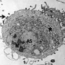
FV3-infizierte Zelle mit Kern (N), Chromatinkondensation, viraler Assemblierungsstelle (AS), parakristallinen [en] Anordnungen (*) und knospenden Virionen (→).
FV3-infizierte Zelle mit verstreuten Virionen
viralen Assemblierungsstelle mit FV3-Virionen in verschiedenen Stadien der Assemblierung. Inset: Membranen (↗), evtl. aus dem ER.
Auch die Spezies Singapore Grouper Iridovirus (SGIV), Erreger der Krankheit Singapore Grouper Iridovirus Disease (SGIVD)[28] beim Rostflecken-Zackenbarsch (Epinephelus tauvina, en. Greasy grouper [en])[29] ist inzwischen gut untersucht. Deren Viruspartikel werden in sog. viral assembly sites (VAS) zusammengebaut (assembliert).[30]
Replikationszyklus von SGIV innerhalb der Wirtszelle im Detail.-->
Kapsid-Morphogenese von Singapore Grouper Iridovirus (SGIV) in einer viral assembly site (VAS)
Kryo-EM-Aufnahme von reifen SGIV-Virionen mit asymmetrischem haarnadelförmigen Komplex auf der einen Seite (schwarzer Pfeil). Für die Sichtbarkeit ist eine in geeignete Orientierung nötig.
Virale Akkumulation von SGIV-Partikeln zu einer parakristallinen [en] Anordnung.
SGIV: Proteine verformen die Membran und bilden eine spezifische spiralförmige Struktur. Die Vakuolenmembran wird so zu einem Membrantubulus, der das Virion im Inneren der Vakuole enthält (Pfeile links).
Das Genom von Ranavirus weist wie bei anderen Iridoviridae terminal redundante DNA auf.[26]
Es wird angenommen, dass die Übertragung von Ranaviren auf mehreren Wegen erfolgt, unter anderem über kontaminiertem Boden, direkten Kontakt, Exposition durch Wasser und Verschlucken von infiziertem Gewebe während der Jagd, Nekrophagie oder Kannibalismus. Ranaviren sind in Gewässern relativ stabil und können außerhalb eines Wirtsorganismus mehrere Wochen oder länger überdauern.[12]
Die Ranaviren scheinen sich aus einem Fischvirus entwickelt zu haben, das anschließend Amphibien und Reptilien infizierte.[31]
Die innere Systematik der Gattung Iridovirus ist mit Stand März 2021 nach ICTV, ergänzt um einige Vorschläge in doppelten Anführungszeichen (nach NCBI, wo nicht anders angegeben):[1]
Es gibt etliche nach ICTV mit Stand März 2021 noch nicht klassifizierten Kandidaten, siehe etwa Halaly et al. (2019).[34]
Ranavirus ist eine Gattung von Riesenviren (Nucleocytoviricota, NCLDVs) aus der Familie der Iridoviridae, Unterfamilie Alphairidovirinae. Ranavirus ist die einzige Gattung in dieser Familie, deren Viren sowohl für Amphibien als auch Reptilien ansteckend sind. Wie auch die beiden anderen Gattungen Lymphocystivirus und Megalocytivirus der Unterfamilie Alphairidovirinae können Viren der Gattung Ranavirus auch Echte Knochenfische (Teleostei) infizieren.

Ranavirus is a genus of viruses, in the family Iridoviridae.[1] There are six other genera of viruses within the family Iridoviridae, but Ranavirus is the only one that includes viruses that are infectious to amphibians and reptiles. Additionally, it is one of the three genera within this family which infect teleost fishes, along with Lymphocystivirus and Megalocytivirus.[2]
The Ranaviruses, like the Megalocytiviruses, are an emerging group of closely related dsDNA viruses which cause systemic infections in a wide variety of wild and cultured fresh and saltwater fishes. As with Megalocytiviruses, Ranavirus outbreaks are therefore of considerable economic importance in aquaculture, as epizootics can result in moderate fish loss or mass mortality events of cultured fishes. Unlike Megalocytiviruses, however, Ranavirus infections in amphibians have been implicated as a contributing factor in the global decline of amphibian populations.[3][4] The impact of Ranaviruses on amphibian populations has been compared to the chytrid fungus Batrachochytrium dendrobatidis, the causative agent of chytridiomycosis.[5][6][7] In the UK, the severity of disease outbreaks is thought to have increased due to climate change.[8]
Rana is derived from the Latin for "frog",[9] reflecting the first isolation of a Ranavirus in 1960s from the Northern leopard frog (Lithobates pipiens).[10][11][12]
The ranaviruses appear to have evolved from a fish virus which subsequently infected amphibians and reptiles.[13]
The genus contains the following species:[24]
The family Iridoviridae is divided into seven genera which include Chloriridovirus, Iridovirus, Lymphocystivirus, Megalocytivirus, and Ranavirus.[1] The genus Ranavirus contains three viruses known to infect amphibians (Ambystoma tigrinum virus (ATV), Bohle iridovirus (BIV), and frog virus 3).[25]
Ranaviruses are large icosahedral DNA viruses measuring approximately 150 nm in diameter with a large single linear dsDNA genome of roughly 105 kbp[26] which codes for around 100 gene products.[27] The main structural component of the protein capsid is the major capsid protein (MCP).
Ranaviral replication is well-studied using Frog virus 3 (FV3).[25][26] Replication of FV3 occurs between 12 and 32 degrees Celsius.[27] Ranaviruses enter the host cell by receptor-mediated endocytosis.[28] Viral particles are uncoated and subsequently move into the cell nucleus, where viral DNA replication begins via a virally encoded DNA polymerase.[29] Viral DNA then abandons the cell nucleus and begins the second stage of DNA replication in the cytoplasm, ultimately forming DNA concatemers.[29] The viral DNA is then packaged via a headful mechanism into infectious virions.[25] The ranavirus genome, like other iridoviral genomes is circularly permuted and exhibits terminally redundant DNA.[29] There is evidence that ranavirus infections target macrophages as a mechanism for gaining entry to cells. [30]
Transmission of ranaviruses is thought to occur by multiple routes, including contaminated soil, direct contact, waterborne exposure, and ingestion of infected tissues during predation, necrophagy or cannibalism.[11][31] Ranaviruses are relatively stable in aquatic environments, persisting several weeks or longer outside a host organism.[11]
Amphibian mass mortality events due to Ranavirus have been reported in Asia, Europe, North America, and South America.[11] Ranaviruses have been isolated from wild populations of amphibians in Australia, but have not been associated with mass mortality on that continent.[11][32][33]
Synthesis of viral proteins begins within hours of viral entry[27] with necrosis or apoptosis occurring as early as a few hours post-infection.[26][34]
There are several hypothesis for seasonal outbreak patterns observed for Ranavirosis mortality events.[35] Ranaviruses grow in vitro between 8-30 °C, however for most isolates, warmer temperature result in faster viral replication.[35] A combination of this optimal growth temperature along with shifts in larval amphibian susceptibility result in seasonal outbreak events most often observed during warm summer months.[36] Amphibian mortality events are often observed as larval amphibians reach late Gosner stages approaching metamorphosis.[37] As larval amphibians reach metamorphic stages of development, their immune system is reorganized prior to the development of adult tissues.[38] During this time period, amphibians are stressed, and their immune systems are down regulated. This decrease in immune function and warmer environmental temperatures allows for greater viral replication and cellular damage to occur. Across 64 mortality events in the United States 54% were found to occur between June-August.[36]
The environmental persistence of Ranaviruses is not understood well, however in realistic environmental conditions the T90 value of an FV3-like virus is 1 day.[39] The duration of persistence is likely affected by temperature and microbial conditions. It is unlikely that ranaviruses persist in the environment outside of host species between outbreak events. Researchers have explored several pathogen reservoirs for the virus which might explain how the virus can persist within an amphibian community. In some amphibian populations, sub-clinically infected individuals may serve as reservoirs for the pathogen.[6] These sub-clinically infected individuals are responsible for reintroduction of the virus to the larval population. With ranaviruses being capable of infected multiple taxa, and with there being differences in susceptibility between taxa, it is likely that sympatric fish and reptile species may serve as reservoirs for virus as well. Interclass transmission has been proven through the use of mesocosm studies.[40]
Gross lesions associated with Ranavirus infection include erythema, generalized swelling, hemorrhage, limb swelling, and swollen and friable livers.[11]
Transmission electron micrograph of a cell infected with ranaviruses, which gather in the cytoplasm and in the assembly bodies next to the contorted nucleus. Ranavirus is a genus of viruses, in the family Iridoviridae. There are six other genera of viruses within the family Iridoviridae, but Ranavirus is the only one that includes viruses that are infectious to amphibians and reptiles. Additionally, it is one of the three genera within this family which infect teleost fishes, along with Lymphocystivirus and Megalocytivirus.
Ranavirus es un género de virus perteneciente a la familia Iridoviridae. Pueden provocar enfermedad en anfibios reptiles y peces. Incluye diferentes especies: virus de la rana tipo 3, iridovirus Bohle, virus de la necrosis hematopoyética epizoótica, virus del bagre europeo, virus del siluro europeo y ranavirus de Santee-Cooper. La especie tipo es el virus de la rana tipo 3 (Frog virus 3) que causa enfermedad en ranas.[1] Se ha sugerido que las infecciones por ranavirus en anfibios han contribuido a la disminución de las poblaciones de estos vertebrados a nivel mundial.
El primer aislamiento se realizó en 1960 de una rana perteneciente a la especie Lithobates pipiens.
Los ranavirus son virus ADN icosaédricos de gran tamaño que miden unos 150 nm de diámetro.
Ranavirus es un género de virus perteneciente a la familia Iridoviridae. Pueden provocar enfermedad en anfibios reptiles y peces. Incluye diferentes especies: virus de la rana tipo 3, iridovirus Bohle, virus de la necrosis hematopoyética epizoótica, virus del bagre europeo, virus del siluro europeo y ranavirus de Santee-Cooper. La especie tipo es el virus de la rana tipo 3 (Frog virus 3) que causa enfermedad en ranas. Se ha sugerido que las infecciones por ranavirus en anfibios han contribuido a la disminución de las poblaciones de estos vertebrados a nivel mundial.
Ranavirus est un genre de virus à ADN qui causent des nécroses hématopoïétiques épizootiques.
Ces virus touchent les animaux aquatiques comme les poissons (dont marins[2]) et les amphibiens (mort par hémorragie internes, avec symptômes d'ulcérations de la peau) ainsi que des reptiles (aux États-Unis, en Australie[3]).
Ils sont source de maladies émergentes[4] a priori impliquées dans le déclin général des amphibiens[4] ; tous les écosystèmes ne sont pas concernés, mais le virus s'étend, imposant des mesures prophylactiques[5]. On en découvre régulièrement de nouveaux isolats[6]
Ces virus ont été isolés depuis la fin des années 1990 chez divers amphibiens[7],[8],[9], on pense que certains d'entre eux sont responsables ou coresponsables de taux de mortalité extrêmement élevés et localisés d'amphibiens[10] en Amérique du Nord, mais aussi en Europe, de l'Espagne [11] au Danemark[12], en passant par le Royaume-Uni[13],[14], la Belgique[15]. On trouve ces virus en Asie aussi, jusqu'au Japon[16].
Depuis la fin des années 2000, des ranavirus déciment des reptiles et amphibiens aux États-Unis[17],[18],[19].
Ranavirus est un genre de virus à ADN qui causent des nécroses hématopoïétiques épizootiques.
Ces virus touchent les animaux aquatiques comme les poissons (dont marins) et les amphibiens (mort par hémorragie internes, avec symptômes d'ulcérations de la peau) ainsi que des reptiles (aux États-Unis, en Australie).
Ils sont source de maladies émergentes a priori impliquées dans le déclin général des amphibiens ; tous les écosystèmes ne sont pas concernés, mais le virus s'étend, imposant des mesures prophylactiques. On en découvre régulièrement de nouveaux isolats
Ranavirus é un xénero de virus da familia Iridoviridae.[1] Hai ademais outros catro xéneros de virus nesa familia, pero Ranavirus é o único que inclúe virus que son infecciosos para anfibios e réptiles. Adicionalmente, é un dos tres xéneros desta familia que infecta peixes teleósteos, xunto con Lymphocystivirus e Megalocytivirus.[2] A familia Iridoviridae é unha das cinco familias de virus de ADN grande nucleocitoplasmáticos.
Os Ranavirus foron illados por primeira vez na década de 1960 da ra Lithobates pipiens.[3][4][5]
Os Ranavirus, como o Megalocytivirus, son un grupo emerxente de virus de ADN bicatenario estreitamente relacionados que causan infeccións sistémicas nunha ampla variedade de peixes de auga salgada e doce silvestres e criados en piscifactoría. Igual que os Megalocytivirus, os abrochos de Ranavirus teñen unha considerable importancia económica en acuicultura, como epizoóticos poden orixinar unha perda moderada de peixes ou episodios de mortaldade en masa en peixes de acicultura. Porén, a diferenza dos Megalocytivirus, as infeccións por Ranavirus en anfibios foron consideradas un factor que contribúe ao declive das poboacións de anfibios global. O impacto dos Ranavirus sobre as poboacións de anfibios foi comparada coa do fungo quitridio Batrachochytrium dendrobatidis, o axente causante da quitridiomicose.[6][7][8]
Os Ranavirus parece que evolucionaron a partir de virus de peixes que despois pasaron a infectar anfibios e réptiles.[9]
Ademais de peixes e anfibios, varias especies de réptiles son afectados por Ranavirus, como:
Grupo: ADN bicatenario (dsDNA)
A familia Iridoviridae está dividida en cinco xéneros, que son Chloriridovirus, Iridovirus, Lymphocystivirus, Megalocytivirus, e Ranavirus.[1] O xénero Ranavirus está composto por 6 especies virais recoñecidas, 3 das cales infectan anfibios, que son: virus de Ambystoma tigrinum (Ambystoma tigrinum virus, ATV), iridovirus Bohle (Bohle iridovirus BIV) e o virus da ra 3 (frog virus 3).[18]
Os Ranavirus son grandes virus de ADN icosaédricos que miden aproximadamente 150 nm de diámetro cun gran xenoma dunha soa molécula de ADN bicatenario linear duns 105 kbp,[19] que codifica uns 100 produtos xénicos.[20] O principal compoñente estrutural da cápside proteica é a proteína maior da cápside (MCP).
A replicación ranaviral foi ben estudada usando a especie tipo do xénero, o virus da ra 3 (Frog virus 3, FV3).[18][19] A replicación do FV3 ocorre entre 12 e 32 graos Celsius.[20] Os Ranavirus entran na célula hóspede por endocitose mediada por receptor.[21] As partículas virais perden a cuberta e seguidamente desprázanse ao núcleo celular, onde empeza a replicación do ADN viral por medio dunha ADN polimerase codificada polo virus.[22] O ADN viral abandona despois o núcleo da célula e empeza a segunda fase da replicación do ADN no citoplasma, formando finalmente concatémeros de ADN.[22] O ADN viral é despois empaquetado en virións infecciosos.[18] O xenoma do Ranavirus, igual que outros xenomas iridovirais está permutado circularmente e presenta un ADN redundante terminalmente.[22]
A transmisión dos Ranavirus crese que ocorre por moitas rutas, incluíndo o solo contaminado, o contacto directo, a exposición á auga e a inxestión de tecidos infectados durante a predación, necrofaxia ou canibalismo.[4] Os Ranavirus son relativamente estables en ambientes acuáticos, e poden persistir varias semanas ou máis fóra dun organismo hóspede.[4]
Informouse de episodios de mortalidade en masa de anfibios debido a Ranavirus en Asia, Europa, Norteamérica e Suramérica.[4] Os Ranavirus foron illados de poboaciósn silvestres de anfibios en Australia, mais non foron asociados coa mortalidade en masa nese continente.[4][23][24]
A síntese de proteínas virais empeza en cuestión de poucas horas despois da entrada viral na célula[20] e a necrose ou apoptose ocorre só unhas poucas horas despois da infección.[19][25]
As principais lesións asociadas coas infeccións de Ranavirus son eritemas, inchamento xeneralizado, hemorraxias, inchamento das extremidades e fígados inchados e fráxiles.[4]
Pode atoparse máis información sobre Ranavirus e outros patóxenos que impactan as poboacións de anfibios, como sobre quitridios Batrachochytrium dendrobatidis e Batrachochytrium salamandrivorans na páxina web do Southeast Partners in Amphibian and Reptile Conservation disease task team. [1]
Ranavirus é un xénero de virus da familia Iridoviridae. Hai ademais outros catro xéneros de virus nesa familia, pero Ranavirus é o único que inclúe virus que son infecciosos para anfibios e réptiles. Adicionalmente, é un dos tres xéneros desta familia que infecta peixes teleósteos, xunto con Lymphocystivirus e Megalocytivirus. A familia Iridoviridae é unha das cinco familias de virus de ADN grande nucleocitoplasmáticos.
Os Ranavirus foron illados por primeira vez na década de 1960 da ra Lithobates pipiens.


.jpg)


