-
Asker, Akershus, Norge
-
Sopark Jantarit, Chutamas Satasook, Louis Deharveng
Zookeys
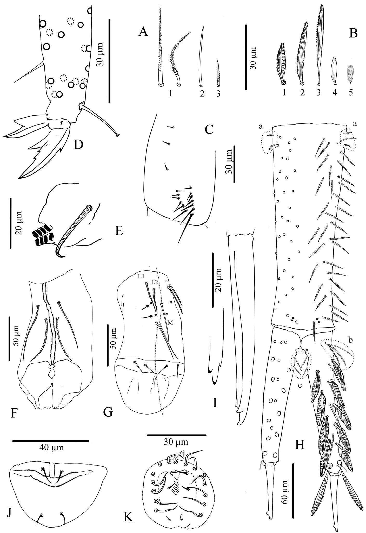
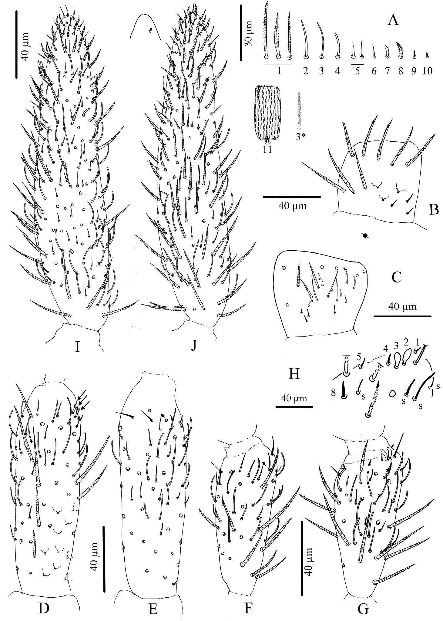
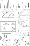
Figure 3.Cyphoderus songkhlaensis sp. n. continued A chaetae of antenna drawn from optical microscope, except 3* derived from SEM image B dorsal side of right Ant.I C ventral side of right Ant.I D dorsal side of right Ant.II; the apical swollen sens of type-7 are indicated by arrows E ventral side of right Ant.II with apical pseudopore F ventral side of right Ant.III with apical pseudopore G dorsal side of right Ant.III H distal organite of Ant.III I ventral side of Ant.IV J dorsal side of Ant.IV with separate view of the subapical organite (left).
-
Asker, Akerhus, Norge
-
Sopark Jantarit, Chutamas Satasook, Louis Deharveng
Zookeys
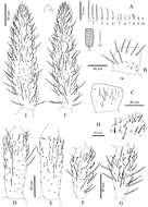
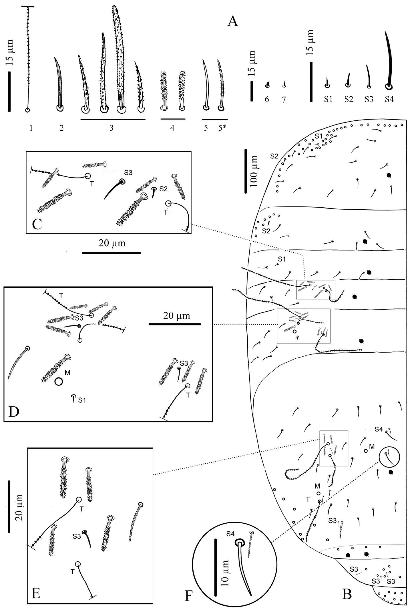
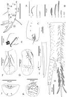
Figure 4.Cyphoderus songkhlaensis sp. n. continued A chaetae of tergites drawn from optical microscope, except 5* derived from SEM image B chaetotaxy of tergites with types of S-chaetae S1 to S4 C trichobothrial complexes of Abd.II D trichobothrial complexes of Abd.III E anterior trichobothrial complexes of Abd.IV F tandem of chaetae on Abd.IV; the smallest is a short type-5 mes and the largest a S4 sens.
-
Asker, Akershus, Norge
-
Sopark Jantarit, Chutamas Satasook, Louis Deharveng
Zookeys
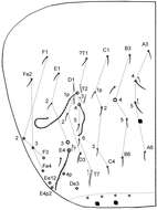
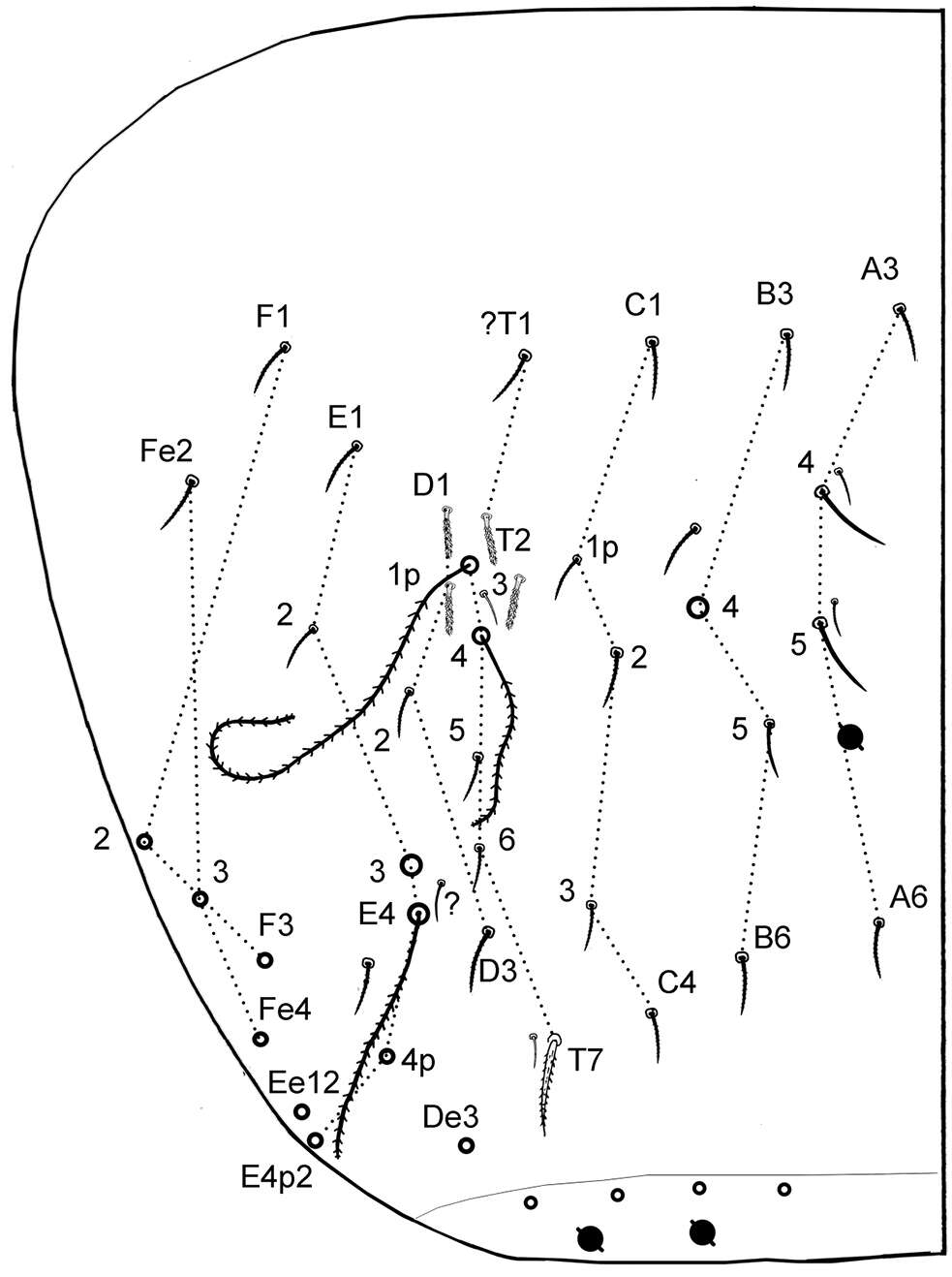
Figure 6.Cyphoderus songkhlaensis sp. n. continued, Szeptycki’s notation of tergal chaetae on Abd.IV (Szeptycki 1979).
-
Sopark Jantarit, Chutamas Satasook, Louis Deharveng
Zookeys
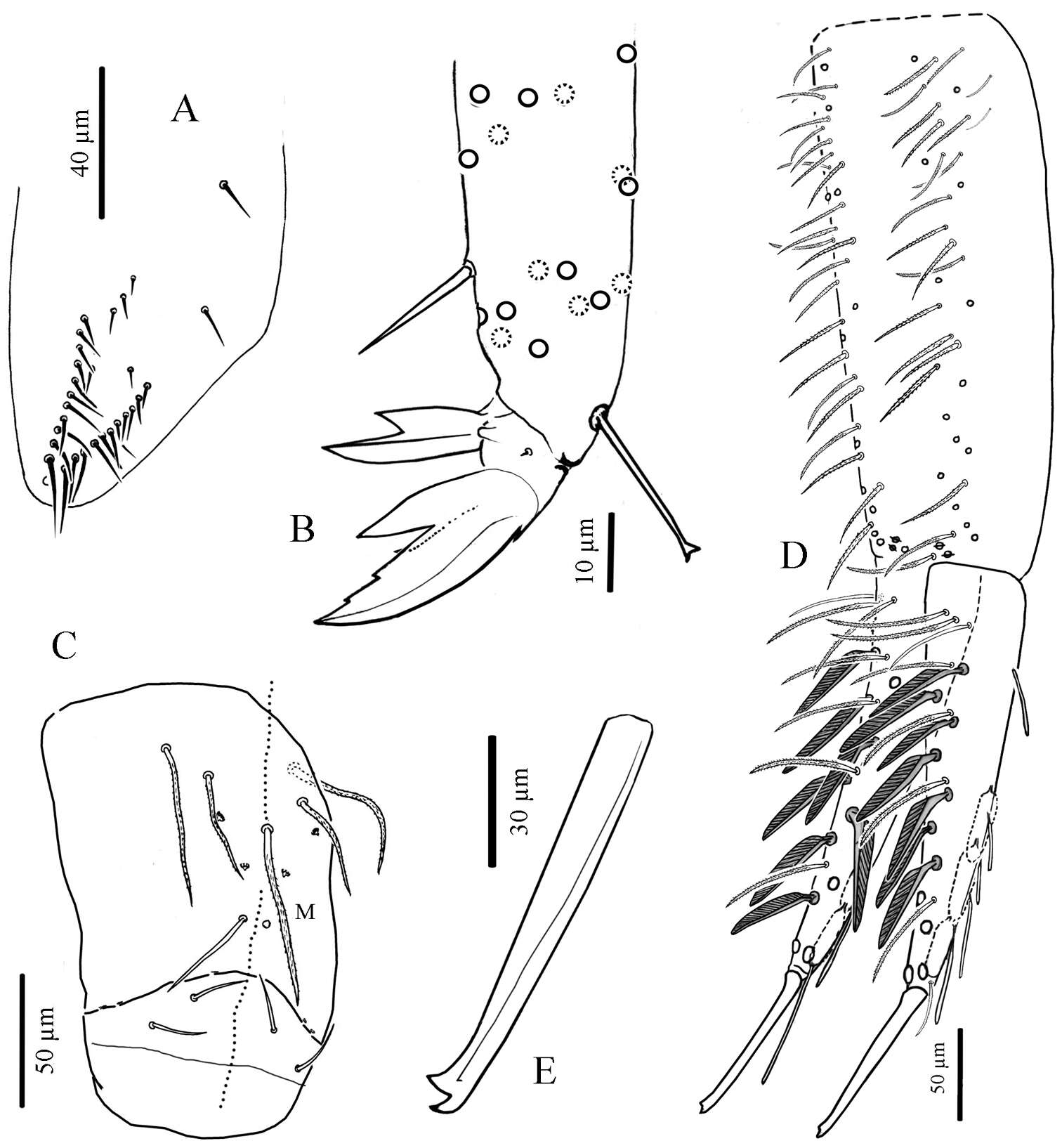
Figure 7.Cyphoderus songkhlaensis sp. n. continued A chaetae of furca B scales of furca C trochanteral organ D claw and distal part of tibiotarsus III E tenaculum F anterior face of the ventral tube G posterior face of the ventral tube; the peg-like setulae are indicated by arrows H furca; encircled by dotted lines are the 2+2 latero-basal mesochaetae of manubrium (a) the 3 outer basal mesochaetae of dens (b) and the 2+2 inner basal mesochaetae of dens (c) (I) mucro in lateral view (right) and in dorsal view (left) showing a third minute external tooth J female genital plate K male genital plate.
-
Sopark Jantarit, Chutamas Satasook, Louis Deharveng
Zookeys
Figure 8.Cyphoderus khaochakanus sp. n. A trochanteral organ B claw and distal part of tibiotarsus III C posterior face of the ventral tube D furca; feathered chaetae in lateral view, only one of the two vanes attached to the rachis is visible E mucro.
-
Sopark Jantarit, Chutamas Satasook, Louis Deharveng
Zookeys
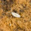
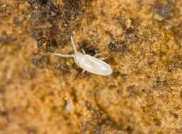
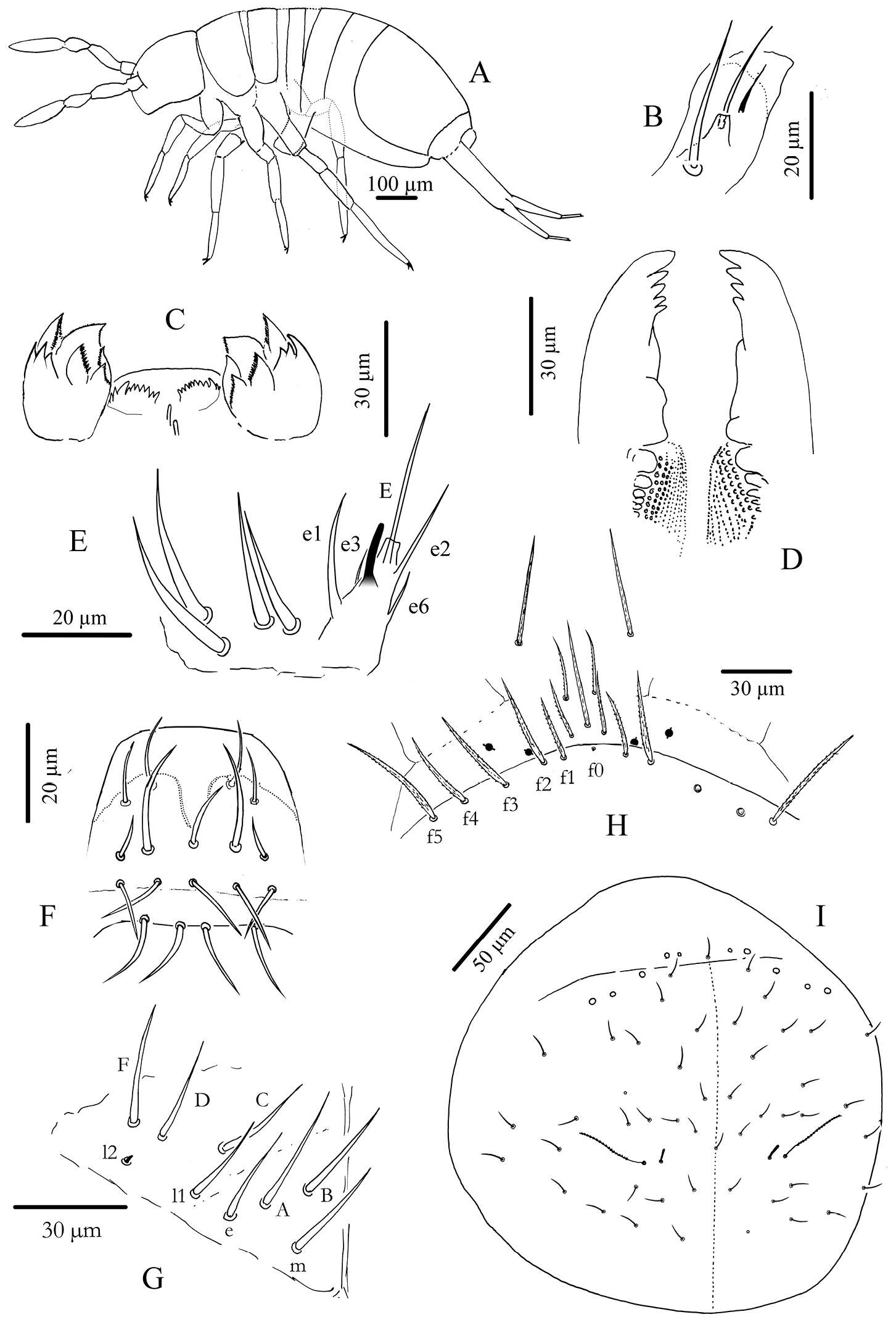
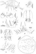
Figure 2.Cyphoderus songkhlaensis sp. n. A habitus B outer maxillary lobe C maxilla head and ventral complex of the labrum D mandible E labial palp: proximal chaetae and external papilla E F labrum, dorsal view G chaetotaxy of labial basis; frontal chaetae H frontal chaetae and pseudopores of head I dorsal chaetotaxy of head.
-
Sopark Jantarit, Chutamas Satasook, Louis Deharveng
Zookeys
Figure 7.Cyphoderus songkhlaensis sp. n. continued A chaetae of furca B scales of furca C trochanteral organ D claw and distal part of tibiotarsus III E tenaculum F anterior face of the ventral tube G posterior face of the ventral tube; the peg-like setulae are indicated by arrows H furca; encircled by dotted lines are the 2+2 latero-basal mesochaetae of manubrium (a) the 3 outer basal mesochaetae of dens (b) and the 2+2 inner basal mesochaetae of dens (c) (I) mucro in lateral view (right) and in dorsal view (left) showing a third minute external tooth J female genital plate K male genital plate.
-
Sopark Jantarit, Chutamas Satasook, Louis Deharveng
Zookeys
Figure 3.Cyphoderus songkhlaensis sp. n. continued A chaetae of antenna drawn from optical microscope, except 3* derived from SEM image B dorsal side of right Ant.I C ventral side of right Ant.I D dorsal side of right Ant.II; the apical swollen sens of type-7 are indicated by arrows E ventral side of right Ant.II with apical pseudopore F ventral side of right Ant.III with apical pseudopore G dorsal side of right Ant.III H distal organite of Ant.III I ventral side of Ant.IV J dorsal side of Ant.IV with separate view of the subapical organite (left).
-
Sopark Jantarit, Chutamas Satasook, Louis Deharveng
Zookeys
Figure 4.Cyphoderus songkhlaensis sp. n. continued A chaetae of tergites drawn from optical microscope, except 5* derived from SEM image B chaetotaxy of tergites with types of S-chaetae S1 to S4 C trichobothrial complexes of Abd.II D trichobothrial complexes of Abd.III E anterior trichobothrial complexes of Abd.IV F tandem of chaetae on Abd.IV; the smallest is a short type-5 mes and the largest a S4 sens.
-
Sopark Jantarit, Chutamas Satasook, Louis Deharveng
Zookeys
Figure 6.Cyphoderus songkhlaensis sp. n. continued, Szeptycki’s notation of tergal chaetae on Abd.IV (Szeptycki 1979).
-
Sopark Jantarit, Chutamas Satasook, Louis Deharveng
Zookeys
Figure 8.Cyphoderus khaochakanus sp. n. A trochanteral organ B claw and distal part of tibiotarsus III C posterior face of the ventral tube D furca; feathered chaetae in lateral view, only one of the two vanes attached to the rachis is visible E mucro.
-
Sopark Jantarit, Chutamas Satasook, Louis Deharveng
Zookeys
Figure 3.Cyphoderus songkhlaensis sp. n. continued A chaetae of antenna drawn from optical microscope, except 3* derived from SEM image B dorsal side of right Ant.I C ventral side of right Ant.I D dorsal side of right Ant.II; the apical swollen sens of type-7 are indicated by arrows E ventral side of right Ant.II with apical pseudopore F ventral side of right Ant.III with apical pseudopore G dorsal side of right Ant.III H distal organite of Ant.III I ventral side of Ant.IV J dorsal side of Ant.IV with separate view of the subapical organite (left).
-
Sopark Jantarit, Chutamas Satasook, Louis Deharveng
Zookeys
Figure 4.Cyphoderus songkhlaensis sp. n. continued A chaetae of tergites drawn from optical microscope, except 5* derived from SEM image B chaetotaxy of tergites with types of S-chaetae S1 to S4 C trichobothrial complexes of Abd.II D trichobothrial complexes of Abd.III E anterior trichobothrial complexes of Abd.IV F tandem of chaetae on Abd.IV; the smallest is a short type-5 mes and the largest a S4 sens.
-
Sopark Jantarit, Chutamas Satasook, Louis Deharveng
Zookeys
Figure 2.Cyphoderus songkhlaensis sp. n. A habitus B outer maxillary lobe C maxilla head and ventral complex of the labrum D mandible E labial palp: proximal chaetae and external papilla E F labrum, dorsal view G chaetotaxy of labial basis; frontal chaetae H frontal chaetae and pseudopores of head I dorsal chaetotaxy of head.
-
Sopark Jantarit, Chutamas Satasook, Louis Deharveng
Zookeys
Figure 7.Cyphoderus songkhlaensis sp. n. continued A chaetae of furca B scales of furca C trochanteral organ D claw and distal part of tibiotarsus III E tenaculum F anterior face of the ventral tube G posterior face of the ventral tube; the peg-like setulae are indicated by arrows H furca; encircled by dotted lines are the 2+2 latero-basal mesochaetae of manubrium (a) the 3 outer basal mesochaetae of dens (b) and the 2+2 inner basal mesochaetae of dens (c) (I) mucro in lateral view (right) and in dorsal view (left) showing a third minute external tooth J female genital plate K male genital plate.