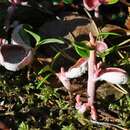
Die Gemeine Preiselbeer-Nacktbasidie (Exobasidium vaccinii) ist eine Brandpilzart aus der Familie der Nacktbasidienverwandten (Exobasidiaceae). Sie lebt als Endoparasit auf Preiselbeeren (Vaccinium vitis-idaea) und infiziert deren Blätter. Symptome des Befalls durch die Gemeine Preiselbeer-Nacktbasidie sind leuchtend rote Blätter und stark verdickte, von Myzel überzogene Gallen auf der Blattunterseite. Die Art ist im gesamten Verbreitungsgebiet der Preiselbeere nachgewiesen.
Die Gemeine Preiselbeer-Nacktbasidie ist mit bloßem Auge zunächst nicht zu erkennen. Anfangssymptome des Befalls erscheinen in Form von leuchtend roten, gelblich umrandeten Flecken auf der Blattoberseite. Später formen sich etwa 1 cm breite, stark verdickte rosa Gallen auf der Blattunterseite aus, auf deren Oberfläche das weiße Myzel durchbricht. Von dort aus überwuchert es schließlich das gesamte Blatt.
Das Myzel von Gemeine Preiselbeer-Nacktbasidie wächst interzellulär und bildet Saugfäden, die in das Speichergewebe des Wirtes wachsen. Die viergesporten Basidien sind 40–50 × 4–5 µm groß, unseptiert und schmalkeulig. Sie werden entweder einzeln oder in Büscheln zwischen den Zellen der Pflanzenepidermis gebildet. Die zylindrischen Sporen sind hyalin, dünnwandig und messen 11–20 × 2–4 µm. Im Reifestadium sind sie ein- bis sechsfach septiert.
Die Gemeine Preiselbeer-Nacktbasidie ist holarktisch verbreitet, sie ist an das Vorkommen der Preiselbeere (Vaccinium vitis-idaea) gebunden.
Einziger Wirt der Gemeinen Preiselbeer-Nacktbasidie ist die Preiselbeere. Der Pilz ernährt sich von den im Speichergewebe der Pflanzen vorhandenen Nährstoffen; zunächst beschränkt auf die Blätter, schließlich dann auf der gesamten Pflanze. Die Übertragung von einer Pflanze zur nächsten erfolgt durch Sporenflug. Die Sporen keimen in 8–11 × 1 µm großen Konidien, aus denen sich dann ein neues Myzel entwickelt.
Die Gemeine Preiselbeer-Nacktbasidie (Exobasidium vaccinii) ist eine Brandpilzart aus der Familie der Nacktbasidienverwandten (Exobasidiaceae). Sie lebt als Endoparasit auf Preiselbeeren (Vaccinium vitis-idaea) und infiziert deren Blätter. Symptome des Befalls durch die Gemeine Preiselbeer-Nacktbasidie sind leuchtend rote Blätter und stark verdickte, von Myzel überzogene Gallen auf der Blattunterseite. Die Art ist im gesamten Verbreitungsgebiet der Preiselbeere nachgewiesen.
Exobasidium vaccinii, commonly known as “red leaf disease,” or “Azalea Gall,” is a biotrophic species of fungus that causes galls on ericaceous plant species, such as blueberry and azalea (Vaccinium and Rhododendron spp.). Exobasidium vaccinii is considered the type species of the Exobasidium genus.[1] As a member of the Ustilagomycota, it is a basidiomycete closely related to smut fungi. Karl Wilhelm Gottlieb Leopold Fuckel first described the species in 1861 under the basionym Fusidium vaccinii,[2] but in 1867 Mikhail Stepanovich Voronin (often cited as “Woronin”) later placed it in the genus Exobasidium.[3] The type specimen is from Germany, and it is held in the Swedish Museum of Natural History.[4] Exobasidium vaccinii, in current definition from John Axel Nannfeldt in 1981, is limited on the host Vaccinium vitis-idaea.[5] This idea is used in most recent papers on E. vaccinii.
In its pathogenic state, E. vacinnii causes discoloration and, depending on the host, may cause hypertrophy and hyperplasia on the leaves and meristem, often forming flower-like structures (i.e. “pseudoflowers”). It may also cause green spots on blueberry fruits, which are sometimes tinted red and have occasional white spore masses.[6] Symptoms within the host plant are often varied compared to other species of Exobasidium, and distinguishing among species has relied traditionally upon spore size.[7]
In a typical disease cycle, leaves on infected shoots will first turn greenish red to bright red when the host species would typically fruit.[8] During the late stage of disease development, the undersurface (abaxial side) of leaves will become covered in a white mass, consisting of sparse hyphae, basidia, basidiospores, secondary spores, and secondary spores forming conidia.[9]
Basidiospores are musiform[7] with a round apex and a distinctive hilar region at the spore base.[9] The spores are hyaline and the dimensions are about 10-13 micrometers long and 3-4 micrometers wide. Some spores have a transverse medial septum separating two nuclei. Woronin first observed Exobasidium’s ability to produce asexual spores in 1867, and over a century later, scanning electron microscopy and transmission electron microscopy has confirmed E. vaccinii’s ability to produce conidia from secondary spores.[9]
There are no known reports of E. vaccinii forming appressoria; however, there are numerous reports of appressoria forming in E. vexans, which is pathogenic on tea,[10] and among other members of the Ustilagomycetes.[11] The intercellular hyphae are septate with short, lobed haustoria. Hyphae and haustoria contacting host cells cause significant amounts of pressure and subsequent distortion in the surrounding tissues. Haustoria contain membranous inclusion bodies and are associated with electron-dense deposits, much like other plant pathogenic fungi.[12]
E. vaccinii is dimorphic and can be grown in culture; in its non-pathogenic state in nature, it likely lives in a yeast-like form in the soil or on the plant similar to many of its smut relatives. In its biotrophic state, E. vaccinii gets its energy from its ericaceous host plants. Most species of native and cultivated rhododendron and azalea are considered susceptible, in addition to high and lowbush blueberry cultivars.[6][8][13] E. vaccinii is distributed across the Northern Hemisphere, including most of eastern North America and western Europe, according to known studies. It has also been reported in parts of Asia on endemic Vaccinium.[14] Endemic species previously reported to be infected with E. vaccinii in Hawaii has been discovered to be a different species, Exobasidium darwinii. [15]
Spores are produced on basidia on the outside of galls, typically in the late spring and early summer. Eventually, the mycelium present in the leaves colonizes the host’s rhizomes, where it becomes systemic; any new shoots growing from these rhizomes are often infected and fail to fruit or flower.[12][16] Systemically infected plants also often experience higher infection rates and gall loads.
Blueberries infected with E. vaccinii remain edible, but the spots result in what may be considered “unsightly” fruits. The disease has been observed to infect up to 25% of certain harvests, rendering the berries unmarketable.[6] Additionally, lower fruit yields in systemically infected plants pose a great risk to commercial growers. Gall formation negatively affects reproductive measures, decreasing flower production, flower size, and fruit yield.[17] A study conducted in Nova Scotia found that the disease decreases flowers by 42% and the number of berries per stem by 74%.[8] Branches of infected shoots will also typically die the following year. Recommendations for preventative control includes pruning infected shoots before the fungus produces spores.
Taxonomic reports on E. vaccinii are ongoing. While Fuckel first described E. vaccinii on a Vaccinium species in Germany, many studies attribute gall formation in multiple species of North American blueberry cultivars and others in native and cultivated azalea.[6][12][17] Most of these records and publications do not have a phylogenetic basis for their identifications and rely on the morphology of the spores; therefore, we cannot confirm their taxonomic placement and host relationship without conducting more in-depth phylogenetic studies. One hypothesis argues that in addition to coevolution, sporulation site plays a significant role in speciation.[18] There is also some disagreement in the literature over whether or not E. vaccinii even causes hypertrophy on certain Vaccinium species.[12] However, taxonomic resolution will require additional phylogenetic studies. Originally E. vaccinii was a broad spectrum group but later studies showed it was a complex made of different species with narrow host ranges. Many species once considered E. vaccinii have been separated as their own. Nannfeldt in 1981 "proposed that Exobasidium species have narrow host ranges that are restricted to one plant species or a group of closely-related species.”[19]
Exobasidium vaccinii, commonly known as “red leaf disease,” or “Azalea Gall,” is a biotrophic species of fungus that causes galls on ericaceous plant species, such as blueberry and azalea (Vaccinium and Rhododendron spp.). Exobasidium vaccinii is considered the type species of the Exobasidium genus. As a member of the Ustilagomycota, it is a basidiomycete closely related to smut fungi. Karl Wilhelm Gottlieb Leopold Fuckel first described the species in 1861 under the basionym Fusidium vaccinii, but in 1867 Mikhail Stepanovich Voronin (often cited as “Woronin”) later placed it in the genus Exobasidium. The type specimen is from Germany, and it is held in the Swedish Museum of Natural History. Exobasidium vaccinii, in current definition from John Axel Nannfeldt in 1981, is limited on the host Vaccinium vitis-idaea. This idea is used in most recent papers on E. vaccinii.
Pohla-paisseen (Exobasidium vaccinii) on kandseente hulka kuuluv seeneliik.
Seent on leitud ka Eestist.[1]
Pohla-paisseen (Exobasidium vaccinii) on kandseente hulka kuuluv seeneliik.
Seent on leitud ka Eestist.
Płaskosz borówki, płaskosz borówkowy (Exobasidium vaccinii (Fuckel) Woronin) – gatunek grzybów z klasy płaskoszy (Exobasidiomycetes)[1]. Jest pasożytem roślin, głównie z rodziny wrzosowatych (Ericaceae)[2].
Pozycja w klasyfikacji według Index Fungorum: Exobasidium, Exobasidiaceae, Exobasidiales, Exobasidiomycetidae, Exobasidiomycetes, Ustilaginomycotina, Basidiomycota, Fungi [1].
Po raz pierwszy zdiagnozował go w 1861 r. K.W.G.L. Fuckel nadając mu nazwę Fusidium vaccinii. Obecną, uznaną przez Index Fungorum nazwę nadał mu M.S. Woronin w 1867 r[1].
Anamorfa: Fusidium vacccini Fuckel[2].
Nazwy polskie taksonów na podstawie ustaleń Komisji do spraw polskiego nazewnictwa grzybów[4].
Na liściach i pąkach kwiatowych porażonych roślin tworzy przylegającą, gęstą grzybnię, często powodując zniekształcenie liści. Przyjmują on łyżeczkowaty kształt. Grzybnia osiąga średnicę do 1 cm, początkowo jest czerwona z żółtawym obrzeżem, potem różowawo-biała, w końcu biała. Dziej się tak dlatego, że w miarę dojrzewania owocnika jego górna powierzchnia pokrywa się białą warstwą hymenium. Podstawki zazwyczaj 3–4- sterygmowe. Bazydiospory o wymiarach 1–19 × 2–4 μm, kiełbaskowate lub lekko zakrzywione, cylindryczne. Dojrzałe posiadają 1–3 przegrody[2].
Płaskosz borówkowy wytwarza także bezpłciowe zarodniki konidialne o wymiarach 8–11 × 1 μm. Zarówno płciowe bazydiospory, jak i bezpłciowe konidia rozprzestrzeniają się unoszone prądami powietrza. Infekcja odbywa się zazwyczaj wiosną na młodych liściach. Po zainfekowaniu rośliny grzybnia patogenu przerasta całe tkanki liści, pąków kwiatowych i łodyg. W porażonej roślinie patogen zimą nie ginie i następnej wiosny rozwija się nadal[2].
Znane jest występowanie tego gatunku w Europie (w tym na Grenlandii) i w Ameryce Północnej (Kanada, USA). Pasożytuje na różnych gatunkach borówek[2], najczęściej na borówce brusznicy[4], ale także na różanecznikach, zwłaszcza z grupy azalii[5].
Wywołuje choroby przynoszące straty gospodarcze[2]. Zwalczanie za pomocą fungicydów i antybiotyków okazało się nieskuteczne. Stwierdzono, że najbardziej jak dotąd skuteczne jest opryskiwanie roztworem chlorku niklu, azotanu i siarczanu niklu w stężeniu 0,3 g /l[2]. Ostatnio na zachodzie Europy zwalcza się chorobę preparatem Bayleton zawierającym triadimefon. W uprawach domowych, gdy choroba zostanie wcześnie wykryta, możliwe jest ręczne jej zwalczanie – należy po prostu wyciąć i zniszczyć (najlepiej spalić) porażone pędy[5].
Płaskosz borówki, płaskosz borówkowy (Exobasidium vaccinii (Fuckel) Woronin) – gatunek grzybów z klasy płaskoszy (Exobasidiomycetes). Jest pasożytem roślin, głównie z rodziny wrzosowatych (Ericaceae).
Exobasidium vaccinii (Fuckel) Woronin 1867
Экзобазидиум брусничный (лат. Exobasidium vaccinii) — вид базидиомикотовых грибов (Basidiomycota) семейства экзобазидиевых (Exobasidiaceae).
Экзобазидиум брусничный имеет бледно-зелёный, розовый, белый или коричневый окрас[1].
Может развиваться на листьях, кончиках веточек, цветочных частях и даже на стручках некоторых растений, в том числе: азалия, камелия[2] и вакциниум[1].
Встречается данный вид грибов с середины лета по начало осени. Зимует гриб внутри поражённого растения[1].
Экзобазидиум брусничный вызывает образование белых подушкообразных наростов на молодых листьях вечнозелёных рододендронов (Rhododendron maximum, Rhododendron catawbiense и их гибридов[1]) и листопадных рододендронов в естественных местах их произрастания[3]. Поражённый данным грибом лист вакциниума с одной стороны более или менее вогнут с красным пятном, на другой стороне листа имеется вздутость светлых тонов (см. биологическое описание); поражённые ягоды также имеют красные пятна, а поражённые веточки становятся припухлыми. Белая светлая вздутость на листе вызвана множеством микроскопических грибных структур, которые производят споры, способные заразить больше растений во влажную погоду[1].
Формы:
Варитет:
Экзобазидиум брусничный (лат. Exobasidium vaccinii) — вид базидиомикотовых грибов (Basidiomycota) семейства экзобазидиевых (Exobasidiaceae).
