-
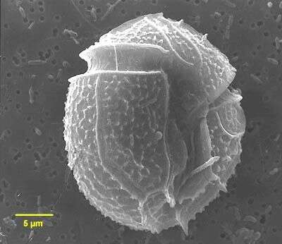
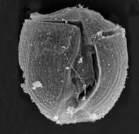
Amphidiniopsis korewalensis cells round to oval from the ventral side, dorso-ventrally flattened, 24 - 35 microns long, 20-30 microns wide, 15 - 23 microns broad. Thecal plates present: an apical pore, 4 apical plates, 3 anterior intercalary plates, 6 precingular plates, an x plate, 4 cingular plates, possibly 4 sulcal plates, 6 postcingular plates, 2 posterior plates. Epicone small and relatively flat, hypocone large and rounded. Average epicone/hypocone ratio 0.2-0.25. A small apical hook points to the left lateral cell side, over the round apical pore. Plates quite thin and boundaries not visible with the light microscope, covered in scattered small pores, approximately 0.2 microns diameter. Small protrusions often present on the posterior sulcal plate. Possesses no chloroplast or eyespot. Nucleus large (20-25 x 12-15 microns), elliptical and positioned centrally. Numerous colourless globules present
-
Amphidiniopsis observed in marine muds and sandy sediments in the vicinity of Broome, Western Australia in September 2003. This image was taken using scanning electron microscopy. This work was supported by the Australian Biological Resources Study.
-
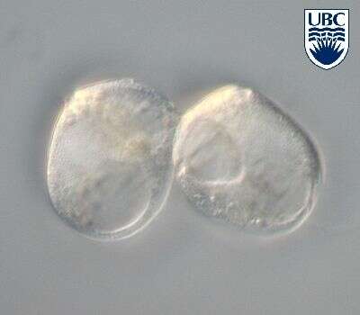
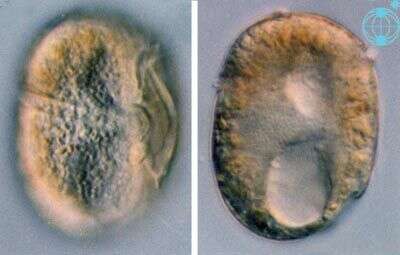
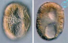
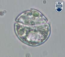
Amphidiniopsis cf. kofoidii Woloszynska 1928. Two cells in lateral view.
-
Amphidiniopsis cf. kofoidii, left lateral view, mid cell focus. Note the large nucleus in the right bottom part of the cell image.
-
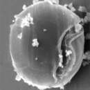
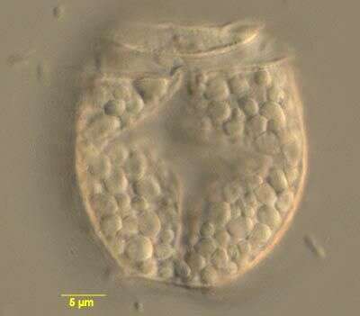
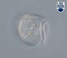
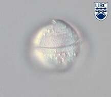
Amphidiniopsis spec. 1, so far undescribed taxon. Sulcus (longitudinal furrow) visible. Also note the cingulum (transverse furrow) in the upper part of the cell.
-
Amphidiniopsis spec. 1. Note growth band between thecal plates, seen as faintly striated horizontal band at the bottom end of the cell.
-
Amphidiniopsis spec. 1. Right lateral view, mid cell focus, showing the nucleus in the left midpart of the cell image. Two pusules are visible on the right side of the cell picture as well as a tiny posterior sulcul spine.
-
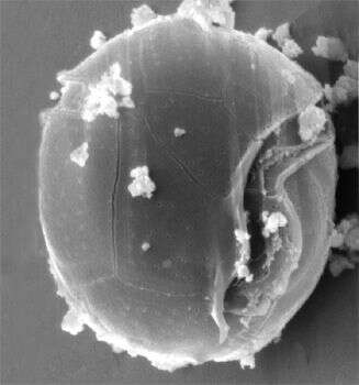
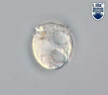
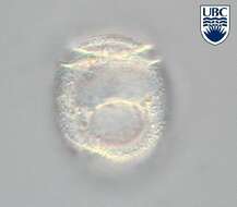
Amphidiniopsis spec. 2. Note the sulcus (longitudinal furrow) and the cingulum (transverse furrow) in the upper part of the cell.
-
Amphidiniopsis spec. 2. Mid cell focus, with large kidney shaped nucleus in the upper part of the cell above a large pusule.
-
Amphidiniopsis spec. 2.
-
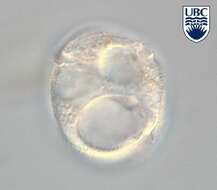
Amphidiniopsis spec. 3.
-
Amphidiniopsis spec. 3.
-
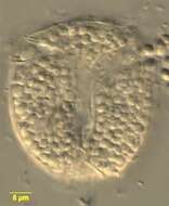
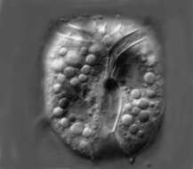
Amphidiniopsis hexagona cells are round, dorso-ventrally flattened, 28 - 42 microns long, 25 - 36 microns wide, approximately 17 microns broad. Thecal plates present: an apical pore, 4 apical plates, 2-3 anterior intercalary plates, 7 precingular plates, possibly 3 cingular plates, 6 sulcal plates, 5 postcingular plates, 2 posterior plates. Two forms were found, one with 3 anterior intercalary plates, the other with two. Two small hooks present at base of sulcus, one on the end of the right sulcal plate (Sd), the other on the end of the left sulcal plate (Ss). Thecal plates relatively thick, covered in small bumps and scattered pores, 0.1 - 0.2 microns diameter. Cingular plates covered by transverse ridges. Plate boundaries may have transverse striation. Possesses no chloroplast or eyespot. Nucleus large, (20 x 10 microns), oval and positioned centrally.
-
Amphidiniopsis hexagona observed in marine muds and sandy sediments in the vicinity of Broome, Western Australia in September 2003. This image was taken using differential interference contrast optics. This work was supported by the Australian Biological Resources Study.
-
Amphidiniopsis hexagona, from the dorsal side, observed in marine muds and sandy sediments in the vicinity of Broome, Western Australia in September 2003. This image was taken using differential interference contrast optics. This work was supported by the Australian Biological Resources Study.
-
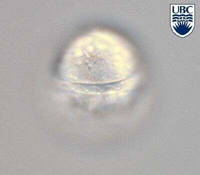
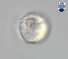
Amphidiniopsis (am-fee-din-ee-op-sis) swedmarkii (Balech) Dodge 1982. The image shows the ventral view of a cell, with two antapical spines visible. The cingulum is near the anterior end of the cell and it is slightly ascending.
-
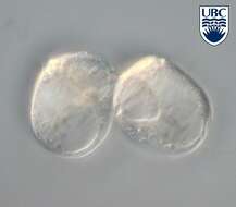
Thecadinium (theek-a-din-ee-um) dragescoi Balech 1956. The image on the left shows a cell in ventral view. The cingulum is not completely encircling the cell, and is descending. There are no plastids present. The image on the left shows a cell in a mid-focal plane. The cell is thecate, but has no processes.
-
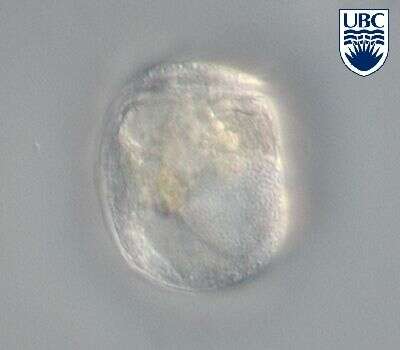
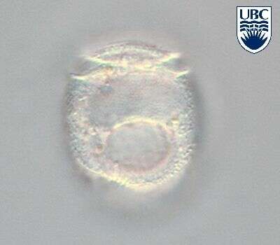
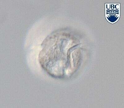
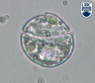
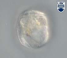
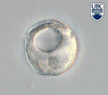
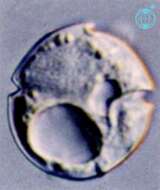
Herdmania (herd-mane-ee-a) litoralis Dodge 1981. This is an armoured dinoflagellate with a superficial similarity to Katodinium. The cingulum is located around the middle of the cell - although it does not completely encircle the cell. There is an apical hook pointed to the left of the cell (this cell is viewed from its ventral surface), the nucleus is in the upper right portion of the cell (upper left in the image), the large empty-looking structure in the hypotheca (posterior part of the cell) is the pusule, an organelle of uncertain function.
-
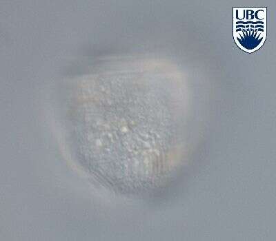
Herdmania litoralis, from the dorsal side, observed in marine muds and sandy sediments in the vicinity of Broome, Western Australia in September 2003. This image was taken using differential interference contrast optics. This work was supported by the Australian Biological Resources Study.
-
Herdmania litoralis, from the dorsal side, observed in marine muds and sandy sediments in the vicinity of Broome, Western Australia in September 2003. This image was taken using differential interference contrast optics. This work was supported by the Australian Biological Resources Study.
-
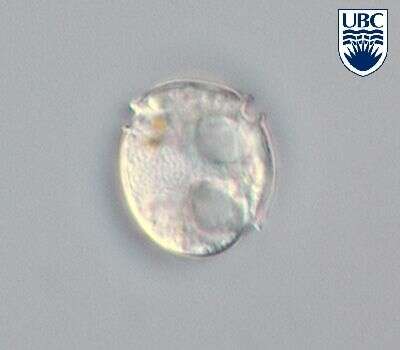
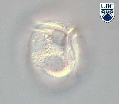
Herdmania litoralis Dodge 1981
-
Herdmania litoralis Dodge 1981
-
Herdmania litoralis Dodge 1981
-
Herdmania litoralis Dodge 1981