-
-
-
-
-
-
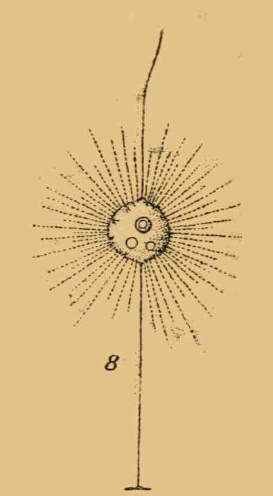
Description: Français : Actinomonas
Actinomonadaceae. Date: 1910. Source: Kryptogamenflora der Mark Brandenburg, Leipzig, 1810 :
voir en ligne. Author: Dritter Band.
-
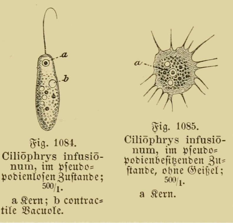
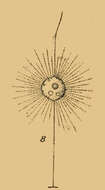
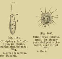
Description: Français : Ciliophrys infusionum,
Dictyochophyceae, famille des
Ciliophryaceae Fig. 1084. Ciliophrys infusionum, im pseudopodien losen zustande ; 500/I. (À l'état sans pseudopodes [avec flagelle]) Fig. 1085. Ciliophrys infusionum, im pseudopodien befissenden zustande, ohne Geissel ; 500/I. (À l'état pseudopodial, sans flagelle) a : noyau ; b : vacuole contractile. Date: 1886. Source: Synopsis der thierkunde. bd. 2, 1886, page 1117 :
lire en ligne. Author: Dr. Johannes Leunis.
-
Description: Dictyocha speculum Ehrenberg 1839 (Distephanus speculum (Ehrenberg) Haeckel 1887); Silicoflagellatа English: North-West
Black Sea, surface water Русский: Северо-Запад
Чёрного моря, поверхностные воды. Date: 18 March 2008. Source: Own work. Author:
Minami Himemiya.
-
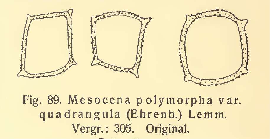
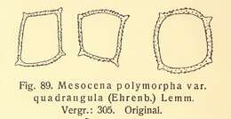
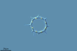
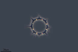
Description: Français : Mesocena polymorpha var quadrangula (Ehrenberg) Lemmann. Date: 1908. Source: Nordisches Plankton. Bot. teil., Verlag & Tischer, Kiel & Leipzig, 47 pages, 1908. p. 26 :
lire en ligne. Author: K. Brandt & C. Apstein.
-
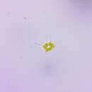
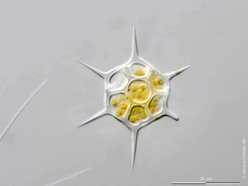
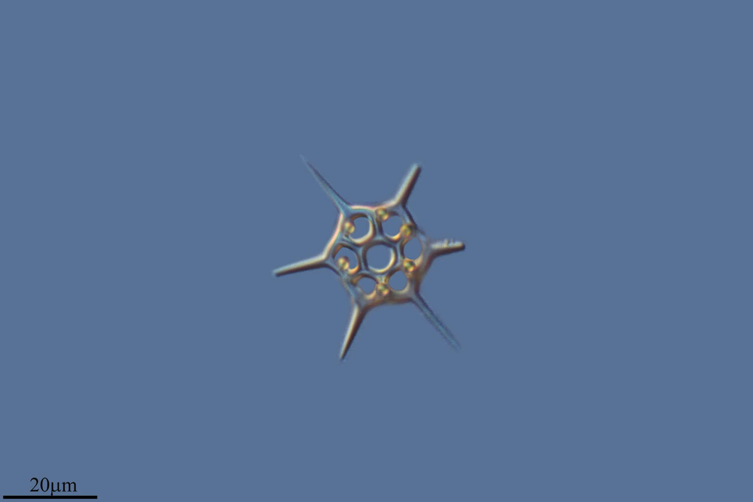
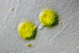
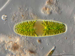
Dictyocha speculum Scale bar indicates 25 µm. The specimen was gathered in the Kieler Förde (German Baltic Sea). Sampling date 5/2018. The image was built up using several photomicrographic frames with manual stacking technique. Images were taken using Zeiss Axioplan with Olympus OM-D M5 MKII. Image under Creative Commons License V 3.0 (CC BY-NC-SA). Place name: Baltic Sea, Kieler Förde, Kiel Fjord (Germany) Latitude: 54.3894126 Longitude: 10.1749055 Multiebenen-Abbildung, manuell gestapelt. Der Messbalken markiert eine Länge von 25 µm. Probe aus der Kieler Förde. Datum der Aufsammlung: 5/2018. Mikrotechnik: Zeiss Axioplan, Kamera: Olympus OM-D M5 MKII. Creative Commons License V 3.0 (CC BY-NC-SA). For permission to use of (high-resolution) images please contact postmaster@protisten.de.
-
Dictyocha speculum Scale bar indicates 25 µm. The specimen was gathered in the Kieler Förde (Baltic Sea). Sampling date 2/2022. The image was built up using several photomicrographic frames with manual stacking technique. Images were taken using Zeiss Axioplan with Olympus OM-D M5 MKII. Image under Creative Commons License V 3.0 (CC BY-NC-SA). Place name: Baltic Sea, Kieler Förde, Kiel Fjord (Germany) Latitude: 54.3894126 Longitude: 10.1749055 Multiebenen-Abbildung, manuell gestapelt. Der Messbalken markiert eine Länge von 25 µm. Probe aus der Kieler Förde. Datum der Aufsammlung: 2/2022. Mikrotechnik: Zeiss Axioplan, Kamera: Olympus OM-D M5 MKII. Creative Commons License V 3.0 (CC BY-NC-SA). For permission to use of (high-resolution) images please contact postmaster@protisten.de.
-
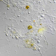
Ciliophrys infusionum The specimen was gathered in the pond Birkensee near Rödelsee (Lower Franconia, Germany). Copyright Dr. Rainer Meisch, Würzburg, Germany.Images were taken using Zeiss Axioplan with Canon DSLR Image under Creative Commons License V 3.0 (CC BY-NC-SA). Place name: Pond Birkensee near Rödelsee (Lower Franconia, Germany) Latitude: 49.71819841 Longitude: 10.27807474 Probe aus dem Birkensee bei Rödelsee (Unterfranken). Datum der Aufsammlung: 7/2018. Copyright Dr. Rainer Meisch, Würzburg. Mikrotechnik: Zeiss Axioplan, Kamera: Canon DSLR. Creative Commons License V 3.0 (CC BY-NC-SA). For permission to use of (high-resolution) images please contact postmaster@protisten.de.
-
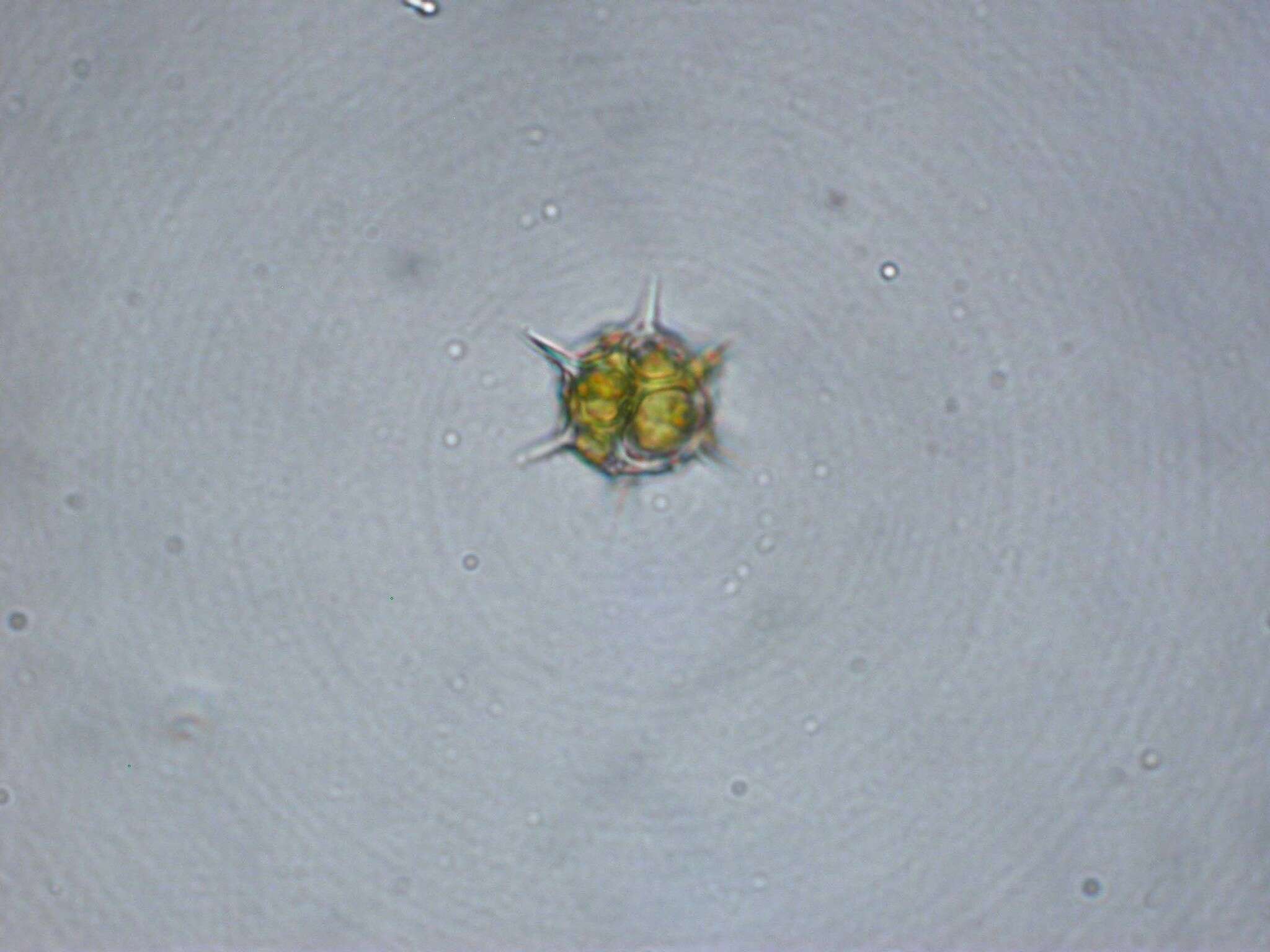
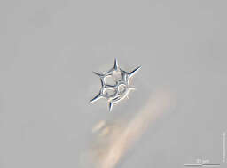
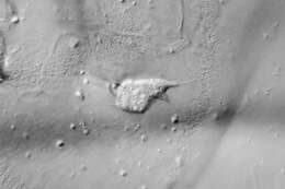
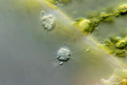
Ciliophrys infusionum Series of final stage of a binary fission. Sample from seaweed meadows from Bodden, the brackish waters lying between the isles of Hiddensee and Ruegen (German Baltic Sea). Sampling date 10/2007. The image was built up using several photomicrographic frames with manual stacking technique. Images were taken using Zeiss Standard with Olympus C7070 CCD camera. Image under Creative Commons License V 3.0 (CC BY-NC-ND). Place name: Hiddensee Bodden (Germany) Latitude: 54.582633 Longitude: 13.115051 Serie von Endstadium einer Zellteilung. Probe aus den Seegraswiesen im Hiddenseer Bodden. Datum der Aufsammlung: 10/2007. Mikrotechnik: Zeiss Standard, Kamera: Olympus C7070 WZ. Creative Commons License V 3.0 (CC BY-NC-ND). For permission to use of (high-resolution) images please contact postmaster@protisten.de.
-
Ciliophrys infusionum The specimen was gathered in the pond Birkensee near Rödelsee (Lower Franconia, Germany). Copyright Dr. Rainer Meisch, Würzburg, Germany.Images were taken using Zeiss Axioplan with Canon DSLR Image under Creative Commons License V 3.0 (CC BY-NC-SA). Place name: Pond Birkensee near Rödelsee (Lower Franconia, Germany) Latitude: 49.71819841 Longitude: 10.27807474 Probe aus dem Birkensee bei Rödelsee (Unterfranken). Datum der Aufsammlung: 7/2018. Copyright Dr. Rainer Meisch, Würzburg. Mikrotechnik: Zeiss Axioplan, Kamera: Canon DSLR. Creative Commons License V 3.0 (CC BY-NC-SA). For permission to use of (high-resolution) images please contact postmaster@protisten.de.
-
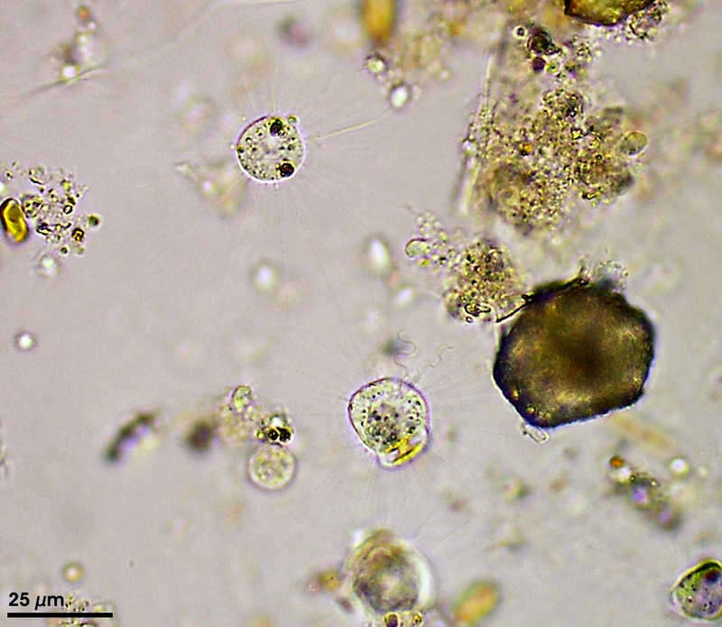
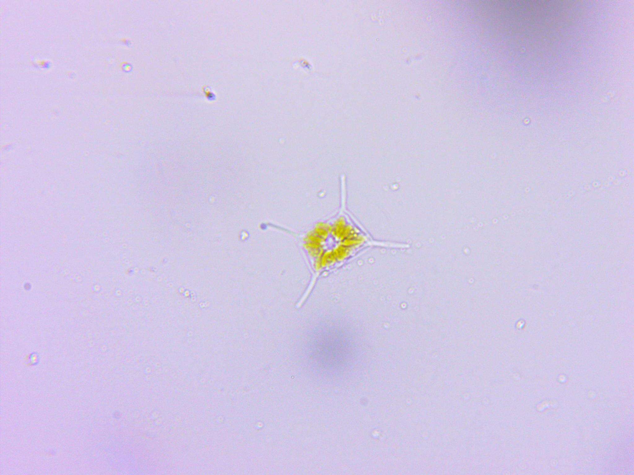
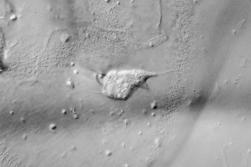
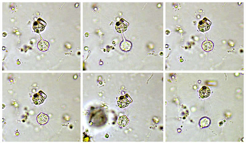
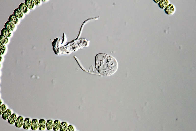
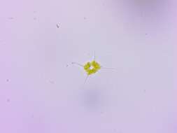
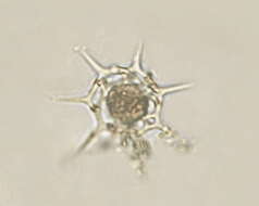
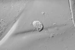
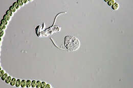
Ciliophrys infusionum Synonym: Ciliophrys marina.Cells in heliozoic state usually has one flagellum and numerous axopods. This specimen is starting with binary fission, the flagellum has already been duplicated. The scale bar indicates 25 µm. Collected from Bodden, the brackish waters lying between the isles of Hiddensee and Ruegen (German Baltic Sea). Images were taken using Zeiss Universal with Olympus C7070 CCD camera. Image under Creative Commons License V 3.0 (CC BY-NC-SA). Place name: Hiddensee Bodden (Germany) Latitude: 54.582633 Longitude: 13.115051 Synonym: Ciliophrys marina.Zellen im Sonnentierchen-Zustand haben in der Regel ein Flagellum und zahlreiche Axopodien. Dieses Exemplar ist bei der Teilung, sie hat die Geißel bereits dupliziert. Der Messbalken markiert eine Länge von 25 µm. Probe aus dem Hiddenseer Bodden, der Brackwasserfläche zwischen den Inseln Hiddensee und Rügen. Mikrotechnik: Zeiss Universal, Kamera: Olympus C7070. Creative Commons License V 3.0 (CC BY-NC-SA). For permission to use of (high-resolution) images please contact postmaster@protisten.de.
-
Ciliophrys infusionum The specimen was gathered in the pond Birkensee near Rödelsee (Lower Franconia, Germany). Copyright Dr. Rainer Meisch, Würzburg, Germany.Images were taken using Zeiss Axioplan with Canon DSLR Image under Creative Commons License V 3.0 (CC BY-NC-SA). Place name: Pond Birkensee near Rödelsee (Lower Franconia, Germany) Latitude: 49.71819841 Longitude: 10.27807474 Probe aus dem Birkensee bei Rödelsee (Unterfranken). Datum der Aufsammlung: 7/2018. Copyright Dr. Rainer Meisch, Würzburg. Mikrotechnik: Zeiss Axioplan, Kamera: Canon DSLR. Creative Commons License V 3.0 (CC BY-NC-SA). For permission to use of (high-resolution) images please contact postmaster@protisten.de.
-
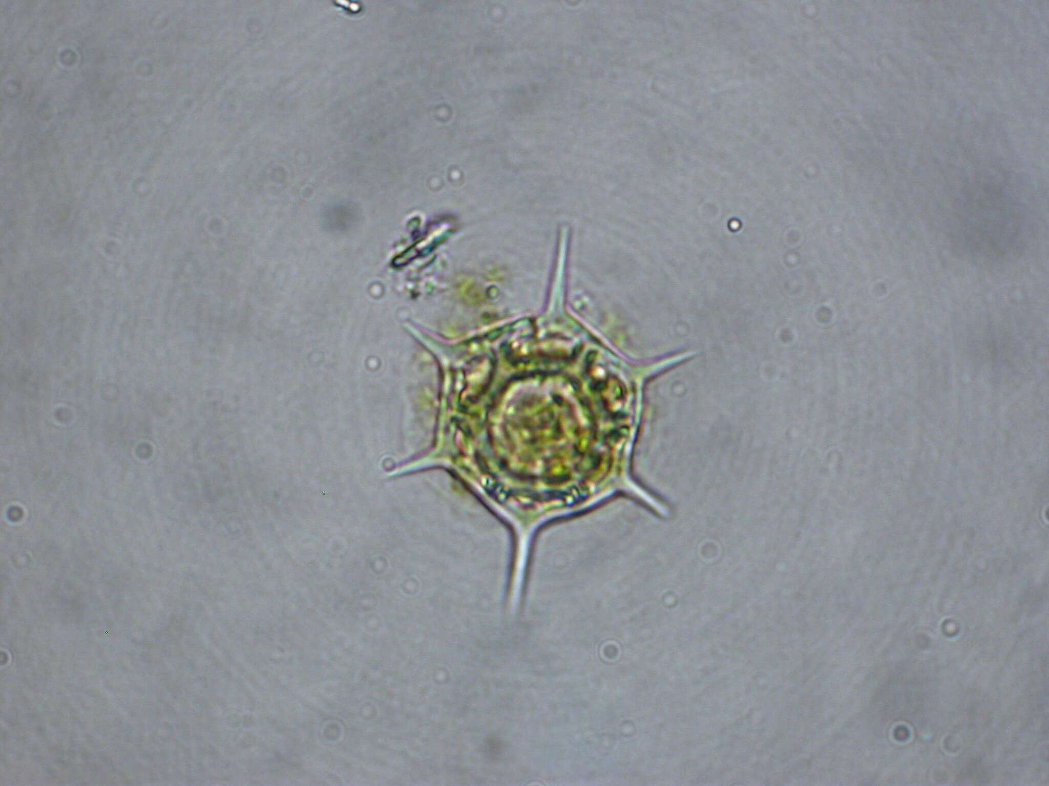
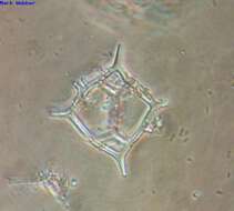
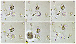
Ciliophrys infusionum Scale bar indicates 50 µm. Sample from wetland Lauchseemoor, Fieberbrunn, Tyrol, Austria. Sampling date 06/2023. The image was built up using several photomicrographic frames with manual stacking technique. Images were taken using Zeiss Axioplan with Olympus OM-D M5 MKII. Image under Creative Commons License V 3.0 (CC BY-NC-SA). Place name: Wetland Lauchseemoor, Fieberbrunn (Tyrol, Austria) Latitude: 47.46954439 Longitude: 12.53826499 Multiebenen-Abbildung, manuell gestapelt. Der Messbalken markiert eine Länge von 50 µm. Probe aus dem Lauchseemoor bei Fieberbrunn, Tirol. Datum der Aufsammlung: 06/2023. Mikrotechnik: Zeiss Axioplan, Kamera: Olympus OM-D M5 MKII. Creative Commons License V 3.0 (CC BY-NC-SA). For permission to use of (high-resolution) images please contact postmaster@protisten.de.
-
Reboredo, Galicia, Espaa
-
Armacao De Pera, Faro, Portugal
-
Armao de Pra, Faro, Portugal