-
Pra, Faro, Portugal
-
Ribadelago de Franco, Castille and Leon, Spain
-
Ribadelago de Franco, Castilla y Len, Espaa
-
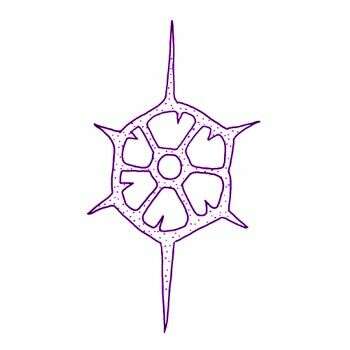
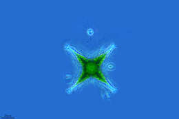
Distephanus speculum (Ehrenberg, 1837) Haeckel, 1899. Silicoflagellate about 18 microns with siliceous skeleton with six major sides, from the corners of which projects spines. Bars also arise inwards and may fuse to form an apical ring. The cytoplasm extends over the skeleton, fine pseudopodia usually project from the tops of the spines. Many small plastids, single emergent flagellum. Marine.
-
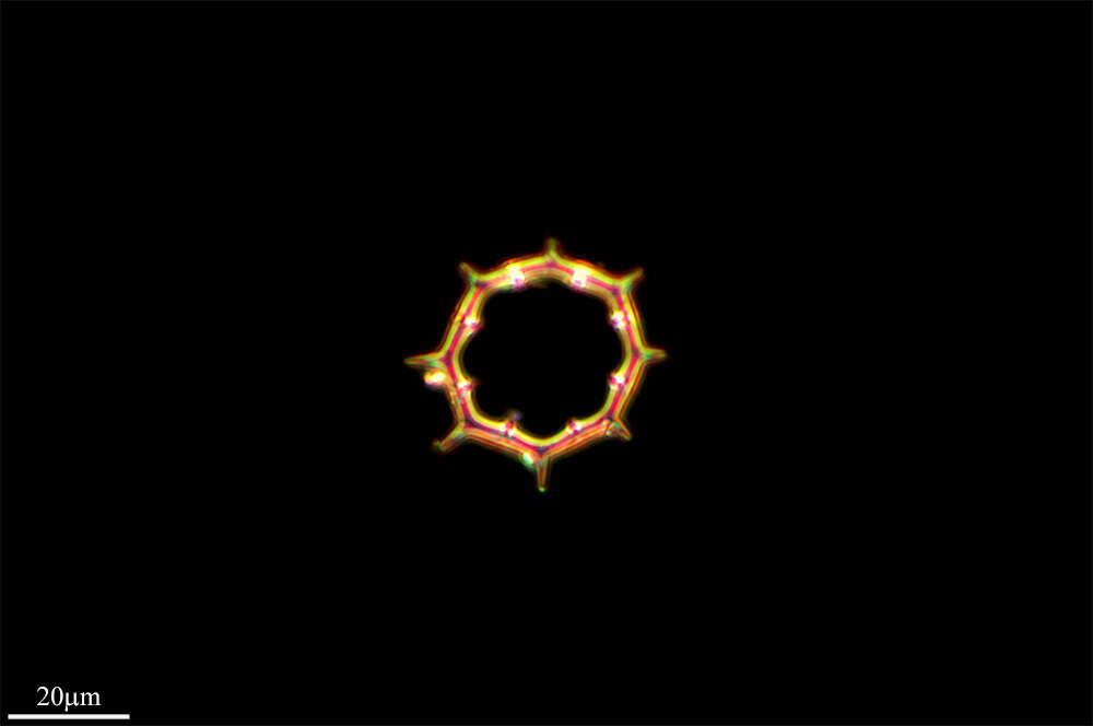
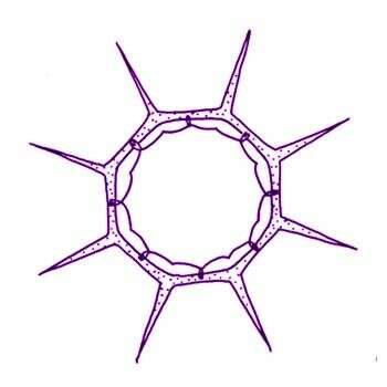
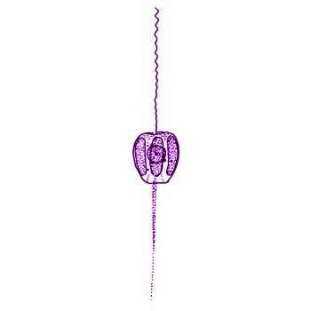
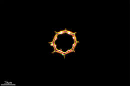
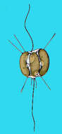
Distephanus speculum octonarius (Ehrenberg, 1844) Joergensen The skeleton is composed of an octagonal basal ring with eight radial spines projecting from the outer surface of the angles. A smaller apical ring, also octagonal, is held out from one side of the skeleton by eight supporting bars. A small protuberances is present mid-way along each of the eight sides of the apical ring.
-
Distephanus speculum bioctonarius (Ehrenberg) Silicoflagellate with siliceous skeleton with eight major sides, and from the corners of which projects eight spines, bars also arise inwards and form a delicate apical ring. Marine, from the Great Barrier Reef. Diameter of skeleton without spines, about 30 microns, spines are up to 13 microns long.
-
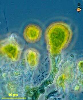
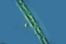
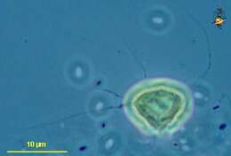
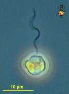
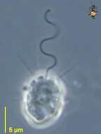
Rhizochromulina (rye-sew-crumb-you-line-a) marina, a dictyochophyte (stramenopile) alga with fine slightly stiffened radiating pseudopodia. Single cell. Phase contrast microscopy.
data on this strain.
-
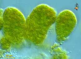
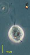
Rhizochromulina (rye-sew-crumb-you-line-a), a stramenopile flagellate with fine slightly stiffened radiating pseudopodia. Mass of cells. Differential interference microscopy.
data on this strain.
-
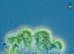
Rhizochromulina (rye-sew-crumb-you-line-a) marina, a dictyochophyte (stramenopile) alga with fine slightly stiffened radiating pseudopodia. Mass of cells. Phase contrast microscopy.
data on this strain.
-
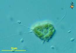
Rhizochromulina (rye-sew-crumb-you-line-a) marina, a dictyochophyte (stramenopile) alga with fine slightly stiffened radiating pseudopodia. Single cell showing plastids. Differential interference microscopy.
data on this strain.
-
Rhizochromulina (rye-sew-crumb-you-line-a) marina, a dictyochophyte (stramenopile) alga with fine slightly stiffened radiating pseudopodia. Phase contrast microscopy.
data on this strain.
-
-
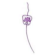
Pedinella hexacostata Vysotsky, 1888. Cells contain six chloroplasts arranged in a row around the equator of the cell.
-
Pseudopedinella (sue-dough-pea-don-ell-a) a dictyochophyte and pedinellid (stramenochrome) alga, related to the silica flagellates. This cell viewed from the apex and shows the 6 plastids forming a ring. This cell viewed from the side. Phase contrast microscopy.
data on this strain.
-
Pseudopedinella (sue-dough-pea-don-ell-a) a dictyochophyte and pedinellid (stramenochrome) alga, related to the silica flagellates. This cell viewed from the apex and shows the 6 plastids forming a ring. To a paid-up member of the NRA, this resembles six bullets in the cylinder of a revolver. This cell viewed from the apex. Phase contrast microscopy.
data on this strain.
-
Pseudopedinella pyriformis Carter, 1937. Cells are 5-8 microns long, and at anterior end 4-9 microns wide and at posterior end 3-6.5 microns wide with 6 chloroplasts. Flagellum 3-5 times longer than the cell. . Green flagellate
-
Pseudopedinella elastica Skuja, 1948. Cells are 14-17 m long, 13-14 m wide with chloroplasts. Flagellum about 3-6 times cell length. Green
-

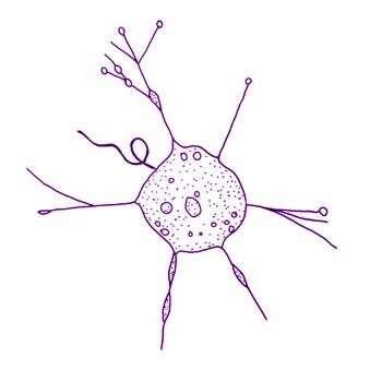
Apedinella radians (Lohmann) Campell. Cells are apple-shaped (7-10.5 microns wide, 6-9.5 microns long) with a single flagellum bearing mastigonemes emerging from an apical pit. Cells are radially symmetrical with six peripheral chloroplasts, each with a prominent pyrenoid on the innerside. The nucleus occupies a central position in the cell and is roughly spherical. Mitochondrial profiles run longitudinally through the cell between the chloroplasts, coalescing anteriorly to form one large mitochondrial reticulum which encircles the apical pit. In the anterior region of the cell the mitochondrial reticulum is closely associated with the filamentous cytoskeleton. A single dictyosome is located at the posterior region of the cell. The cells have two scale types: ovoid body scales and elongate spine-scales. The ovoid scales are of two size classes (0.6-0.8 x 0.4-0.5 microns and 1.5-2 x 1.1-1.3 microns) and cover the entire cell. Cells also bear six slender spine-scales, (9-13 microns long) which, according to Throndsen, are cellulosic. The spine-scales are evenly distributed around the lateral circumference and have their attachment points between the chloroplasts just anterior to the equator of the cell. Each spine-scale has a triangular base bearing a long tapering spine. The proximal side is concave with a central evagination at the base to which is attached an extracellular striated fibrous connector or micro ligament. The other two points of the triangular base appear to rest on the cell surface, that is, the covering of ovoid body scales. The microligament attaches the spine-scale to the plasma membrane. Plaques are located beneath the areas where the micro ligaments contact the plasma membrane. Each plaque consists of two discrete electron dense plates, the innermost being the thinnest, and has the following dimensions: height 0.25 microns, length 0.39 microns, width 0.07 microns The distal face of each plaque is somewhat concave following the contours of the plasma membrane.
-
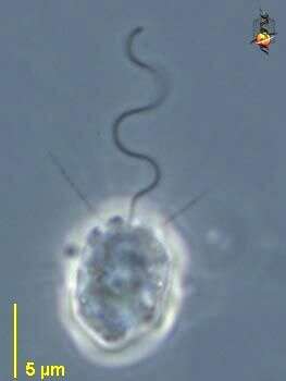
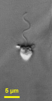
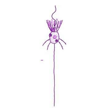
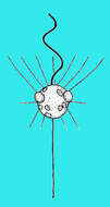
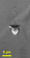
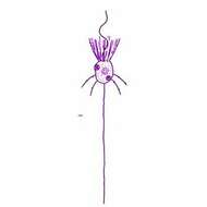
Actinomonas (act-in-o-moan-ass) is a colourless pedinellid, single anterior flagellum surrounded by a wreath of (12) arms, two of which can be seen to either side of the flagellum. Often with additional arms extending from other parts of the body. Not rare. very (very) similar to Pteridomonas. Flagellum photographically frozen in sine-wave beat pattern that is typical of stramenopiles. Phase contrast.
-
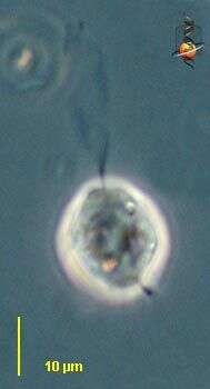
Actinomonas (act-in-o-moan-ass) is one of the pedinellid stramenopiles, a small flagellate with an apical flagellum (upper) and stalk (lower). Usually with a number of fine arms around the flagellum. These arms intercept bacteria and other suspended particles. They may also be withdrawn, as is the case here. The flagellum has not been frozen by the photography and what we see is the beat envelope. Phase contrast.
-
-
Actinomonas (ack-tin-owe-moan-ass) mirabilis is to all intents and purposes indistinguishable from Pteridomonas danica (there are small ultrastructural differences). Cells are 4 to 6 microns long and have one flagellum emerging from a small depression in the anterior end of the cell. The cells have a ring of arms around the flagellum and below the equator of the cell, the arms around the flagellum are evenly spaced. The anterior part of the cell is slightly broader than the posterior part. The single thickened flagellum is about 3 - 5 times the cell length and has an undulating beat. The cells usually swim rapidly, but occasionally attach to the substrate with a long posterior stalk trailing. Small particles are seen on the cell surface. Sometimes commonly observed.
-
Actinomonas mirabilis Kent, 1880. Colourless pedinellid, cell body measuring 5-8 microns, and often stalked. Arms present, up to two rings of 12 arms each around the anterior flagellum and extending from other parts of the body. When disturbed may swim with arms trailing from posterior.
-
Actinomonas vernalis Stokes, 1885. Cells subspherical, the frontal border slightly emarginate, somewhat changeable in shape, free-swimming or temporarily attached by a short pedicel, flagellum active throughout its length. The flagellum equals or somewhat exceeds the diameter of the body in length, cytoplasm transparent, slightly granular, pseudopodia few in number, radiating from any part of the periphery, simple or variously branched, often capitate, sometimes curved, their length exceeding the diameter of the body, several small contractile vacuoles distributed near the periphery, nucleus spherical, subcentral. Diameter of body 16.9-21.2 microns Status of taxon dubious.