-
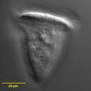
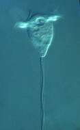
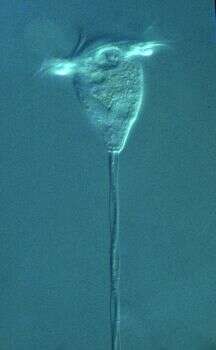
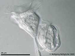
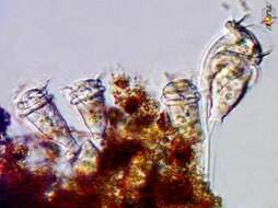
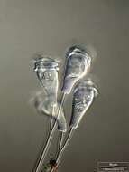
Late state of binary fission of Vorticella. The adoral membranelles are retracted and the new cell (the swarmer) starts to develop a collar of cilia for locomotion. The dots in the stalk are mitochondria, lying apon the spasmoneme, the "muscle" of the stalk. The mitochondria support relaxation after contraction of the stalk via spasmoneme. Sample from a pond on the island of Hiddensee (Baltic Sea, Germany). This image was taken using Zeiss Universal with Olympus C7070 CCD camera.
-
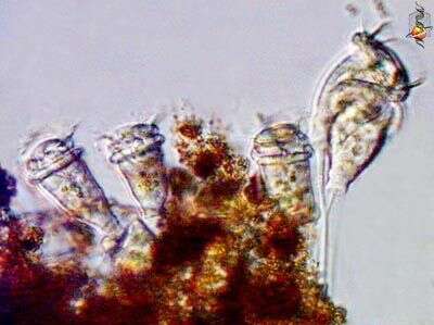
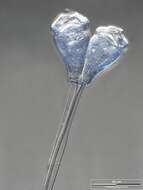
Late state of binary fission of Vorticella. The swarmer has developed a collar of cilia for locomotion. Moment shortly before the swarmer disconnected itself from the stalk of mother cell. Sample from a pond on the island of Hiddensee (Baltic Sea, Germany). This image was taken using Zeiss Universal with Olympus C7070 CCD camera.
-
-
-
-
-
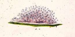
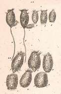
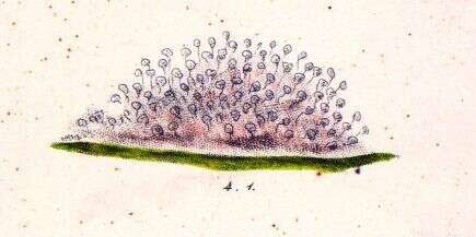
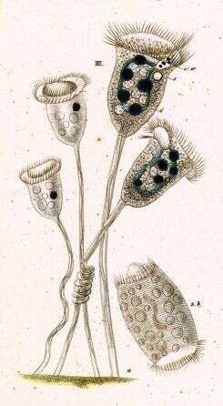
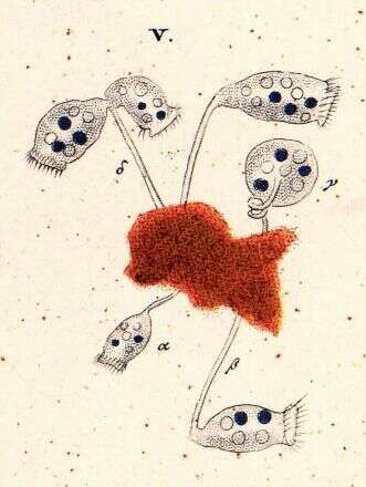
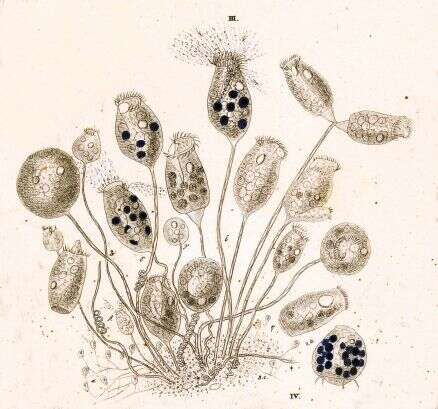
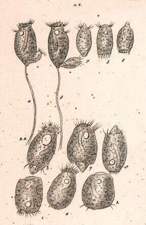
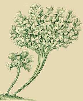
Vorticella convallaria.
-
The heros of the process are the peritrich ciliates, like this Vorticella. Primarily through their efforts, the coliform and other suspended bacteria are filtered out of the sewage in the activated sludge tank. If the rate of throughout is increased to a rate where the growth of peritrichs is insufficient to keep up, then the ciliates are removed and the quality of the effluent plummets.
-
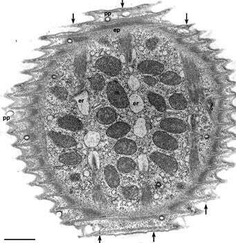
Electron micrograph of the surface of Vorticella convallaria. Like other peritrichs the pellicle is sculpted into ridges and grooves that circle the cell perpendicular to the aboral/adoral axis of the cell body. Pellicular pores (pp) penetrate the pellicle as cylindrical indentations of the plasma membrane (see Fig.3) that end as clathrin-coated pits. A system of alveoli underlies the plasma membrane and rod-like densities occupy the cytosolic tips of the pellicular ridges. A fibrous epiplasm (ep) covers the inside of the alveoli. Distinct myonemal bands run in the aboral to adoral direction (indicated by arrows) just inside the epiplasm. These bands are associated with the endoplasmic reticulum (er) by unique specializations of the ER (see Fig 17). In this EM preparation the ER has artifactually segmented into vesicles caused by the fixation process. Mitochondria (m) occupy the space between the bands. EM taken on 4/15/71 by R. Allen with Hitachi HU11A TEM. Neg. 9,250X. Bar = 1 micron.
This image is available in Richard Allen's collection.
-
-
-
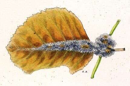
Phase contrast micrograph of a cell associated with detritus that attached to a submerged slide in the Lake.
-
-
-
-
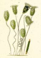
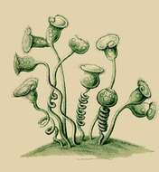
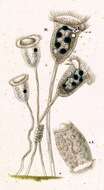
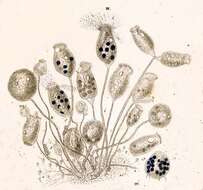
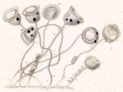
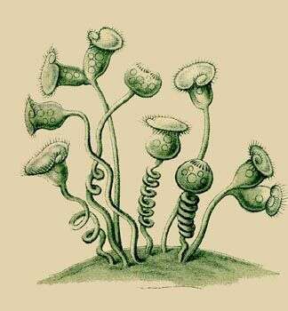
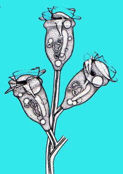
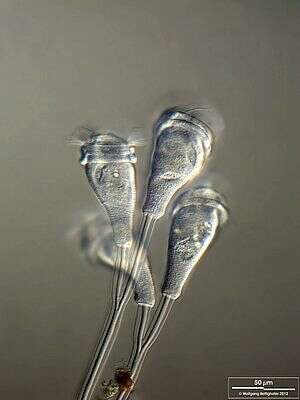
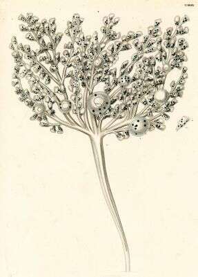
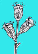
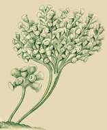
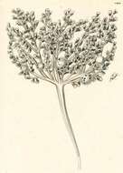
Zoothamnium (zoo-tham-knee-um) is a colonial peritrich ciliate. The feeding cells in sessile peritrich ciliates have lost all of the somatic cilia and only have the feeding cilia. The feeding cilia form a wreath which extends around the front of the cell and descends into a narrowing buccal cavity. This cavity ends at the cytostome where food is packaged into food vacuoles. If the cells become unhappy, they produce a temporary wreath of basal cilia (trochal cilia), break away from their stalk and use these to swim. The contractile elements of all associated cells of Zoothamnium colonies are interconnected so that if one cell contracts, all will tend to contract together. Differential interference contrast.
-
-
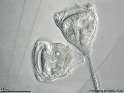
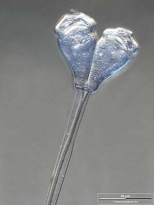
Small colony, two cells, attached to long stalk. The contractile spasmoneme is common to both cells.
-
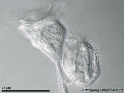
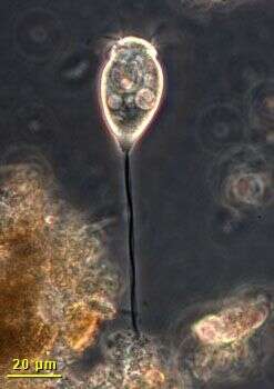
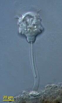
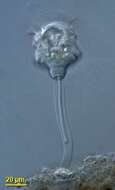
This is a single cell of the usually colonial Zoothamnium, showing the contractile spasmoneme within the stalk.
-
Scale bar indicates 50 µm. Collected from Bodden, the brackish waters lying between the isles of Hiddensee and Ruegen (German Baltic Sea). The image was built up using several photomicrographic frames with manual stacking technique. This image was taken using Zeiss Universal with Olympus C7070 CCD camera.
-
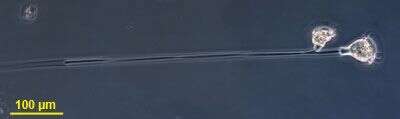
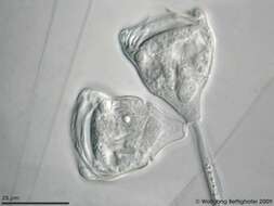
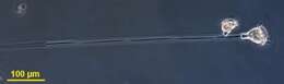
Cells shortly after binary fission. Scale bar indicates 100 µm. Collected from Bodden, the brackish waters lying between the isles of Hiddensee and Ruegen (German Baltic Sea). The image was built up using several photomicrographic frames with manual stacking technique. This image was taken using Zeiss Universal with Olympus C7070 CCD camera.
-
-
-